Introduction
Lung cancer, which originates from the lung,
represents an aggressive and life-threatening type of cancer
worldwide. In 2017, lung cancer was the primary cause of
cancer-associated mortality in the USA (1). Similarly, in 2015, lung cancer was the
primary cause of cancer-associated death in men aged >75 years
in China (2). Therefore, development
of a therapy for lung cancer is urgently required to improve human
health. Although substantial achievements have been made in lung
cancer therapy, treatment failure and decreased survival rates are
often encountered, owing to progression, recurrence, metastasis and
multi-drug resistance of lung cancer (3,4). Lung
cancer initiating cells are a specific subpopulation of lung cancer
cells, which contribute to the progression, recurrence, metastasis,
and multi-drug resistance of lung cancer (3,4).
Therefore, targeting and eliminating lung cancer initiating cells
is likely to contribute to curing lung cancer from the origin.
CD133 is a marker for lung cancer initiating cells
(5). Bertolini et al
(6), showed that CD133+
lung cancer cells isolated from lung cancer tissues showed
significantly increased aggressive properties compared with their
counterparts, CD133− lung cancer cells, as reflected by
their increased proliferative, clonogenic and tumorigenic
properties. All-trans retinoic acid (RA), an active metabolite of
vitamin A under the family retinoid, is a promising drug that can
cause the differentiation, proliferation inhibition and apoptosis
of cancer cells in various types of cancer (7,8). An
RA-based differentiation therapy is regarded as a significant
advance in cancer therapy, and RA has become the first choice drug
for the treatment of acute promyelocytic leukemia (APL) (7). RA has also been demonstrated to be
effective in treating APL as an adjuvant (8). In addition, RA has shown therapeutic
potential against cancer initiating cells in several types of
cancer, including breast cancer (9–11).
Although RA has been reported to exert promising
therapeutic effects against lung cancer in previous studies, there
have been no reports on the therapeutic effect of RA on lung cancer
initiating cells (12–14). Furthermore, the aqueous solubility of
RA is poor, resulting in it being a less promising candidate drug
with low bioavailability and poor therapeutic effect in vivo
(9). It is known that
nanoparticle-based strategies can markedly improve the
bioavailability and therapeutic index of conventional therapeutics,
by improving the solubility of poorly soluble drugs and providing
targeted delivery of drugs (15–17).
Several studies have developed RA-loaded nanoparticles to
facilitate the preclinical application of RA in cancer therapy
(9,18). In these studies, the solubility and
bioavailability of RA were markedly increased, and the RA-loaded
nanoparticles exhibited superior therapeutic efficacy against
cancer compared with that of RA (9,18).
Lipid-polymer hybrid nanoparticles of biodegradable
polymers and lipids represent superior candidate drug delivery
systems, as they combine the advantages of liposomes and polymer
nanoparticles (19,20). Liposomes feature superior
biocompatibility, and easy modification of the hydrophilic polymer,
and targeting molecules including aptamers (21,22). The
advantages of polymer nanoparticles include controlled and
sustained release, high drug loading and superior stability
(19,20). Therefore, the advantages of
lipid-polymer hybrid nanoparticles include superior
biocompatibility and stability, easy modification, and controlled
and sustained release (19).
To promote the efficacy of chemotherapy drugs to
cancer cells, considerable interest has been paid to
aptamer-targeted nanoparticles (21–23). It
is well-known that aptamer-targeted nanoparticles have improved the
therapeutic effect of chemotherapy in various types of cancer
(24,25). As CD133 is generally accepted as a
marker of lung cancer initiating cells, it was hypothesized in the
present study that CD133 aptamers can be used to promote the
delivery of RA-loaded nanoparticles to lung cancer initiating
cells. The purpose of the present study was to target lung cancer
initiating cells through the construction of RA-loaded lipid-PLGA
nanoparticles with CD133 aptamers (RA-LPNPs-CD133).
Materials and methods
Culture of H446 and A549 lung cancer
cell lines
H446 and A549 cells are two human lung cancer cell
lines. These cells were purchased from American Type Culture
Collection (Manassas, VA, USA) and maintained at 37°C under 5%
CO2 in Roswell Park Memorial Institute 1640 (RMPI-1640)
medium (HyClone; GE Healthcare Life Sciences, Logan, UT, USA). The
RPMI-1640 medium was supplemented with 10% fetal bovine serum
(FBS), streptomycin (100 µg/ml), and penicillin (100 U/ml). The
cell culture medium containing 10% FBS was replaced at 3–4 day
intervals to ensure the cell growth was optimal.
Reagents and kits
Poly(DL-lactide-co-glycolide) (PLGA) with a molar
ratio of lactide:glycolide of 50:50 (40–75 kDa), polyvinyl alcohol
(PVA; 30–70 kDa), RA and organic reagents were all purchased from
Sigma-Aldrich; EMD Millipore (Billerica, MA, USA). The lipids,
including [(1,2-distearoyl-sn-glycero-3-
phosphoethanolamine-N-maleimide- polyethylene glycol-2000;
DSPE-PEG-Mal,
1,2-dioleoyl-sn-glycero-3-phosphoethanolamine-N-carboxyfluorescein;
PECF)], phosphatidylcholine, and cholesterol were purchased from
Avanti Polar Lipids (Alabaster, AL, USA). The CD133 MicroBead kit
was provided by Miltenyi Biotec, Inc. (Shanghai, China). The
ultra-low attachment surface 6-well dishes were purchased from
Corning Life Sciences (Tewksbury, MA, USA). R&D Systems, Inc.
(Minneapolis, MN, USA) provided recombinant anti-human CD133 Alexa
Fluor® 488-conjugated antibody (cat. no. FAB11331G). The
synthesis of CD133 aptamers with the sequence of
5′-SH-CCCUCCUACAUAGGG-3′ was performed by Ruibo Co., Ltd.
(Guangzhou, China). The Pierce BCA Protein Assay kit, FBS, B27,
epidermal growth factor (EGF), basic fibroblast growth factor
(bFGF), insulin-transferrin-selenium (ITS),
4′,6′-diamidino-2-phenylindole dihydrochloride (DAPI) and the cell
dissociation reagent (StemPro® Accutase®)
were purchased from Thermo Fisher Scientific, Inc. (Waltham, MA,
USA).
Expression of CD133 in lung cancer
cell lines
The flow cytometry-based approach was used to
perform the analysis of CD133 in lung cancer cell lines. In short,
the lung cancer cells were dissociated into single cells, and the
dissociated cells were treated with the fluorescent antibody
(anti-human CD133 Alexa Fluor® 488-conjugated antibody
at 1 µg/ml) for 0.5 h at 4°C. The cells were then washed three
times with phosphate-buffered saline (PBS) to wash away
unconjugated fluorescent antibody. At the end of the assay, the
washed cells were suspended in PBS, and a FACSCalibur flow
cytometer (FCM; BD Biosciences, Franklin Lakes, NJ, USA) was used
to analyze the proportion of positively stained cells and the mean
fluorescence intensity of the cells.
Magnetic cell sorting-based
separation
As the expression of CD133 in lung cancer cell lines
was low, magnetic cell sorting-based separation was performed to
separate the CD133+ cells from lung cancer cells
according to the manufacturer's protocol provided with the CD133
MicroBead kit from Shanghai Miltenyi Biotec, Inc. Following
separation, the FACSCalibur FCM was used to analyze the proportion
of positively stained cells, as described above.
Formation of tumorspheres of lung
cancer cells
Tumorsphere formation of the lung cancer cells was
assessed to evaluate the self-renewal ability of the lung cancer
initiating cells when the single cells were suspended in serum-free
medium. In short, lung cancer cells suspended in stem cell medium
were cultured in Corning® ultra-low attachment surface
6-well dishes. The density of the suspended cells was 5,000
cells/well, and the components of the stem cell medium included
DMEM/F12, B27 (1×), ITS (1X), EGF and bFGF (both cytokines were at
a concentration of 20 ng/ml). The cells were cultured in the stem
cell medium for 7 days at 37°C, following which the tumorspheres
were counted under an Olympus CKX41 conventional light microscope.
For the second-passage tumorspheres, those of the first passage
were washed with PBS and dissociated with a cell dissociation
reagent (StemPro® Accutase®), and then
propagated.
Tumorigenicity of lung cancer cells in
mice
The tumorigenicity assay was performed by the
inoculation of lung cancer cells into male BALB/c nude mice (aged
4–5 weeks old, ~20 g; 6 per group; 240 mice in total). The mice
were obtained from the Shanghai Experimental Animal Center
(Shanghai, China). Following delivery of mice, the mice were raised
in a clean environment without pathogens, and were acclimated for
~7 days. Animals were housed in separate cages (3–4 animals per
cage) and maintained under a controlled atmosphere (humidity,
50±7%; temperature, 21±1°C) and with a 12 h light/dark cycle. Mice
were allowed free access to food and water. All animal procedures
were performed in line with the regulations of the Animal
Administrative Committee of the Second Military Medical University
(Shanghai, China). In brief, CD133+ and
CD133− lung cancer cells were isolated using the
magnetic bead-based approach. The collected cells were then mixed
with BD Matrigel™. The mixture of cells
(2×10⁷−2×10⁴/ml) and gel (0.2 ml per mouse)
was implanted subcutaneously into the right flank of mice. The
tumor formation was recorded during an observation period of 15
weeks.
Fabrication and characteristics of
RA-loaded lipid-PLGA nanoparticles
The RA-loaded lipid-PLGA nanoparticles were
fabricated as described below. In brief, 0.5 mg RA and 5 mg PLGA
were completely dissolved in acetone to form an oil phase. The oil
solution was injected into 2% PVA solution, followed by
homogenization. The mini-emulsion was then poured into 0.2% PVA
solution, and rapidly mixed for 6 h to remove remaining acetone by
evaporation. Subsequently, the nanoparticles were recovered using
ultracentrifugation (80,000 g) for 30 min at 25°C. At the same
time, a lipid film composed of phosphatidylcholine, DSPE-PEG-Mal
and cholesterol (57:3:40 molar ratio) was formed in a round bottom
flask with a vacuum rotary evaporator. When the lipid film was
formed, the prepared nanoparticles were added to hydrate the film.
A hand held extruder (Avanti Polar Lipids) with 200-nm membranes
was used to extrude the obtained lipid-polymer suspension to
produce small and homogeneous nanoparticles. The resultant
lipid-polymer nanoparticles were washed by centrifugation at a
speed of 3,000 × g for 30 min at 25°C with Amicon centrifugal
filters (MWCO 100 kDa) with distilled water. The CD133 aptamers
with thiol groups were incubated with the nanoparticles (molar
ratio of aptamers to DSPE-PEG-Mal of 1:2) for 6 h at room
temperature, with the aim of conjugating the aptamers with thiol
groups to nanoparticles. Subsequently, the unconjugated aptamers
were removed using Amicon centrifugal filters [molecular weight
cut-off (MWCO) 100 kDa]. Nontargeted nanoparticles were developed
using the method described above but without the addition of
aptamers. Blank nanoparticles were also developed using the method
described above without the initial addition of RA. The fluorescent
PECF-labeled nanoparticles were established in a similar manner but
with 0.1% (molar ratio) of PECF added to the lipid film.
The nanoparticles were as follows: RA-loaded
lipid-PLGA nanoparticles (RA-LPNPs); RA-loaded lipid-PLGA
nanoparticles with CD133 aptamers (RA-LPNPs-CD133); blank
lipid-PLGA nanoparticles with CD133 aptamers (LPNPs-CD133).
Aptamer conjugation efficiency of
lipid-PLGA nanoparticles
The aptamer conjugation efficiency of the prepared
nanoparticles was defined as the quantity of conjugated aptamers on
the nanoparticles relative to the total quantity of added aptamers
in the preparation procedure. A simple and rapid
ultrafiltration-based approach was used to evaluate the aptamer
conjugation efficiency of the prepared nanoparticles. Briefly, the
aptamers were incubated with the nanoparticles, and the mixture of
aptamers/nanoparticles was centrifuged with the Amicon centrifugal
filters (MWCO 100 kDa) at 3,000 × g for 30 min at 25°C to remove
the unconjugated aptamers. The unconjugated aptamers were measured
using UV at 260 nm. Finally, the aptamer conjugation efficiency of
the nanoparticles was evaluated using the following equation:
(Mt-Mu)/Mt, where Mt is
the mass of total aptamers and MU is the mass of
unconjugated aptamers.
Size, ζ-potential, morphology and drug
loading of lipid-PLGA nanoparticles
The particle size and ζ-potential were evaluated
using a Zetasizer Nano ZS90 instrument (Malvern Instruments, Ltd.,
Malvern, UK), following the diluting 100 µl of nanoparticles in 1.9
ml of pure water. Furthermore, the morphology of the nanoparticles
was investigated using transmission electron microscopy (TEM) with
the Hitachi H-600 microscope (Hitachi, Ltd., Tokyo, Japan) as
described below. The nanoparticles were negatively stained with 2%
phosphotungstic acid and dried on mesh copper TEM grids. The
nanoparticles were then observed under TEM. The RA encapsulation
efficiency and loading of the nanoparticles were determined by
reverse-phased high performance liquid chromatography (HPLC) with
an ODS column (Diamonsil®; packing carriers: 5 µm;
length and width: 250×4.6 mm). The nanoparticles (5 mg) were fully
dissolved in 1 ml dichloromethane. The dichloromethane solution was
evaporated in a vacuum evaporator and 10 ml methanol was added to
form a clear solution sample for HPLC. The analysis was performed
with the Agilent HPLC system (Agilent Technologies, Inc., Cotati,
CA, USA). The mobile phase was acetic acid/acetonitrile/water
(0.5/95/4.5, volume/volume), and the flow rate was fixed at 1.0
ml/min. The detection wavelength of RA was set at 350 nm.
RA release from nanoparticles
The release of RA from lipid-PLGA nanoparticles was
evaluated as described below. First, the lipid-PLGA nanoparticles
at a concentration of 1 mg/ml were suspended in PBS or PBS with 10%
FBS in a centrifuge tube. The nanoparticles were then placed on an
orbital shaker at 37°C with gentle agitation at a speed of 80 × g.
Subsequently, at different designated time points, the centrifuge
tubes were centrifuged at 15,000 × g for 30 min at 25°C. The
supernatant was then removed from the centrifuge tubes, and
measured by HPLC as described above. The cumulative RA release rate
from the nanoparticles was calculated using the following formula:
(Mi/Mt) ×100%, where Mi is the
mass of released RA, and Mt is the total mass of RA.
In vitro targeting of fluorescent
nanoparticles to lung cancer cells
The in vitro targeting of fluorescent
nanoparticles to lung cancer cells was evaluated by flow cytometry.
Briefly, the lung cancer cells were isolated as described above,
and were then cultured on 12-well cell culture plates overnight at
37°C. The density of lung cancer cells was 5×10⁵ cells
per well. The medium was then replaced with a fresh medium
containing PECF or PECF-labeled nanoparticles (1 µg/ml PECF). After
2 h at 37°C, the cells were washed with PBS to remove unbound
nanoparticles, and dissociated into single cells. The cells were
suspended in PBS and analyzed using a FACSCalibur FCM.
Fluorescent immunohistochemistry
The lung cancer cells were isolated using the
magnetic cell sorting-based separation method described above.
Subsequently, the cells were seeded on coverslips in 12-well tissue
culture plates overnight. The cells were seeded at a density of
5×10⁵ cells per well. Following overnight incubation,
the medium was replaced with fresh cell medium containing PECF or
PECF-labeled nanoparticles (1 µg/ml PECF). After 4 h, the cell
medium was aspirated and removed, and the cells were washed with
PBST (PBS containing 0.1% Tween-20) twice. Following washing with
PBST, 4% paraformaldehyde was added to fix the cells for 20 min at
4°C. The cells were then incubated with the nuclear staining
reagent DAPI. Finally, the Carl Zeiss LSM510 confocal microscope
(Carl Zeiss AG, Oberkochen, Germany) was used to visualize the
immunofluorescence of the cells. The blue fluorescence of DAPI and
the green fluorescence of PECF were observed and merged.
Cytotoxic effects of nanoparticles on
lung cancer cell lines
The cytotoxic effects of nanoparticles were examined
using the Cell Counting Kit-8 (CCK-8) assay. Briefly, the lung
cancer cells were washed, trypsinized, and seeded at a density of
4×10³ cells/well in 96-well cell culture plates
overnight. Following overnight incubation, the medium was replaced
with a fresh medium containing RA or nanoparticles at a series of
concentrations (0.04, 0.13, 0.41, 1.23, 3.7, 11.1, 33.3, 100.0,
300.0 and 900.0 µg/ml) at 37°C. After 72 h, the medium was replaced
with fresh medium. The cell viability was examined using the CCK-8
assay with a microplate reader (Multiskan MK3). Finally, the data
was processed using GraphPad Prism 5.0 (GraphPad Software, Inc., La
Jolla, CA, USA) to calculate the IC50 values.
Impact of nanoparticles on the
proportion of lung cancer initiating cells
Tumorsphere formation and the proportion of
CD133+ cells were examined to evaluate the impact of
nanoparticles on the proportion of lung cancer initiating cells in
the lung cancer cell population. In brief, the lung cancer cells
were washed, trypsinized into single cells, and inoculated
overnight in 12-well cell culture plates. The density of lung
cancer cells was 5×10⁴ cells per well. Following
overnight incubation, the removal of old cell culture medium was
performed by aspiration and washing with PBS. Following washing,
the cells were treated with fresh medium dissolved with the
nanoparticles (at a concentration equivalent to 15 µg/ml RA). After
24 h, removal of the old medium was performed by aspiration, and
the cells were washed. Fresh medium was then added to the cells and
incubated for 72 h. The cells were then washed and trypsinized to
single cells, and the formation of tumorspheres measured as
described in the above section. Alternatively, flow cytometry was
performed to measure the percentage of CD133+ cells of
the trypsinized cells.
Statistical analysis
The differences between two groups was measured with
Student's non-paired t-test, and the differences among three or
more groups were measured by one-way analysis of variance, and
Newman-Keuls post hoc test was used. P<0.05 was considered to
indicate a statistically significant difference. All data are
presented as the mean ± standard deviation, unless otherwise
stated.
Results
Fabrication and characterization of
lipid-PLGA nanoparticles
The RA-loaded lipid-PLGA nanoparticles were
fabricated in two steps. In the first step, PLGA nanoparticles were
initially developed using the emulsion/solvent evaporation
approach. In the second step, the prepared PLGA nanoparticles were
coated with lipid-based film to form the RA-loaded lipid-PLGA
nanoparticles. Finally, the thiolated aptamers were conjugated to
the lipid-PLGA nanoparticles via the reaction of sulfydryl groups
on the aptamers with maleimide groups of the lipid-PLGA
nanoparticles. The size, ζ-potential and drug loading of
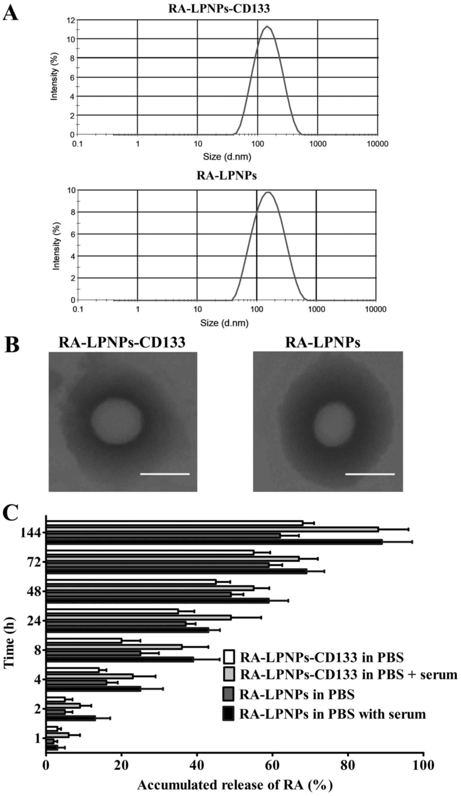
nanoparticles are shown in Table I.
The RA-LPNPs, as nanoparticles without conjugated aptamers, had a
small size of 123.8 nm. The conjugation of aptamers did not
significantly alter the size of the nanoparticles, as reflected by
the fact that the RA-LPNPs-CD133 and LPNPs-CD133 had sizes of 129.9
and 122.5 nm, respectively. The ζ-potential of the nanoparticles
was negative at approximately-18 mV. The drug loading of RA-LPNPs
and RA-LPNPs-CD133 was 10.2 and 9.8%, respectively. The
encapsulation efficiency of RA-LPNPs and RA-LPNPs-CD133 was 82.2
and 83.2%, respectively. The conjugation efficiency of aptamers on
RA-LPNPs-CD133 was 25%. The size distribution of the nanoparticles
is shown in Fig. 1A. On being
negatively stained with phosphotungstic acid, TEM showed that the
RA-LPNPs and RA-LPNPs-CD133 were spherical in shape (Fig. 1B). The RA release assay, shown in
Fig. 1C, indicated that all
nanoparticles exhibited sustained release of RA during the 144-h
period. The RA release of all nanoparticles was markedly higher in
the PBS + serum (10% FBS) group, compared with that in the PBS
group (P<0.05), indicating that serum destabilized the
nanoparticles and facilitated the release of RA.
 | Table I.Characteristics of nanoparticles. |
Table I.
Characteristics of nanoparticles.
| Nanoparticle | Size (nm) | ζ-potential
(mv) | PDI | Drug loading
(%) | EE (%) |
|---|
| RA-LPNPs | 123.8±15.2 | −18.8±5.6 | 0.14±0.04 | 10.2±4.8 | 82.2±6.5 |
| RA-LPNPs-CD133 | 129.9±13.2 | −19.7±5.5 | 0.15±0.05 | 9.8±5.9 | 83.2±9.4 |
| LPNPs-CD133 | 122.5±16.6 | −17.3±4.9 | 0.16±0.07 | – | – |
CD133+ subpopulation of
lung cancer cells exhibit properties of lung cancer initiating
cells
The isolation of CD133+ cells was
performed using the CD133 MicroBead kit for cell sorting, and the
results showed that a high percentage of CD133+ cells
(>99%) was obtained. Tumorsphere formation is a common approach
to identify the self-renewal ability of cancer initiating cells
(16). As shown in Fig. 2A, the tumorsphere number of
CD13+ H446 cells was significantly higher than that of
CD133− H446 cells (1st passage: P<0.01; 2nd passage:
P<0.001). In the A549 cells, similar results were achieved (1st
passage: P<0.01; 2nd passage: P<0.001; Fig. 2B).
The in vivo tumorigenicity assay showed that
CD133+ cells had markedly enhanced capacity in terms of
lung cancer formation compared with CD133− cells
(Fig. 2C and D). Compared with the
CD133− cells, the tumor volume derived from
CD133+ cells was significantly larger at day 28 for the
H446 cells and at day 24 for the A549 cells (P<0.05). The
tumorigenicity in mice also showed that CD133+ cells had
markedly enhanced capacity in terms of lung cancer formation
(Fig. 2E). Notably, a 100% incidence
of tumors (10/10) in mice was found in CD133+ H446 cells
with a cell count ≥2×10⁴ cells. By contrast, only a 60%
incidence of tumors (6/10) in mice was found in CD133−
H446 cells, even the cell number was 2×10⁶, indicating
that CD133+ H446 cells had significantly increased
tumorigenic potential in comparison with CD133− H446
cells. Similarly, CD133+ A549 cells had significantly
increased tumorigenic ability in comparison with CD133−
A549 cells. The CD133+ A549 cells produced tumors in
mice with a 100% incidence when the cell number was
≥2×10⁴ cells, whereas CD133− A549 cells only
produced tumors with a 20% incidence (2/10) at 2×10⁴
CD133− A549 cells. Taken together, the tumorigenicity of
CD133+ lung cancer cells was markedly higher than that
of CD133− lung cancer cells.
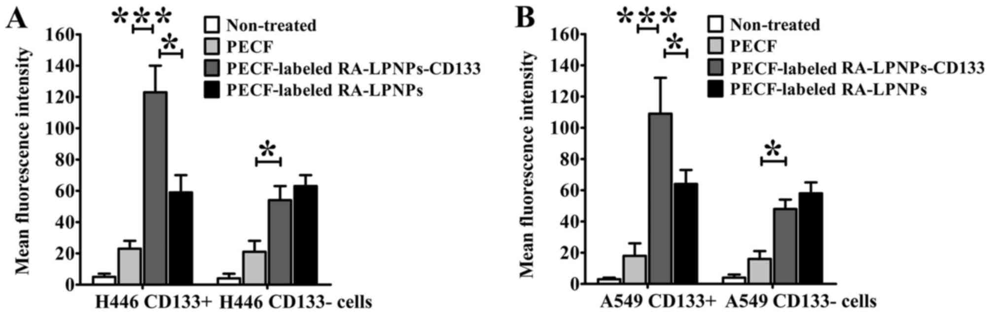
In vitro targeting and uptake of
fluorescent nanoparticles in lung cancer cells
The green fluorescent lipid, PECF, is widely used in
the evaluation of the cellular uptake of nanoparticles. As shown in
Fig. 3A, the uptake of PECF-labeled
RA-LPNPs-CD133 in the H446 CD133+ cells was prominently
higher compared with that of PECF (P<0.001) and PECF-labeled
RA-LPNPs (P<0.05). However, in the H446 CD133− cells,
uptake of PECF-labeled RA-LPNPs-CD133 was similar to that of
PECF-labeled RA-LPNPs, although uptake was increased compared with
that of free PECF (P<0.05). For the A549 cells, similar results
were observed (Fig. 3B). The
PECF-labeled RA-LPNPs-CD133 showed increased uptake compared with
the PECF-labeled RA-LPNPs (P<0.05) and free PECF (P<0.001) in
the A549 CD133+ cells, with similar uptake of
PECF-labeled RA-LPNPs in A549 CD133− cells.
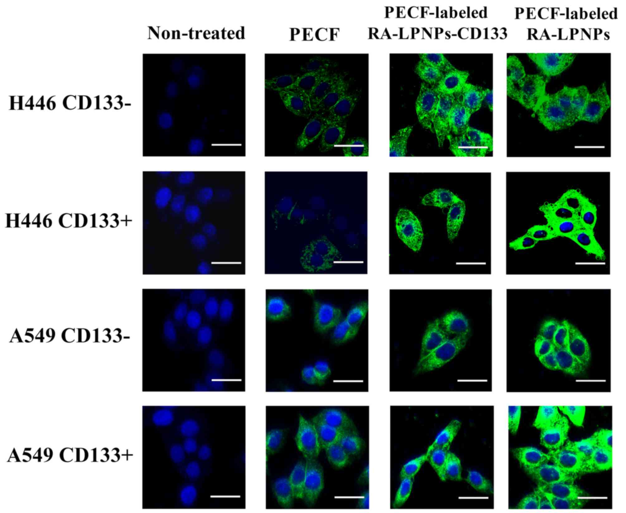
The in vitro cellular uptake of PECF-labeled
nanoparticles was also evaluated by confocal microscopy. As shown
in Fig. 4, H446 CD133+
cells treated with PECF labeled RA-LPNPs-CD133 showed significant
internalization of nanoparticles. By contrast, non-targeted
PECF-labeled RA-LPNPs showed no significant internalization in H446
CD133+ cells, as reflected by the reduced green
fluorescence intensity compared with PECF-labeled RA-LPNPs-CD133.
As expected, PECF exhibited the lowest green fluorescence
intensity, suggesting that nanoparticles markedly facilitated the
uptake of free drugs. In the H446 CD133− cells,
PECF-labeled RA-LPNPs-CD133 did not differ in green fluorescence
intensity from PECF-labeled RA-LPNPs. Similar results were obtained
in A549 cells. Taken together, PECF-labeled RA-LPNPs-CD133 showed
specific increased uptake in CD133+ lung cancer
cells.
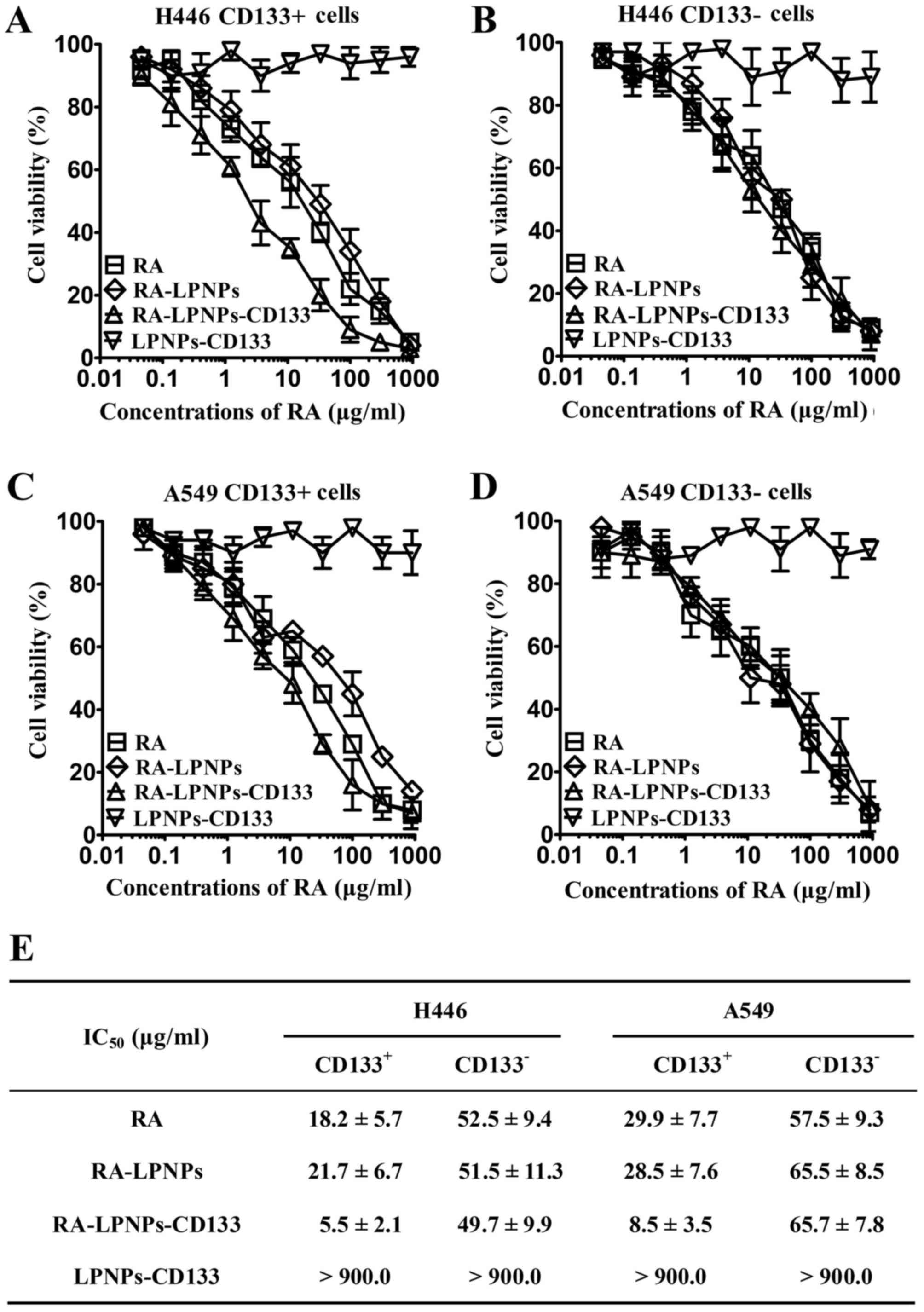
Cytotoxic effects of RA and various
nanoparticles against lung cancer cells
As shown in Fig.
5A-D, LPNPs-CD133, the blank lipid-PLGA nanoparticles with
CD133 aptamers, exhibited no marked cytotoxic effects towards lung
cancer cells, as reflected by the almost horizontal curve induced
by LPNPs-CD133. By contrast, dose-dependent cytotoxicity was
observed for RA, RA-LPNPs and RA-LPNPs-CD133, as reflected by their
dose-dependent curves of inverse sigmoid. The IC50
values of the RA and other nanoparticles are listed in Fig. 5E. In the CD133+ H446
cells, RA-LPNPs exhibited similar cytotoxic effect as RA (21.7
µg/ml for RA-LPNPs, vs. 18.2 µg/ml for RA). Compared with RA-LPNPs
and RA, RA-LPNPs-CD133 possessed significantly increased cytotoxic
effects (5.5 µg/ml) (P<0.001). However, the IC50 of
RA-LPNPs-CD133 (49.7 µg/ml), RA-LPNPs (51.5 µg/ml) and RA (52.5
µg/ml) did not differ markedly in the CD133− H446 cells.
For the A549 cells, similar results were observed. In the
CD133+ A549 cells, the cytotoxic effect of
RA-LPNPs-CD133 (IC50 8.5 µg/ml) was also markedly higher
compared with that in other groups, including RA-LPNPs (28.5 µg/ml)
and RA (29.9 µg/ml) (P<0.001), whereas its cytotoxic effect
(65.7 µg/ml) in CD133− A549 cells was similar to
RA-LPNPs (65.5 µg/ml) and RA (57.5 µg/ml). Taken together,
RA-LPNPs-CD133 exhibited preferential cytotoxic effects towards
CD133+ lung cancer cells.
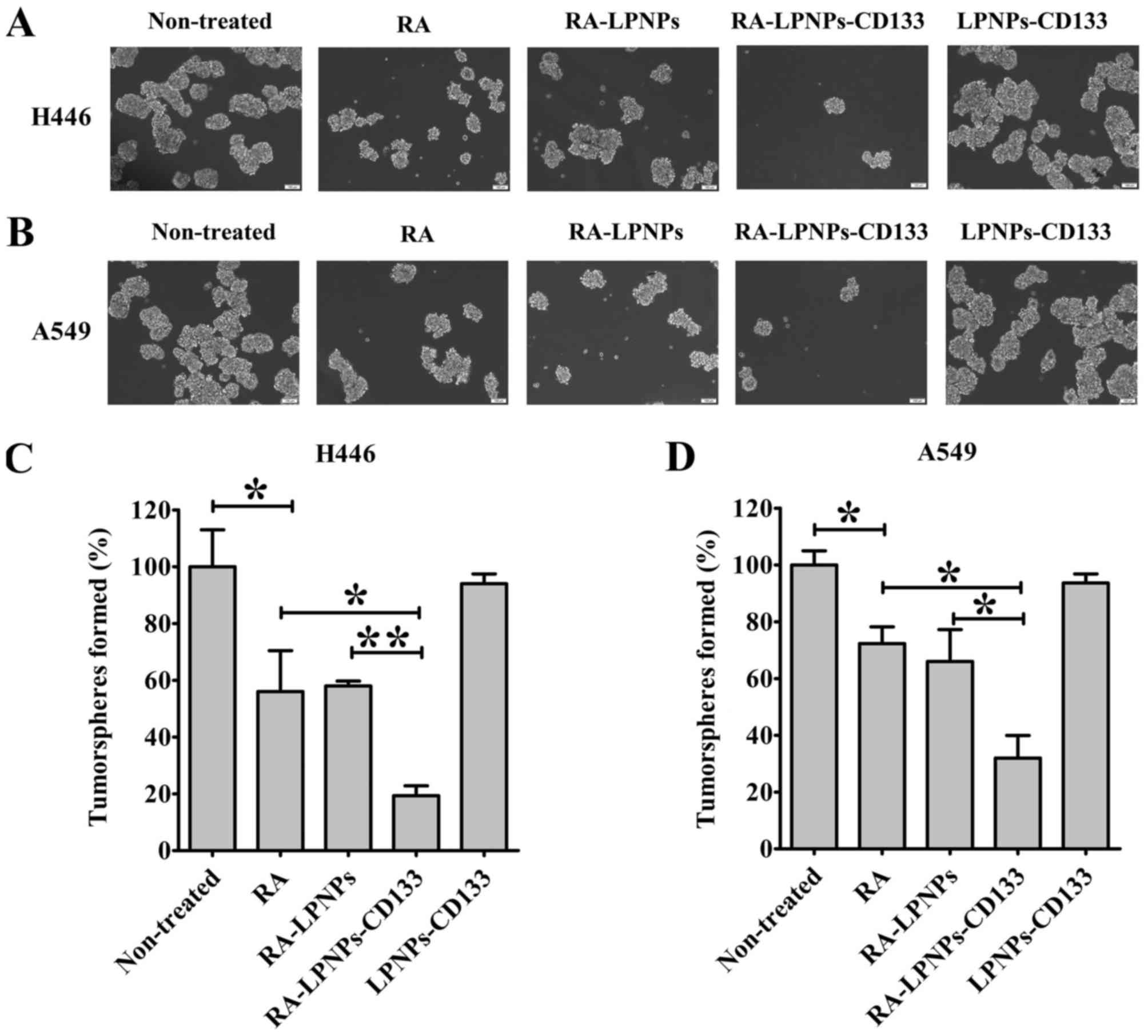
Impact of nanoparticles on lung cancer
initiating cells
The impact of nanoparticles on the percentage of
lung cancer initiating cells was investigated in the H446 and A549
lung cancer cell lines (Fig. 6A and
B). In the H446 cells, treatment with RA markedly inhibited the
number of tumorspheres (P<0.05; Fig.
6C). The inhibitory effect of RA was similar to that of
RA-LPNPs, as reflected by their equal effects on the inhibition of
tumorsphere formation. Notably, the number of tumorspheres
following RA-LPNPs-CD133 treatment was significantly decreased
compared with that following treatment with RA and RA-LPNPs
(P<0.05). Treatment with LPNPs-CD133 had no effect on the number
of tumorspheres. Similar results were observed in the A549 cells
(Fig. 6D). Although the number of
tumorspheres was not affected by treatment with LPNPs-CD133,
treatment with RA-LPNPs-CD133 exhibited the most marked inhibitory
activity on the number of tumorspheres in A549 cells. Consistent
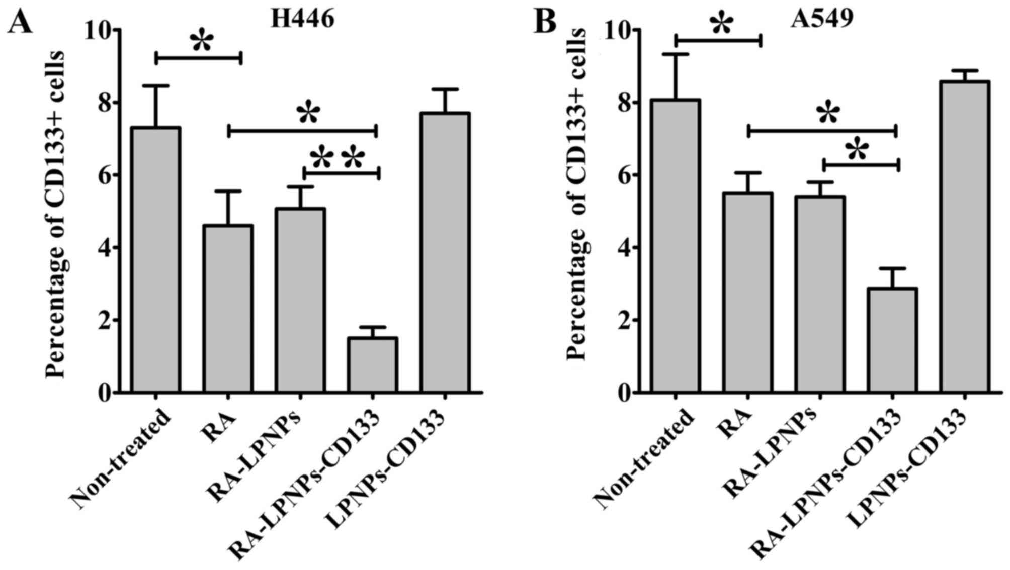
with the above results, the percentage of CD133+ H446
cells was significantly reduced following RA treatment (P<0.05;
Fig. 7A). RA and RA-LPNPs exhibited
similar inhibitory effects towards the percentage of
CD133+ H446 cells. The percentage of CD133+
H446 cells was the lowest among all groups following treatment with
RA-LPNPs-CD133. As shown in Fig. 7B,
similar results were obtained in the case of A549 cells. Taken
together, RA-LPNPs-CD133 exhibited the optimal efficacy towards
lung cancer initiating cells.
Discussion
Lung cancer initiating cells are regarded as the
initial cause of lung cancer. Therefore, the eradication of lung
cancer initiating cells is considered an effective treatment. CD133
is a marker of lung cancer initiating cells. In the present study,
RA-loaded lipid-PLGA nanoparticles conjugated with CD133 aptamers,
RA-LPNPs-CD133, were constructed to target lung cancer initiating
cells. RA-LPNPs-CD133 exhibited significantly higher therapeutic
effects towards lung cancer initiating cells than did free RA and
non-targeted nanoparticles.
Nanoparticles of biodegradable polymers represent
superior drug delivery systems. Their advantages include controlled
and sustained release, high drug loading capacity and superior
stability (24). Lipid-PLGA
nanoparticles represent one of the most commonly used nanoparticles
of biodegradable polymers, due to their superior biocompatibility
and flexibility in modulation of drug release (19). Commonly, poly(ethylene glycol) (PEG)
chains can be incorporated as copolymers throughout the
nanoparticles to increase the hydrophilicity, modification
flexibility and circulation time of the nanoparticles (20). However, PEGylation of PLGA
nanoparticles requires the synthesis of PLGA-PEG copolymers. In
preparing lipid-PLGA nanoparticles in the present study, the
DSPE-PEG molecule was inserted into the nanoparticles by physical
mixing, avoiding the complicated synthesis of PLGA-PEG copolymers.
Furthermore, unlike biodegradable organic nanoparticles, inorganic
nanoparticles cannot be degraded, and may cause damage to humans
(26,27). Therefore, the potential clinical use
of inorganic nanoparticles is limited by their poor safety
(27). By contrast, biodegradable
organic nanoparticles are more promising in clinical application
due to their superior safety profile (26,27). In
the present study, the components of RA-LPNPs-CD133 included
lipids, PLGA and a CD133 antibody, which are safe USA Food and Drug
Administration (FDA)-approved biomaterials. As for RA, the FDA has
approved the combination use of arsenic trioxide (Trisenox)
injection plus RA, as a first-line treatment for low-risk acute
promyelocytic leukemia. In the present study, LPNPs-CD133,
comprising blank lipid-PLGA nanoparticles with CD133 aptamers,
exhibited no marked cytotoxic effects towards lung cancer cells, as
reflected by the almost horizontal curve induced by LPNPs-CD133.
The preliminary safety data in the present study demonstrated that
the nanoparticles represent a safe drug delivery system. Therefore,
CD133-SA-NP is expected to be safe in clinic use, and this safety
is likely to facilitate its clinical translation.
RA is a promising drug that has shown potential
therapeutic effects in various types of cancer, including bladder
cancer (7). Its anticancer
mechanisms include promising effects on the growth, differentiation
and apoptosis of cancer cells (7,8). It is
noteworthy that RA has shown therapeutic potential against cancer
initiating cells in several types of cancer (9–11). To
the best of our knowledge, there have been no reports on the
therapeutic effect of RA on lung cancer initiating cells. In the
present study, it was confirmed that RA preferentially eliminated
CD133+ lung cancer initiating cells. In the tumorsphere
formation assay, RA also reduced the tumorsphere numbers in lung
cancer cells. Consistently, the percentage of CD133+
cells in lung cancer cells was significantly decreased following RA
treatment. To the best of our knowledge, the present study is the
first to demonstrate that RA exhibits potential activity towards
lung cancer initiating cells.
Ligand-conjugated nanoparticles are a promising tool
against various types of cancer, as they can significantly enhance
the therapeutic efficacy of chemotherapy drugs (21–23).
Notably, three types of ligand-conjugated nanoparticles have been
successfully translated into early-phase clinical trials (28,29). In
the present study, the selection of CD133 aptamers was critical for
the specific targeting of the developed nanoparticles to lung
cancer initiating cells. The results showed that, in
CD133+ lung cancer initiating cells, RA-LPNPs-CD133
exhibited significantly increased targeting compared with RA-LPNPs,
resulting in increased cytotoxic effects and inhibitory effects on
tumorspheres. However, in CD133− lung cancer cells, the
cytotoxicity and tumorsphere inhibitory effects of RA-LPNPs-CD133
did not differ from those of RA-LPNPs. These data firmly
demonstrated that RA-LPNPs-CD133 was able to exert increased
targeting and therapeutic effects towards lung cancer initiating
cells and that CD133 aptamers promoted the targeting of
nanoparticles to lung cancer initiating cells. To the best of our
knowledge, the present study is the first to report the promotion
of RA delivery via nanoparticles to lung cancer initiating cells by
the utilization of CD133 aptamers.
In conclusion, the present study provides the first
report of the anticancer activity of RA against lung cancer
initiating cells. RA-LPNPs-CD133 selectively targeted
CD133+ lung cancer initiating cells. Therefore,
RA-LPNPs-CD133 represents a promising approach for therapy
targeting lung cancer initiating cells.
Acknowledgements
Not applicable.
Funding
The present study was supported by the Xiangyang
Municipal Fund (grant no. 20140915).
Authors' contributions
QL and LH contributed to the design of the study and
wrote the manuscript. YZ and JZ performed the experiments. JS
analyzed the data. All authors have read and approved the
manuscript.
Availability of data and materials
All data generated or analyzed during the present
study are included in the published article.
Ethics approval and consent to
participate
The animal experimental protocols were approved by
the Animal Administrative Committee of the Second Military Medical
University (Shanghai, China).
Patient consent for publication
Not applicable.
Authors' information
YZ, JZ, LH and QL: Department of Oncology, Xiangyang
Central Hospital, Affiliated Hospital of Hubei University of Arts
and Science, 136 Jingzhou Street, Xiangyang, Hubei 441000, P.R.
China; JS: Department of Pharmacy, The Second Military Medical
University, 325 Guohe Road, Shanghai 200433, P.R. China.
Competing interests
The authors declare that they have no competing
interests.
References
|
1
|
Siegel RL, Miller KD and Jemal A: Cancer
statistics, 2017. CA Cancer J Clin. 67:7–30. 2017. View Article : Google Scholar : PubMed/NCBI
|
|
2
|
Chen W, Zheng R, Baade PD, Zhang S, Zeng
H, Bray F, Jemal A, Yu XQ and He J: Cancer statistics in China,
2015. CA Cancer J Clin. 66:115–132. 2016. View Article : Google Scholar : PubMed/NCBI
|
|
3
|
Eramo A, Lotti F, Sette G, Pilozzi E,
Biffoni M, Di Virgilio A, Conticello C, Ruco L, Peschle C and De
Maria R: Identification and expansion of the tumorigenic lung
cancer stem cell population. Cell Death Differ. 15:504–514. 2008.
View Article : Google Scholar : PubMed/NCBI
|
|
4
|
Kim JJ and Tannock IF: Repopulation of
cancer cells during therapy: An important cause of treatment
failure. Nat Rev Cancer. 5:516–525. 2005. View Article : Google Scholar : PubMed/NCBI
|
|
5
|
Zakaria N, Satar NA, Halim Abu NH, Ngalim
SH, Yusoff NM, Lin J and Yahaya BH: Targeting lung cancer stem
cells: Research and clinical impacts. Front Oncol. 7:802017.
View Article : Google Scholar : PubMed/NCBI
|
|
6
|
Bertolini G, Roz L, Perego P, Tortoreto M,
Fontanella E, Gatti L, Pratesi G, Fabbri A, Andriani F, Tinelli S,
et al: Highly tumorigenic lung cancer CD133+ cells display
stem-like features and are spared by cisplatin treatment. Proc Natl
Acad Sci USA. 106:16281–16286. 2009. View Article : Google Scholar : PubMed/NCBI
|
|
7
|
Siddikuzzaman Guruvayoorappan C and Berlin
Grace VM: All trans retinoic acid and cancer. Immunopharmacol
Immunotoxicol. 33:241–249. 2011. View Article : Google Scholar : PubMed/NCBI
|
|
8
|
Montesinos P, Bergua JM, Vellenga E, Rayón
C, Parody R, de la Serna J, León A, Esteve J, Milone G, Debén G, et
al: Differentiation syndrome in patients with acute promyelocytic
leukemia treated with all-trans retinoic acid and anthracycline
chemotherapy: Characteristics, outcome, and prognostic factors.
Blood. 113:775–783. 2009. View Article : Google Scholar : PubMed/NCBI
|
|
9
|
Li RJ, Ying X, Zhang Y, Ju RJ, Wang XX,
Yao HJ, Men Y, Tian W, Yu Y, Zhang L, et al: All-trans retinoic
acid stealth liposomes prevent the relapse of breast cancer arising
from the cancer stem cells. J Control Release. 149:281–291. 2011.
View Article : Google Scholar : PubMed/NCBI
|
|
10
|
Karsy M, Albert L, Tobias ME, Murali R and
Jhanwar-Uniyal M: All-trans retinoic acid modulates cancer stem
cells of glioblastoma multiforme in an MAPK-dependent manner.
Anticancer Res. 30:4915–4920. 2010.PubMed/NCBI
|
|
11
|
Han D, Rodriguez-Bravo V, Charytonowicz E,
Demicco E, Domingo-Domenech J, Maki RG and Cordon-Cardo C:
Targeting sarcoma tumor-initiating cells through differentiation
therapy. Stem Cell Res. 21:117–123. 2017. View Article : Google Scholar : PubMed/NCBI
|
|
12
|
Li B, Gao MH, Chu XM, Teng L, Lv CY, Yang
P and Yin QF: The synergistic antitumor effects of all-trans
retinoic acid and C-phycocyanin on the lung cancer A549 cells in
vitro and in vivo. Eur J Pharmacol. 749:107–114. 2015. View Article : Google Scholar : PubMed/NCBI
|
|
13
|
Greve G, Schiffmann I and Lübbert M:
Epigenetic priming of non-small cell lung cancer cell lines to the
antiproliferative and differentiating effects of all-trans retinoic
acid. J Cancer Res Clin Oncol. 141:2171–2180. 2015. View Article : Google Scholar : PubMed/NCBI
|
|
14
|
Li HX, Zhao W, Shi Y, Li YN, Zhang LS,
Zhang HQ and Wang D: Retinoic acid amide inhibits JAK/STAT pathway
in lung cancer which leads to apoptosis. Tumour Biol. 36:8671–8678.
2015. View Article : Google Scholar : PubMed/NCBI
|
|
15
|
Chen D, Xie F, Sun D, Yin C, Gao J and
Zhong Y: Nanomedicine-mediated combination drug therapy in tumor.
Open Pharmaceutical Sci J. 4:1–10. 2017. View Article : Google Scholar
|
|
16
|
Xie FY, Xu WH, Yin C, Zhang GQ, Zhong YQ
and Gao J: Nanomedicine strategies for sustained, controlled, and
targeted treatment of cancer stem cells of the digestive system.
World J Gastrointest Oncol. 8:735–744. 2016. View Article : Google Scholar : PubMed/NCBI
|
|
17
|
Gao J, Feng SS and Guo Y: Nanomedicine
against multidrug resistance in cancer treatment. Nanomedicine
(Lond). 7:465–468. 2012. View Article : Google Scholar : PubMed/NCBI
|
|
18
|
Cristiano MC, Cosco D, Celia C, Tudose A,
Mare R, Paolino D and Fresta M: Anticancer activity of all-trans
retinoic acid-loaded liposomes on human thyroid carcinoma cells.
Colloids Surf B Biointerfaces. 150:408–416. 2017. View Article : Google Scholar : PubMed/NCBI
|
|
19
|
Gao J, Xia Y, Chen H, Yu Y, Song J, Li W,
Qian W, Wang H, Dai J and Guo Y: Polymer-lipid hybrid nanoparticles
conjugated with anti-EGF receptor antibody for targeted drug
delivery to hepatocellular carcinoma. Nanomedicine (Lond).
9:279–293. 2014. View Article : Google Scholar : PubMed/NCBI
|
|
20
|
Kapoor DN, Bhatia A, Kaur R, Sharma R,
Kaur G and Dhawan S: PLGA: A unique polymer for drug delivery. Ther
Deliv. 6:41–58. 2015. View Article : Google Scholar : PubMed/NCBI
|
|
21
|
Gao J, Chen H, Song H, Su X, Niu F, Li W,
Li B, Dai J, Wang H and Guo Y: Antibody-targeted immunoliposomes
for cancer treatment. Mini Rev Med Chem. 13:2026–2035. 2013.
View Article : Google Scholar : PubMed/NCBI
|
|
22
|
Gao J, Feng SS and Guo Y: Antibody
engineering promotes nanomedicine for cancer treatment.
Nanomedicine (Lond). 5:1141–1145. 2010. View Article : Google Scholar : PubMed/NCBI
|
|
23
|
Wang J, Wu Z, Pan G, Ni J, Xie F, Jiang B,
Wei L, Gao J and Zou W: Enhanced doxorubicin delivery to
hepatocellular carcinoma cells via CD147 antibody-conjugated
immunoliposomes. Nanomedicine. 14:1949–1691. 2018. View Article : Google Scholar : PubMed/NCBI
|
|
24
|
Herr JK, Smith JE, Medley CD, Shangguan D
and Tan W: Aptamer-conjugated nanoparticles for selective
collection and detection of cancer cells. Anal Chem. 78:2918–2924.
2006. View Article : Google Scholar : PubMed/NCBI
|
|
25
|
Xiao Z and Farokhzad OC:
Aptamer-functionalized nanoparticles for medical applications:
Challenges and opportunities. ACS Nano. 6:3670–3676. 2012.
View Article : Google Scholar : PubMed/NCBI
|
|
26
|
Auffan M, Rose J, Bottero JY, Lowry GV,
Jolivet JP and Wiesner MR: Towards a definition of inorganic
nanoparticles from an environmental, health and safety perspective.
Nat Nanotechnol. 4:634–641. 2009. View Article : Google Scholar : PubMed/NCBI
|
|
27
|
Cushing BL, Kolesnichenko VL and O'Connor
CJ: Recent advances in the liquid-phase syntheses of inorganic
nanoparticles. Chem Rev. 104:3893–3946. 2004. View Article : Google Scholar : PubMed/NCBI
|
|
28
|
Mamot C, Ritschard R, Wicki A, Stehle G,
Dieterle T, Bubendorf L, Hilker C, Deuster S, Herrmann R and
Rochlitz C: Tolerability, safety, pharmacokinetics, and efficacy of
doxorubicin-loaded anti-EGFR immunoliposomes in advanced solid
tumours: A phase 1 dose-escalation study. Lancet Oncol.
13:1234–1241. 2012. View Article : Google Scholar : PubMed/NCBI
|
|
29
|
Miller K, Cortes J, Hurvitz SA, Krop IE,
Tripathy D, Verma S, Riahi K, Reynolds JG, Wickham TJ, Molnar I and
Yardley DA: HERMIONE: A randomized Phase 2 trial of MM-302 plus
trastuzumab versus chemotherapy of physician's choice plus
trastuzumab in patients with previously treated,
anthracycline-naïve, HER2-positive, locally advanced/metastatic
breast cancer. BMC Cancer. 16:3522016. View Article : Google Scholar : PubMed/NCBI
|