Introduction
Coronary heart disease is a common disease,
involving coronary stenosis or occlusion of the lumen, triggering a
series of symptoms, which poses a threat to the patient's life and
health. Coronary heart disease is closely related to inflammation
and immune and plays a role in the occurrence and development of
coronary heart disease. The pathological basis of coronary heart
disease is atherosclerosis, which is formed by long-term vascular
inflammation and fibrosis together (1). Previous findings showed that there were
autoimmune cell imbalance and immune function decrease in coronary
heart disease patients (2).
miRNAs, as a research hotspot, are evolutionarily
highly conserved, endogenous single-stranded, non-coding small
molecule RNAs, which can regulate the expression of protein-coding
genes after transcription. A large number of studies have proved
that miRNAs play an important role in cell differentiation,
apoptotic cells, angiogenesis, lipid metabolism, inflammation,
immune and other physiological and pathological process (3–5). miR-155
is located on human chromosome 21 and plays an important role in
hematopoiesis, immunity, inflammation, cancer and cardiovascular
disease (6–8). A previous study found that miR-155 was
closely related to immune inflammatory response in the process of
atherosclerosis (9,10). Peripheral blood mononuclear cells
(PBMCs) are mononuclear leucocytes isolated from peripheral blood,
which is composed of T and B lymphocytes, macrophages, and natural
killer cells (NKs) and plays a crucial role in the immune response
(11).
The aim of the current study was to observe the
changes of miRNA-155 in plasma and PBMCs of coronary heart disease
patients and to investigate the relationship of miRNA-155 with the
apoptosis rate of PBMCs and the severity of coronary arteries, in
order to provide the basis for further study on the effect of
miRNA-155 in coronary heart disease.
Materials and methods
Seventy cases with chest pain were randomly selected
from January 2014 to December 2016 in the Department of Cardiology
in The First Affiliated Hospital of Sun Yat-sen University
(Guangzhou, China). The data and venous blood were collected and 3
groups were separated by clinical symptoms and signs, laboratory
examinations and coronary angiography results as referenced in the
Chinese Medical Association Cardiovascular Branch standards. There
were 21 cases of acute myocardial infarction (AMI group: history of
typical chest pain, dynamic changes of electrocardiogram, elevated
myocardial enzymes and troponin, at least one vessel stenosis or
occlusion of coronary angiography), 23 cases in the angina pectoris
group (AP group, chest pain history, ECG changes, rest or
medication relief, coronary angiography to determine coronary
artery stenosis) and 26 cases in the control group (CT group, chest
pain but no coronary lesions).
All the patients enrolled in this study were
first-episode and the following diseases were excluded: Atrial
fibrillation, pacemaker implantation, valvular heart disease,
malignancy, thromboembolic disease, severe liver-kidney dysfunction
and stroke. There were no recent infection and no immune-related
history. The study was approved by the Ethics Committee of The
First Affiliated Hospital of Sun Yat-sen University and the
patients signed informed consent form. The time of blood collection
from all the patients was within 24 h after chest pain attack.
Coronary angiography and Gensini score (9): The coronary angiography of all the
cases were carried out by the same group of doctors through the
radial artery or femoral artery approach, and all segments of the
coronary arteries were fully displayed. Coronary lesions were
quantitatively scored according to the coronary angiographic
recording segment criteria set forth in the AHA (9), then the Gensini score and the degree of
coronary stenosis were recorded.
Isolation of PBMCs
Venous blood (10 ml) was collected from all the
patients in the early morning, which was anticoagulated by Heparin,
and the Lymphocyte separation fluid was added. The blood was
centrifuged at 1,600 × g for 20 min at 4°C and the cells were
divided into four layers: First layer, plasma or tissue homogenate
layer; second layer, cyan milky lymphocyte or monocyte layer; third
layer, transparent separation layer; and fourth layer, red blood
cell layer. The cells in the second layer were harvested and washed
twice with 5 volumes of normal saline prior to centrifugation at
1,600 × g for 10 min at 4°C. The cell pellet was resuspended in
phosphate-buffered saline (PBS) and divided into two portions. One
of the portions was added to a cell culture flask and cultured in
RPMI-1640 medium (Gibco, Grand Island, NY, USA) containing 10%
fetal bovine serum, with the cell concentration of
1×10⁶/ml, and the other was used for RNA extraction.
Apoptosis detection by Annexin V-FITC/propidium
iodide (PI). PBMCs were seeded in 6-well plates and cultured in an
incubator for 48 h. The cells were harvested and resuspended at a
density of 1×10⁶/ml in binding buffer. The PBMCs were
labeled by Annexin V-FITC and PI according to instructions of
Annexin V-FITC/PI Dual Staining kit (Invitrogen, Carlsbad, CA,
USA). Firstly, 5 µl Annexin V-FITC was added and incubated for 10
min in the dark. Then the tube was centrifuged, the supernatant
discarded and the buffer re-added to resuspend. Secondly, 10 µl PI
staining solution was added, mixed and dark-stained for 15 min at
4°C. Finally, the samples were detected by flow cytometry
(cytometry; Beckman Coulter, Brea, CA, USA). Experiments were
repeated at least three times.
Detection of miR-155 in plasma and
PBMCs
Peripheral blood (6 ml) was collected in EDTA
anticoagulant tube, with the patients fasting 8 h, prior to
centrifugation at 1,600 × g for 10 min at 4°C twice. Supernatant of
500 µl was collected as a plasma sample. Total RNA was extracted
from the plasma sample and PBMCs cells using TRIzol LS reagent
according to the manufacturer's protocol and stored at −80°C for
reverse transcription. Total RNA (20 ng) from each sample was used
for reverse transcription according to the manufacturer's protocol
of the TaqMan microRNA Reverse Transcription kit (Applied
Biosystems; Thermo Fisher Scientific, Inc., Waltham, MA, USA).
Primers used for the TaqMan microRNA assays (Applied
Biosystems; Thermo Fisher Scientific) were: Control U6 primers,
upstream, 5′-GCTTCGGCAGCACATATACTAAAAT-3′ and downstream,
5′-CGCTTCACGAATTTGCGTGTCAT-3′. miRNA-155 primers, upstream,
5′-GGAGGTTAATGCTAATCGTGATAG-3′ and downstream,
5′-GTGCAGGGTCCGAGGT-3′.
The reaction conditions were as follows: 95°C for 15
sec and 40 cycles of 60°C for 30 sec. We repeated each sample in
each group in triplicate. Reverse transcription product (2 µl) was
used for TaqMan probes reverse transcription-quantitative
polymerization chain reaction (RT-qPCR) and PCR double-specific
primers were amplified using the TaqMan universal Master Mix II, no
UNG (Applied Biosystems; Thermo Fisher Scientific) by the ABI 7500
Thermal Cycler to detect Cq value of the target gene and control
gene using 2−ΔΔCq method according to the following
formula: ΔΔCq (target gene) = Cq (target gene) - Cq (control gene)
(12).
Statistical analysis
The experimental data were expressed as mean ±
standard deviation and analyzed using SPSS 18.0 software. Student's
t-test, and Chi-square test were used for comparisons between two
groups. For pairwise comparisons ANOVA and LSD post hoc test were
used. Spearman's linear correlation analysis were used to analyze
the data. P<0.05 or P<0.01 indicated a statistically
significant difference.
Results
General situation comparison
There was no statistically significant difference of
age, sex, disease history and medication (P>0.05) among the
patients in the three groups (Table
I).
 | Table I.General information for patients. |
Table I.
General information for patients.
| Group | No. | Age | Male/female | Hypertension | Diabetes | Hyperlipidemia | Smoke | β-blocker | ACEI | CCB | Statins |
|---|
| AMI | 21 | 59±7 | 15/6 | 13 | 5 | 13 | 6 | 14 | 15 | 9 | 17 |
| AP | 23 | 58±9 | 17/5 | 13 | 4 | 11 | 5 | 13 | 10 | 11 | 15 |
| CT | 26 | 60±8 | 16/10 | 10 | 3 | 7 | 5 | 7 | 8 | 8 | 10 |
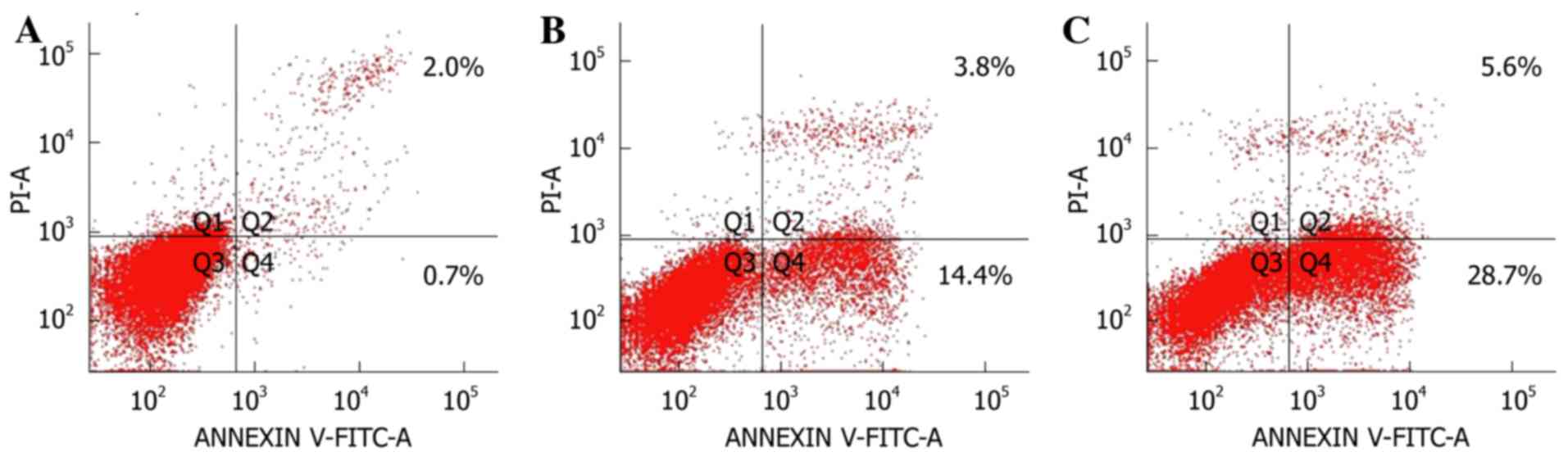
Apoptosis of PBMCs in each group
The apoptotic rates of PBMCs in the three groups
were detected by flow cytometry: 34.3±5.4% in the AMI group,
18.2±1.6% in the AP group and 2.7±1.22% in the CT group. The
apoptosis rate of PBMCs in the AMI group was significantly higher
than that in the AP and CT groups (P<0.05). The apoptosis rate
of PBMCs in the AP group was significantly higher than that in CT
group, and the difference was statistically significant (P<0.05)
(Fig. 1).
Coronary stenosis and Gensini score in
each group
The degree of coronary artery stenosis and Gensini
score in the AMI group were significantly higher than those in the
AP and CT groups (P<0.05). The degree of coronary artery
stenosis and Gensini score in the AP group were significantly
higher than those in CT group (P<0.05) (Table II).
| Table II.miR-155, apoptosis rate of PBMCs,
coronary artery stenosis and Gensini score in patients. |
Table II.
miR-155, apoptosis rate of PBMCs,
coronary artery stenosis and Gensini score in patients.
| Group | No. | Apoptosis rate of
PBMCs (%) | Degree of coronary
artery stenosis (%) | Gensini score | miR-155 (plasma) | miR-155 (PBMCs) |
|---|
| AMI | 21 | 34.3±5.4a,b |
87.61±8.78a,b |
49.15±12.87a,b |
0.69±0.10a,b |
0.56±0.08a,b |
| AP | 23 | 18.2±1.6a |
62.40±5.98a |
33.71±8.87a |
1.55±0.29a |
1.34±0.25a |
| CT | 26 | 2.7±1.22 | 16.6±9.4 | 0.70±1.95 | 3.00±0.80 | 2.32 ±0.53 |
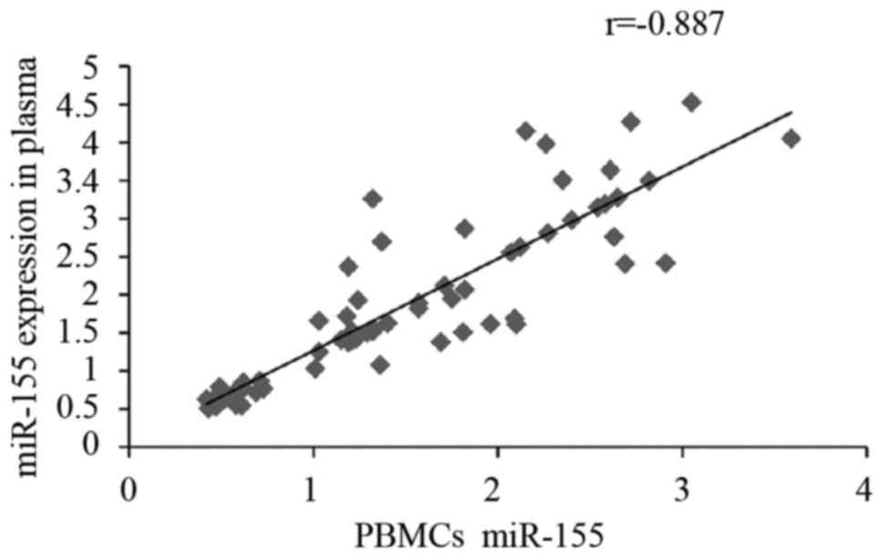
Comparison of miR-155 levels in each
group
The level of miR-155 in AMI group was significantly
lower than that in the AP and CT groups (P<0.05) and the level
of miR-155 in AP group was significantly higher than that in CT
group (P<0.05). There was a positive correlation between miR-155
levels in plasma and PBMCs in three groups (r=−0.887, P<0.001)
(Table II, Fig. 2).
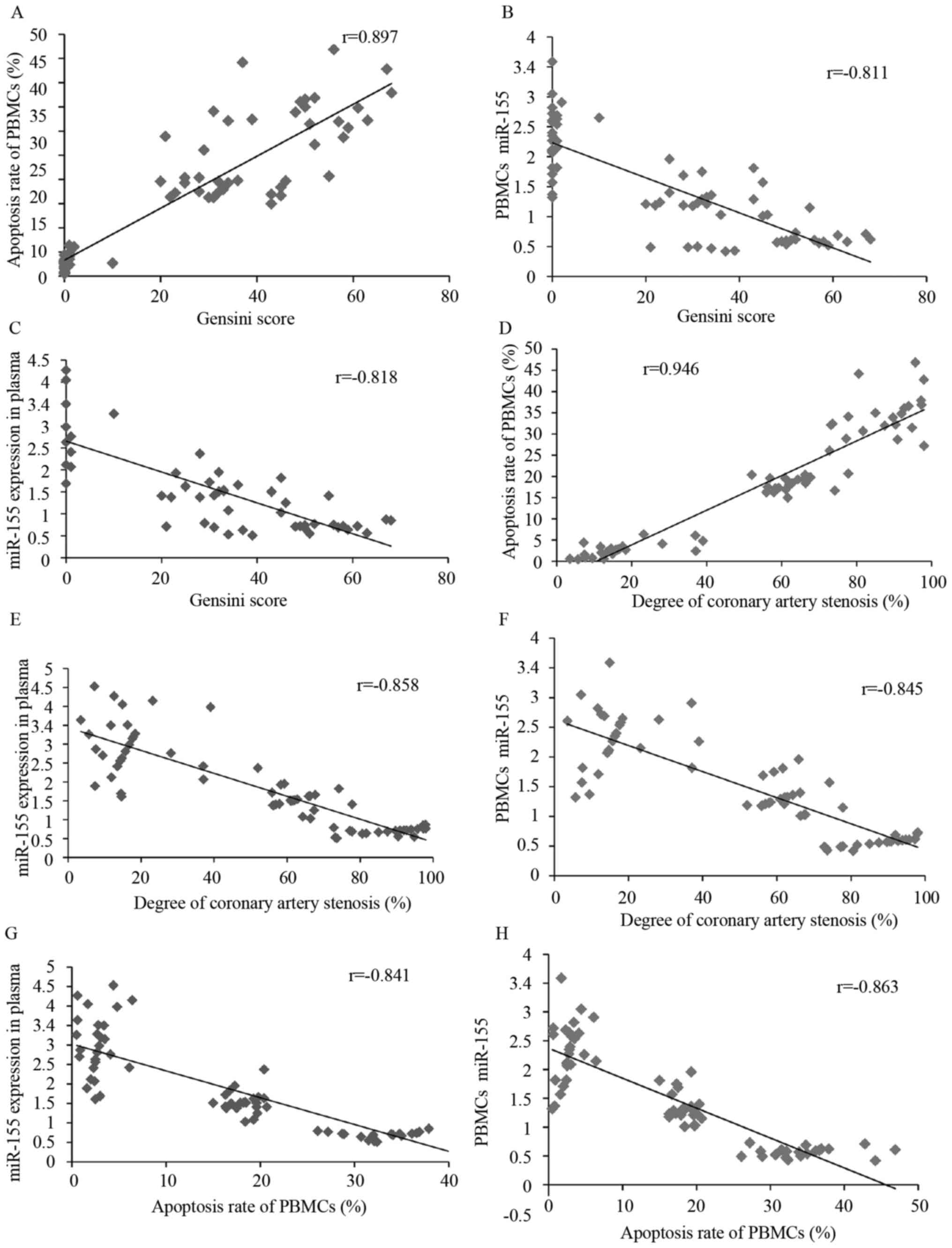
Correlation of miR-155, apoptosis rate of PBMCs with
coronary artery stenosis and Gensini score. As shown in Fig. 3, miR-155 expression in plasma of
patients with coronary heart disease was negatively correlated with
the Gensini score, the degree of coronary artery stenosis and the
apoptosis rate of PBMCs (r=−0.818, P<0.001; r=−0.858,
P<0.001; r=−0.841, P<0.001). The expression level of miR-155
in PBMCs of patients with coronary heart disease was negatively
correlated with the Gensini score, the degree of coronary artery
stenosis and the apoptosis rate of PBMCs (r=−0.811, P<0.001;
r=−0.845, P<0.001; r=−0.863, P<0.001). The apoptosis rate of
PBMCs in patients with coronary heart disease was positively
correlated with the Gensini score and the degree of coronary artery
stenosis (r=0.897, P<0.001; r=0.946, P<0.001).
Discussion
The pathogenesis of coronary heart disease is
complicated. A variety of factors lead to the damage of intima,
long-term vasculitic inflammation and fibrogenesis causing the
formation of atherosclerotic plaque. Inflammatory reaction promotes
the formation and development of atherosclerotic plaque, which
plays an important role in the process of coronary heart disease
(13). The Gensini score was used to
assess the severity of coronary lesions by giving quantified
weights of different sites of stenosis and the degree of stenosis.
The higher the score, the more severe the coronary artery disease.
This method has been widely used clinically to assess the severity
Degree of coronary artery disease (9,14,15).
miR-155 is a multi-functional miRNAs located in
human chromosome 21 and expressed in many tissues and cells,
involved in many physiological and pathological processes such as
immunity, inflammation, cell differentiation, cardiovascular
diseases and tumors. miR-155 is closely related to inflammation and
immunity, and can regulate the activation of immune cells and the
release of immune factors (6–8). Studies
have shown that miR-155 regulates the transcription of angiotensin
II-1 receptors and affects the migration of endothelial cells and
thus the progression of atherosclerosis (16). However, the current expression of
miR-155 in coronary heart disease is controversial. Studies have
shown that miR-155 is upregulated in atherosclerotic mouse models
(17,18), but other studies have shown that
miR-155 is significantly decreased in patients with coronary heart
disease compared with non-CAD patients (9,10,19). The
results in our study have shown that the expression of miR-155 in
plasma and PBMCs in patients with myocardial infarction was
significantly lower than that with angina pectoris. The expression
level of miR-155 in patients with angina pectoris was significantly
lower than that with non-coronary heart disease. Moreover, the
expression of miR-155 in plasma and PBMCs was highly negatively
correlated with the severity of coronary heart disease. This study
is consistent with some studies (9–11,19).
It is generally accepted that atherosclerosis is an
inflammatory disease, as abnormal activation of immune cells,
excessive inflammatory mediators released, excessive inflammatory
reactions, damage to coronary endothelial cells, causing platelet
aggregation, thrombosis, which accelerated the formation of
coronary atherosclerotic plaques, severe even porridge plaque
rupture and induced myocardial infarction or acute angina attacks
(20). Immune cells in patients with
coronary heart disease repeatedly activated, apoptosis, resulting
in decreased immune cells, impaired immune function, varying degree
of decreased immune function in coronary heart disease patients.
Previous findings have shown that miR-155 regulates the
differentiation of T lymphocyte subsets by regulating the
expression of two target genes of SMAD2 and SOCS1 in coronary heart
disease (21). SMAD2 can induce the
differentiation of Th17 cells and the production of interleukin-17A
(IL-17A), while SOCS1 inhibition of Th17 cell differentiation by
inhibiting IL-6/STAT3 signaling pathway (22), suggesting that miR-155 expression is
negatively correlated with Th17 differentiation. However, little is
known about the expression of miR-155 and the apoptosis of PBMCs.
The present study has shown that the apoptosis rate of PBMCs in
patients with myocardial infarction is significantly higher than
that with angina pectoris, while the apoptosis rate of PBMCs in
patients with angina pectoris is significantly higher than that in
control group (P<0.05). The apoptosis rate of PBMCs is highly
positive correlation with the severity degree of coronary heart
disease, while the apoptosis rate of PBMCs and the miR-155
expression in plasma and PBMCs was negatively correlated.
In conclusion, the apoptosis rate of PBMCs in
patients with coronary heart disease increased, and the expression
of miR-155 in plasma and PBMCs decreased, which were all related to
the severity of coronary heart disease.
Acknowledgements
Not applicable.
Funding
No funding was received.
Availability of data and materials
The datasets used and/or analyzed during the current
study are available from the corresponding author on reasonable
request.
Authors' contributions
DZ and ZY performed RT-PCR. JZ, JS and YS were
responsible for the isolation of PBMCs. JJ, HY and JL assisted in
the apoptosis rate detection. JF and ZW contributed to statistical
analysis. All authors read and approved the final manuscript.
Ethics approval and consent to
participate
The study was approved by the Ethics Committee of
The First Affiliated Hospital of Sun Yat-sen University (Guangzhou,
China) and the patients signed informed consent form.
Patient consent for publication
Not applicable.
Competing interests
The authors declare that they have no competing
interests.
References
|
1
|
Vasilets LM, Grigoriadi NE, Karpunina NS,
Tuev AV and Rotanova EA: Immune status of patients with persistent
atrial fibrillation and coronary heart disease. Klin Med (Mosk).
91:32–34. 2013.(In Russian). PubMed/NCBI
|
|
2
|
Zheng H, Tu Y and Teng ZH: Effect of
immune response mediated by antigen-specific T cells on plaque
stability in coronary heart disease. Nan Fang Yi Ke Da Xue Xue Bao.
301610–1611. (1614)2010.(In Chinese). PubMed/NCBI
|
|
3
|
Fu L, Jin L, Yan L, Shi J, Wang H, Zhou B
and Wu X: Comprehensive review of genetic association studies and
meta-analysis on miRNA polymorphisms and rheumatoid arthritis and
systemic lupus erythematosus susceptibility. Hum Immunol. 77:1–6.
2016. View Article : Google Scholar : PubMed/NCBI
|
|
4
|
Mizuguchi Y, Takizawa T, Yoshida H and
Uchida E: Dysregulated miRNA in progression of hepatocellular
carcinoma: A systematic review. Hepatol Res. 46:391–406. 2016.
View Article : Google Scholar : PubMed/NCBI
|
|
5
|
Wen MM: Getting miRNA therapeutics into
the target cells for neurodegenerative diseases: A Mini-Review.
Front Mol Neurosci. 9:1292016. View Article : Google Scholar : PubMed/NCBI
|
|
6
|
Yu DD, Lv MM, Chen WX, Zhong SL, Zhang XH,
Chen L, Ma TF, Tang JH and Zhao JH: Role of miR-155 in drug
resistance of breast cancer. Tumour Biol. 36:1395–1401. 2015.
View Article : Google Scholar : PubMed/NCBI
|
|
7
|
Lind EF and Ohashi PS: Mir-155, a central
modulator of T-cell responses. Eur J Immunol. 44:11–15. 2014.
View Article : Google Scholar : PubMed/NCBI
|
|
8
|
Vigorito E, Kohlhaas S, Lu D and Leyland
R: miR-155: An ancient regulator of the immune system. Immunol Rev.
253:146–157. 2013. View Article : Google Scholar : PubMed/NCBI
|
|
9
|
Zhu GF, Yang LX, Guo RW, Liu H, Shi YK, Ye
JS and Yang ZH: microRNA-155 is inversely associated with severity
of coronary stenotic lesions calculated by the Gensini score. Coron
Artery Dis. 25:304–310. 2014. View Article : Google Scholar : PubMed/NCBI
|
|
10
|
Fichtlscherer S, De Rosa S, Fox H,
Schwietz T, Fischer A, Liebetrau C, Weber M, Hamm CW, Röxe T,
Müller-Ardogan M, et al: Circulating microRNAs in patients with
coronary artery disease. Circ Res. 107:677–684. 2010. View Article : Google Scholar : PubMed/NCBI
|
|
11
|
Zhang YH, Xia LH, Jin JM, Zong M, Chen M
and Zhang B: Expression level of miR-155 in peripheral blood. Asian
Pac J Trop Med. 8:214–219. 2015. View Article : Google Scholar : PubMed/NCBI
|
|
12
|
Livak KJ and Schmittgen TD: Analysis of
relative gene expression data using real-time quantitative PCR and
the 2(-Delta Delta C(T)) method. Methods. 25:402–408. 2001.
View Article : Google Scholar : PubMed/NCBI
|
|
13
|
Wirtz PH and von Känel R: Psychological
stress, inflammation, and coronary heart disease. Curr Cardiol Rep.
19:1112017. View Article : Google Scholar : PubMed/NCBI
|
|
14
|
He LY, Zhao JF, Han JL, Shen SS and Chen
XJ: Correlation between serum free fatty acids levels and Gensini
score in elderly patients with coronary heart disease. J Geriatr
Cardiol. 11:57–62. 2014.PubMed/NCBI
|
|
15
|
Che J, Li G, Wang W, Li Q, Liu H, Chen K
and Liu T: Serum autoantibodies against human oxidized low-density
lipoproteins are inversely associated with severity of coronary
stenotic lesions calculated by Gensini score. Cardiol J.
18:364–370. 2011.PubMed/NCBI
|
|
16
|
Jia QW, Chen ZH, Ding XQ, Liu JY, Ge PC,
An FH, Li LH, Wang LS, Ma WZ, Yang ZJ, et al: Predictive effects of
circulating miR-221, miR-130a and miR-155 for coronary heart
disease: A multi-ethnic study in China. Cell Physiol Biochem.
42:808–823. 2017. View Article : Google Scholar : PubMed/NCBI
|
|
17
|
Zhu J, Chen T, Yang L, Li Z, Wong MM,
Zheng X, Pan X, Zhang L and Yan H: Regulation of microRNA-155 in
atherosclerotic inflammatory responses by targeting MAP3K10. PLoS
One. 7:e465512012. View Article : Google Scholar : PubMed/NCBI
|
|
18
|
Tian FJ, An LN, Wang GK, Zhu JQ, Li Q,
Zhang YY, Zeng A, Zou J, Zhu RF, Han XS, et al: Elevated
microRNA-155 promotes foam cell formation by targeting HBP1 in
atherogenesis. Cardiovasc Res. 103:100–110. 2014. View Article : Google Scholar : PubMed/NCBI
|
|
19
|
Pan RY, Liu P, Zhou HT, Sun WX, Song J,
Shu J, Cui GJ, Yang ZJ and Jia EZ: Circular RNAs promote TRPM3
expression by inhibiting hsa-miR-130a-3p in coronary artery disease
patients. Oncotarget. 8:60280–60290. 2017. View Article : Google Scholar : PubMed/NCBI
|
|
20
|
Farrokhian A, Raygan F, Soltani A,
Tajabadi-Ebrahimi M, Esfahani Sharifi M, Karami AA and Asemi Z: The
effects of synbiotic supplementation on carotid intima-media
thickness, biomarkers of inflammation, and oxidative stress in
people with overweight, diabetes, and coronary heart disease: A
randomized, double-blind, placebo-controlled trial. Probiotics
Antimicrob Proteins. Oct 27–2017.(Epub ahead of print). doi:
10.1007/s12602-017-9343-1. View Article : Google Scholar : PubMed/NCBI
|
|
21
|
Louafi F, Martinez-Nunez RT and
Sanchez-Elsner T: MicroRNA-155 targets SMAD2 and modulates the
response of macrophages to transforming growth factor-{beta}. J
Biol Chem. 285:41328–41336. 2010. View Article : Google Scholar : PubMed/NCBI
|
|
22
|
Jiang S, Zhang HW, Lu MH, He XH, Li Y, Gu
H, Liu MF and Wang ED: MicroRNA-155 functions as an OncomiR in
breast cancer by targeting the suppressor of cytokine signaling 1
gene. Cancer Res. 70:3119–3127. 2010. View Article : Google Scholar : PubMed/NCBI
|