Introduction
Due to an aging population and an increasing number
of traffic accidents, the incidence of bone fractures continues to
rise, which was reported as being 6.42% in people aged over 50
(1). Open reduction and internal
fixation has become a common surgical treatment for fractures
(2). Internal fixation has
substantial healing advantages as it fixes the fracture and
maintains the morphological structure and biomechanics of the
broken bones, allows for improved pain control, and prevents pain
and joint stiffness by facilitating motion in the postoperative
phase (3). Although the vast
majority of fractures treated with modern internal fixation
techniques will heal, complications do occasionally occur (4). To easily mount the implant, the soft
tissue and periosteum are extensively dissected, which increases
the likelihood of infection, neurovascular injury and skin flap
necrosis (5,6). All of the aforementioned complications
may cause visible and serious consequences, including delayed union
and nonunion (7). Such complications
may be life-altering due to the symptoms and also the economic
burden they place on the patient (8). Furthermore, they may cause a
significant financial impact on health care systems (9,10).
Therefore, accelerating early fracture healing is necessary and
important to prevent short- and long-term complications and to
minimize the occurrence of delayed union and nonunion.
Bone is much denser than muscle, fat or hematoma,
and so most forms of therapeutic heating are unable to penetrate
it; however, microwaves are able to penetrate bone to reach
therapeutic values (11).
High-frequency microwaves are used as a clinical hyperthermia
method to heat deep tissues (12).
Microwave energy may be directly focused on localized areas without
affecting the temperature in adjacent tissues (13). Hyperthermia induced by microwaves
increases local blood perfusion and consequently nutrient and
oxygen supply to the heated region. It also alleviates pain, speeds
up metabolism, enhances apoptosis and stimulates new bone formation
following injury (14–16).
The use of microwave diathermy in fracture healing
is not a novel concept. There has been an abundance of relevant
reports and research since 1993, when Leon et al (17,18) and
Chang et al (19)
systematically demonstrated that microwave diathermy promoted bone
deposition in fracture healing both in vitro and in
vivo. As a considerable amount of microwave energy may be
reflected or refracted at the interfaces between tissue and the
metal implant used in orthopedic surgery (20), there is controversy regarding whether
microwaves should be applied to fractures with metal-implanted
internal fixation. Historically, it was considered an absolute
contraindication (21,22), but in recent years opinions have
changed and reports have increasingly questioned whether the
contraindication is based on consensus or scientific evidence.
Internal fixation of fractures using titanium alloys
has gained popularity (23,24). Given that the magnetic permeability
and electric conductivity of titanium alloy is lower than
conventional biomedical metal implants (25,26). Ye
et al (27) performed two
randomized controlled studies and demonstrated that 25 W microwave
(2,450 MHz) improved the healing of fractures with titanium alloy
internal fixation without irreversible heat damage (<41°C) in a
rabbit model. Ye et al (28)
also reported that the temperature of muscle tissue adjacent to
titanium alloy implants is beyond the scope of security (>41°C)
when the power of microwave irradiation is tuned to 50 W. This
report alone is insufficient and further research is required to
reach a new consensus. The aim of the present study was to further
verify the security and feasibility of microwave therapy from
another aspect by comparing the proliferative ability of skeletal
muscle satellite cells (MSCs) under 0, 25 and 50 W of microwave
irradiation.
Materials and methods
Ethics statement
All animal welfare and experimental procedures
involving animals were conducted in strict conformity with the
recommendations in the Guide for the Care and Use of Laboratory
Animals of National Laboratory Animals and protocols were
specifically approved by the Animal Welfare and Ethics Committee of
Shanghai Sixth People's Hospital [Permit no. SYXK (HU) 2011–0128;
Shanghai, China].
Animals and grouping
A total of 45 healthy adult white New Zealand male
rabbits (Shanghai Biomodel Organism, Shanghai, China) aged 16–18
weeks and weighing 2.2–3.0 kg (mean, 2.5 kg) were supplied by the
Animal Laboratory Center of Shanghai Jiao Tong University
Affiliated Sixth People's Hospital. The rabbits were fed with a
standard diet and were allowed access to forage and drinking water.
Rabbits were housed at a constant temperature (25°C) and relative
humidity (45–60%) with a 12 h light/dark cycle.
The rabbits were randomly and evenly assigned into
i) the control group (group A), ii) the 25 W microwave treatment
group (group B) or iii) the 50 W microwave treatment group (group
C) after a week of adjustable feeding. No significant differences
were observed in age or weight between groups following
randomization. All rabbits underwent orthopedic surgery to create a
femoral shaft fracture and titanium alloy internal fixation model.
Following surgery, group A was left untreated and served as the
control. Group B were treated with 25 W microwaves and group C with
50 W microwave irradiation.
Creating an animal fracture model
The entire operation process was performed in
operating room aseptic conditions. Intravenous sodium pentobarbital
(30 mg/kg) anesthesia was administered to the rabbits via the ear
vein. Following preoperative hair removal, the rabbits were fixed
on the experiment table in the dorsal position and the right hind
limb skin was sterilized. The right femur was exposed laterally via
a longitudinal skin incision (3–4 cm), with care taken to minimize
disruption of the periosteum. Under vertical loading in the middle
of the femur, a transverse osteotomy with a 3.0-mm gap was created
and stabilized with an internal fixation system (DePuy Synthes
Biomaterials, West Chester, PA, USA), which consisted of a titanium
alloy plate (4.58±0.24×0.41±0.08 cm) with seven holes and screws.
After washing the incision with pure iodine liquid, the wound was
closed in layers in the usual manner and covered with sterile dry
gauze. Rabbits were transferred to a warm recovery room and
monitored until awakening from anesthesia. No wound complications
or mortalities occurred within 3 days postoperatively, during which
time rabbits received daily intramuscular injections of penicillin
(800,000 units per rabbit per day).
Microwave diathermy
Rabbits in group B began receiving 25 W microwave
treatment on postoperative day 4. Those rabbits were fixed with a
specialized fixture to prevent movement during the course of
therapy. Prior to treatment, a wattmeter (Enraf Nonius B.V.,
Rotterdam, The Netherlands) was utilized to adjust the actual
output power of the microwave to meet the experimental
requirements, and a shelter was used to minimize radiation exposure
to the tissues close to the irradiation target. The microwave
diathermy system had two main components: A 2450-MHz microwave
generator (PM-800; ITO Physiotherapy and Rehabilitation Co., Ltd.,
Tokyo, Japan) and a non-contact applicator (RM-170A; ITO
Physiotherapy and Rehabilitation Co., Ltd.). Microwave treatment
was perpendicularly applied 10 cm away from the right upper thigh
for 15 days for 10 min per day. The same treatment regimen was
provided to group C with a microwave power of 50 W. Group A did not
receive any treatment. Microwave treatments were administered at
the same time each day to eliminate factors that may influence the
interference.
Sampling
After 15 days of treatment, rabbits were humanely
euthanized by lethal injection of sodium pentobarbital under
strictly sterile conditions in the operating room. Hind limbs were
soaked with 75% ethanol disinfectant for 5 min and the skin of the
hind limb was opened with sharp scissors to expose the quadriceps
femoris muscle adjacent to the fracture. By holding the muscle
through its tendons, the muscles were excised and trimmed of fat
and connective tissue carefully to avoid damaging the myofibers.
Each individual muscle sample was divided into two immediately: One
was used for electron microscope observation, and the other was
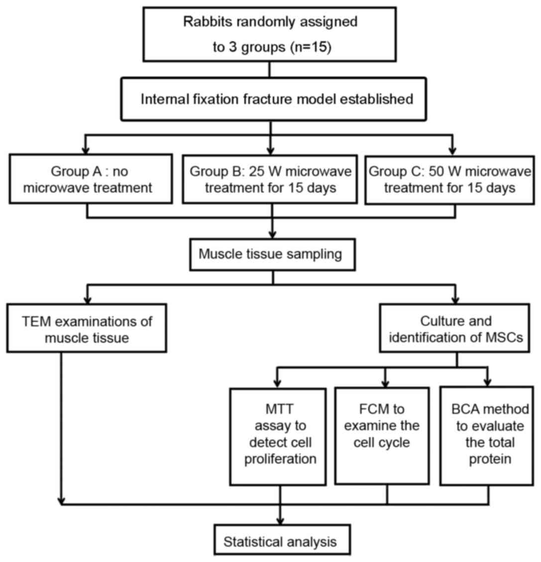
used for cell culture. Fig. 1
illustrates the whole protocol in a simplified sequence flow
diagram.
Transmission electron microscopy (TEM)
examinations of muscle tissue
The muscle tissue was first fixed with 2.5%
glutaraldehyde in phosphate buffer (pH 7.0) overnight at 4°C,
postfixed with 1% OsO4 in phosphate buffer (pH 7.0) for
1 h at 4°C and dehydrated in a graded series of ethanol (30, 50,
70, 80, 90, 95 and 100%) for ~15 min at each step. Following
infiltration with a mixture of acetone and resin, specimens were
placed in capsules containing embedding medium (epoxy resin;
Beijing Solarbio Science & Technology Co., Ltd., Beijing,
China) and heated at 70°C overnight. The ultrathin sections (90 nm
thick) were stained using uranyl acetate and alkaline lead citrate
at room temperature for 15 min and observed with an electron
microscope (EM400T; Phillips Healthcare, Amsterdam, The
Netherlands).
Isolation, purification and culture of
MSCs
MSCs were obtained from the rabbits using the tissue
explant method (29). Briefly, a 2
cm3 skeletal muscle biopsy was harvested from the
quadriceps femoris muscle as described above in sampling. The
isolated muscle biopsy was cut with ophthalmic scissors in dishes
and minced into smaller cubes approximately 1 mm3 along
the length of the fibers. The minced muscles were rinsed well with
phosphate-buffered saline (PBS), and the tissues that floated on
the surface of the liquid were removed. The minced muscles were
placed evenly in an empty culture flask with ophthalmic forceps and
left for 30 min to help the adherence of the explants. A growth
medium (Dulbecco's modified Eagle's medium supplemented with 10%
fetal bovine serum and 100 U/ml penicillin-streptomycin; Gibco;
Thermo Fisher Scientific, Inc., Waltham, MA, USA) was added to the
flask, then cultured in a standard cell incubator at 37°C for 9
days. After using a modified differential adhesion method to remove
fibroblasts, the purified MSCs were transferred to three fresh
poly-L-lysine-coated flasks and cultured for 7 days at 37°C, in an
atmosphere containing 5% CO2 in a standard cell
incubator. The medium was changed every other day. The cells were
removed from the culture flask, dissociated with trypsin (Beijing
Solarbio Science & Technology Co., Ltd.) until small holes
formed in the opaque monolayer. Cells were subsequently diluted,
plated in three fresh flasks and subcultured when the culture
reached ~80% confluence. The morphology and growth features of the
cells were observed daily using an inverted microscope.
Immunocytochemical identification of
satellite cells
Fourth generation cells were chosen as testing cells
and were digested with trypsin, seeded at a density of
1×10⁵ cells/well in 6-well plates precoated with
poly-L-lysine, and subsequently cultured with the growth medium.
When cell confluence reached 80%, they were used for
immunocytochemistry. Briefly, the culture medium was discarded and
cells were fixed with prewarmed 4% paraformaldehyde for 10 min at
room temperature. Cells were extensively washed in PBS three times
for 5 min, blocked with 3% H2O2 diluted with
distilled water for 10 min at room temperature in the dark to
deactivate the endogenous peroxidase, and further blocked with 5%
goat serum (Sigma-Aldrich; Merck KGaA, Darmstadt, Germany) for 30
min at room temperature. Cells were subsequently incubated
overnight at 4°C in primary antibody solution (1:100; mouse
monoclonal anti-α-sarcomeric actin; BM0001; Boster Biological
Technology, Pleasanton, CA, USA), and PBS displaced the primary
antibody as negative criteria. Cells were washed three times in PBS
to remove any unbound antibody, and stained with a Streptavidin
Biotin-peroxidase kit (Boster Biological Technology) for 30 min at
room temperature followed by diaminobenzidine staining using a
Histostain-Plus kit (Beijing Solarbio Science & Technology Co.,
Ltd.) for 15 min at room temperature.
MTT assay
Following trypsinization, cells in the logarithmic
growth phase formed a single cell suspension, and the cell
concentration was adjusted to 5×10⁴ cells/ml. Cells were
seeded into 96-well culture plates (100 µl/well) with six
replicates used for each group. To avoid the edge effect, the
peripheral wells were filled with sterile PBS. Culture medium
without cells was used as the blank control group and cultured for
24, 48 and 72 h, respectively, at 37°C. A total of 100 µl MTT
solution (1 mg/ml; Sigma-Aldrich; Merck KGaA) was added to each
well, followed by incubation for 4 h at 37°C. After centrifugation
for 10 min at 100 × g, the supernatant was discarded and 150 µl
dimethyl sulfoxide (Sigma-Aldrich; Merck KGaA) was added into each
well to fully dissolve the formazan precipitates with gentle
agitation for 10 min at room temperature. The optical density of
each well was measured at a wavelength of 570 nm using a microplate
reader (Multiskan FC; Thermo Fisher Scientific, Inc.). The
background absorbance of the blank group was subtracted, the value
was pooled and averaged, and the cell growth curve was plotted.
Cell cycle analysis by flow
cytometry
Logarithmic growth phase cells were incubated in
6-well plates (2 ml/well) at a density of 1×10⁵ cells/ml
overnight (>16 h) at 37°C, harvested by 0.2% trypsinization,
washed with cold PBS twice and subsequently fixed in ice-cold 70%
ethanol for 2 h at 4°C. The fixed cells were washed with PBS and
incubated with 10 µl RNase A (20 µg/ml; Sigma-Aldrich; Merck KGaA)
in 500 µl PBS at 37°C for 30 min in the dark. The cellular DNA was
stained with 500 µl of propidium iodide (Beijing Solarbio Science
& Technology Co., Ltd.) solution (50 µg/ml in PBS) in the dark
for 30 min at room temperature. The DNA content of the stained
cells was determined using a FACSCalibur flow cytometer (BD
Biosciences, San Jose, CA, USA), and the distribution of each phase
was immediately analyzed using ModFit LT for Windows 4.1 analysis
software (Verity Software House, Inc., Topsham, ME, USA). A total
of 10,000 events were observed per sample.
Bicinchoninic acid (BCA) protein
assay
The total protein content (mg/ml) was measured
according to the method previously described by Huang et al
(30), with slight modifications.
Briefly, the medium was discarded and cells were washed with PBS
three times. Cells were lysed by adding 0.5 ml 0.1% sodium dodecyl
sulfate per well for 30 min at 100°C, and were subsequently
transferred into 96-well plates. A total of 200 µl of the BCA
Protein Assay Reagent (Boster Biological Technology) was added
followed by oscillation for 10 min. Finally, the optical density
value was measured at a 562-nm wavelength using a microplate reader
and a standard curve was plotted with bovine serum albumin (Gibco;
Thermo Fisher Scientific, Inc.) as a standard.
Statistical analysis
Results were presented as the mean ± standard
deviation. All data were processed by SAS 9.1 for Windows (SAS
Institute, Inc., Cary, NC, USA). Following the normality test and
homogeneity test of variances, one-way analysis of variance was
performed to analyze any statistically significant differences
among the groups. Pairwise comparisons among the groups were made
using the least significant difference and Student-Newman-Keuls
methods. P<0.05 was considered to indicate a statistically
significant difference.
Results
General condition of experimental
animals
All rabbits survived to the end of the experiment.
The wounds were free from bleeding, suppuration and necrosis
throughout the postoperative period.
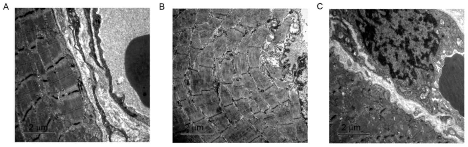
TEM examinations of muscle tissue
The morphology and histology of muscle tissues in
the three groups were observed by TEM. In group A, myocytes and
myomere structures were integral, myofilaments were well organized,
mitochondria appeared normal, and the Z line, M line and H band
remained clear (Fig. 2A). Following
a 15-day microwave treatment, the majority of muscle cells in group
B appeared to have a normal structure, although myofilaments were
slightly disarrayed and occasional swelling of myocytes was
observed in the treatment field (Fig.
2B). Cells in group C exhibited an unusual morphology; the
muscular fibers were twisted and ruptured, the Z line was vague,
local myocytes were partially dissolved and the mitochondria were
swollen (Fig. 2C). Submicroscopic
morphological findings revealed that no marked abnormal
morphological changes occurred in groups A and B; however, the
muscle damage in group C was severe.
Morphological characteristics of
MSCs
A few cells grew from explanted tissue on the second
day of culture in groups A and B. On day 3, the outgrowth of cells
also occurred in group C. The cells were round or spherical,
sparsely distributed in the flask and had a small cell body.
Although the cell morphologies of the three groups were the same, a
greater number of outgrown cells were observed in group B compared
with group A, and the fewest cells were observed in group C
(Fig. 3A-C). On day 7, the speed of
cell growth increased and the number of cells observed increased
markedly. Cells in groups A and C had an ellipsoid or short
shuttle-like form and were large with high refraction. Cells in
group B changed to fusiform and displayed obvious orientation, with
parallel longitudinal axes (Fig.
3D-F). The MSCs were passaged when they approached 80%
confluence, ~9 days later. At that time, the growth rate declined
due to contact inhibition and adjacent cells began to fuse and form
myotube-shaped cells that decreased gradually in width but
increased in length, and exhibited typical spontaneous contraction.
After passage, the cells grew even more rapidly, spread more and
exhibited a spindle shape. They were flat, elongated and evenly
distributed (Fig. 3G-I). Cells grew
steadily after eight passages, and the amount and quality of the
cells in the fourth to sixth passage met the requirements for the
follow-up experiments.
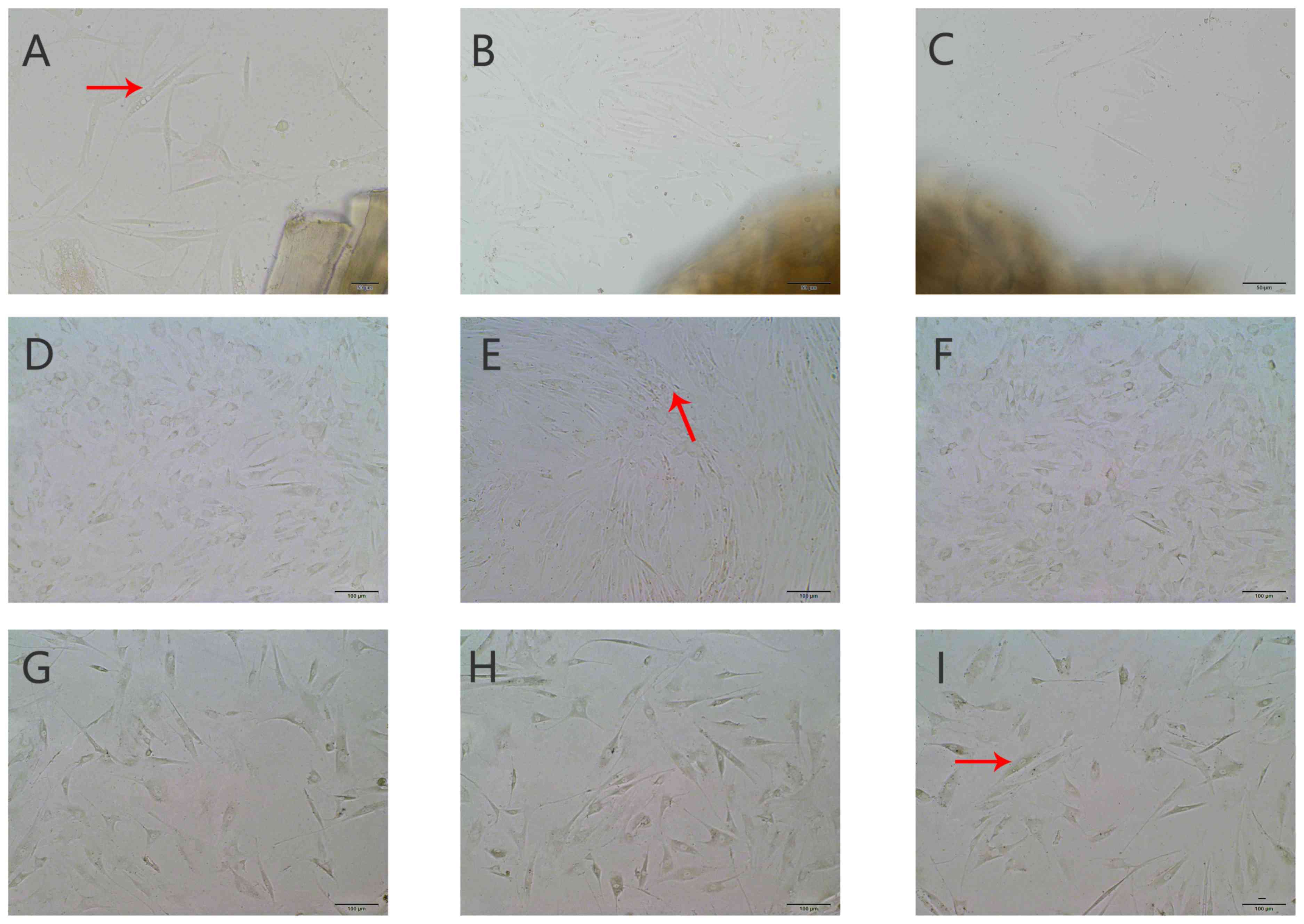 | Figure 3.Morphological observations of muscle
satellite cells in vitro (magnification, ×200). Cell
morphology of primary cell culture on day 3 in groups (A) A, (B) B
and (C) C. Cells were round or spherical (red arrow), a greater
number of outgrown cells were observed in group B compared with
group A, and the fewest cells were observed in group C. Primary
cell culture on day 7 in groups (D) A, (E) B and (F) C. Cells in
groups A and C had an ellipsoid or short shuttle-like form, cells
in group B changed to fusiform (red arrow). Cell sub-culture on day
3 in groups (G) A, (H) B and (I) C, cells exhibited a spindle
shape. They were flat, elongated and evenly distributed (red
arrow). Group A, control group; group B, 25 W microwave treatment;
group C, 50 W microwave treatment. |
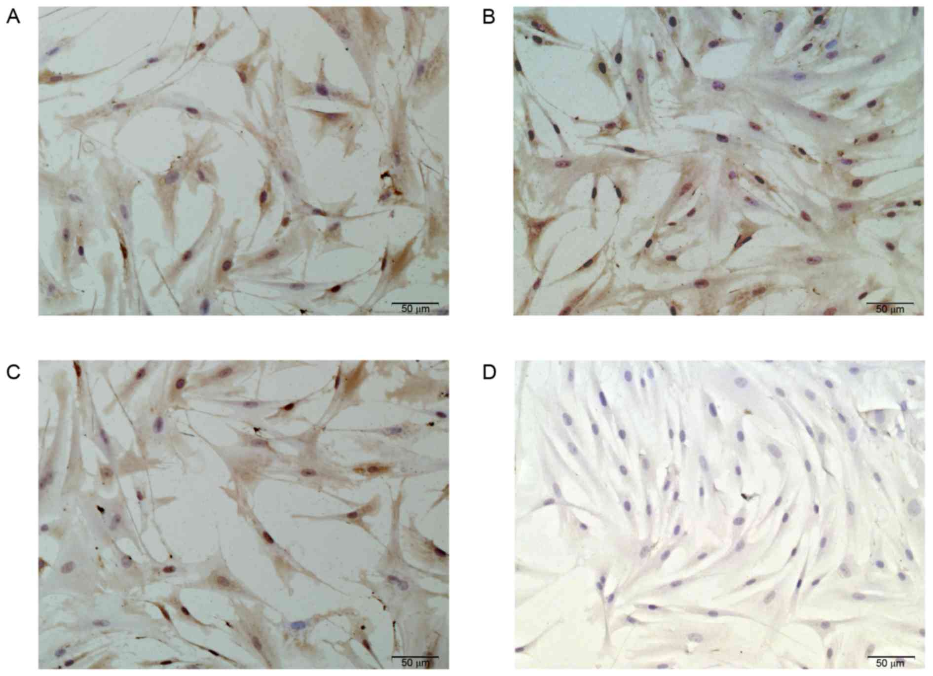
Identification of MSCs
Immunohistochemistry staining results revealed that
the cultured cells in all groups had strong positive staining by
the mouse monoclonal anti-α-sarcomeric actin in the cytoplasm,
while the negative control group had no reaction with PBS. The
cultured cells expressed α-sarcomeric actin, which confirmed the
cells were MSCs. Staining also demonstrated that there was a high
quantity and purity (>95%) of cells (Fig. 4A-D).
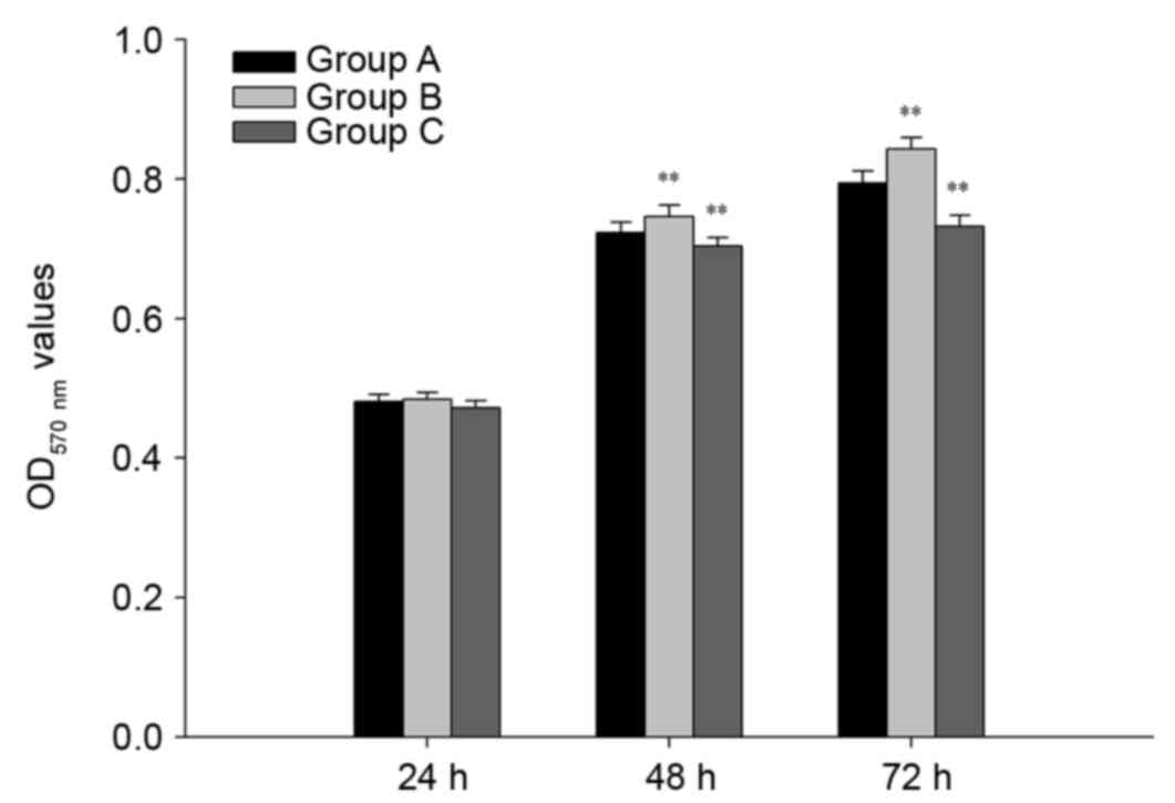
MTT assay
MTT assays were performed to examine the effects of
25 and 50 W microwave irradiation on cell viability in MSCs. The
results were plotted as the MTT curve, which demonstrated that cell
viability of the three groups was enhanced gradually with time
(Fig. 5). At 24 h no statistical
differences were observed in cell viability among the three groups;
however, at 48 and 72 h, the MSC viability in group B was
significantly greater compared with group A (P<0.01).
Furthermore, a significant decrease in cell viability was observed
in group C at 48 and 72 h compared with group A (P<0.01). These
results demonstrated that microwave treatment affects the viability
of MSCs in a time- and dose-dependent manner. The results indicated
that 25 W microwave irradiation had a positive effect on cell
proliferation, whereas 50 W microwave irradiation significantly
inhibited the proliferation of MSCs.
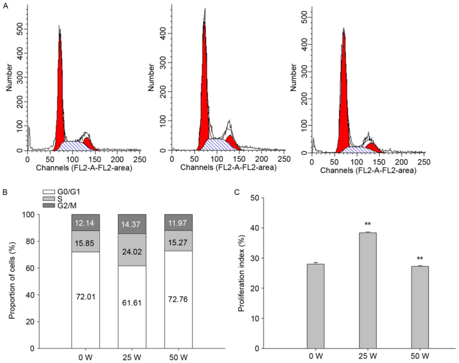
Cell cycle analysis
To investigate the effects of microwave treatment on
the cell cycle of MSCs, the distribution of MSCs in different
phases of the cell cycle was assessed using flow cytometry
(Fig. 6). The percentage of cells in
the G0/G1, G2/M, S-phase, and the proliferation index (PI) were
72.01, 12.14, 15.85 and 27.99% in group A, 61.61, 14.37, 24.02 and
38.39% in group B, and 72.76, 11.97, 15.2 and 27.24% in group C,
respectively. Treatment with 50 W microwave (group C) resulted in a
significant decrease in the PI and cells in the S-phase compared
with group A (P<0.05), suggesting that the proliferation ability
of cells was inhibited. No significant difference was observed in
the G2/M phase cell percentage. Treatment with 25 W microwave
(group B) had the opposite effect to 50 W; the PI and percentage of
cells in the G2/M and S-phase were significantly increased
(P<0.05) compared with group A, indicating that the
proliferative capacity was greater, the proliferation cycle was
shorter and more cells entered the mitotic phase.
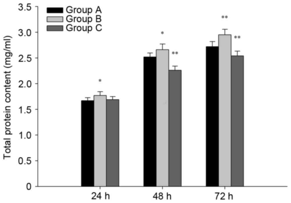
BCA protein assay
The total protein content of MSCs cultured at 24, 48
and 72 h was measured using the BCA protein assay. The results
revealed that the total protein content at 24, 48 and 72 h was
significantly higher in group B compared with the group A
(P<0.05; Fig. 7). At 24 h there
was no statistical difference between groups A and C, whereas the
total protein content in group C at 48 and 72 h was significantly
lower than in group A (P<0.05; Fig.
7). These findings indicated that 25 W microwave treatment may
induce an increase in total protein, whereas 50 W microwave
treatment reduces it. These results were consistent with the cell
proliferation analysis.
Discussion
A large number of animal experimental and clinical
studies have demonstrated that microwaves are able to stimulate the
proliferation and differentiation of osteoblasts (31–33). As
a noninvasive physical factor, microwaves have served an important
role in promoting the rehabilitation of fractures with delayed
union and nonunion in recent years (17,18).
Historically, microwaves have not been used on metallic alloys due
to the theory that when metallic alloys are exposed to microwaves,
the reflection and eddy current generate too much heat to damage
the surrounding tissues, particularly muscle (20,34,35).
Skeletal muscle regenerates poorly, and so such damage would cause
permanent division and proliferation incapacity and irreversible
loss of function. The unique properties of titanium alloy provide
an opportunity to investigate the effects of microwaves. Ye et
al (27,28) demonstrated that 25 W microwave (2,450
MHz) promoted fracture rehabilitation in an animal model with a
titanium alloy implant with no dramatic increase in heat
(<41°C). To the best of our knowledge, the effect of microwaves
on the skeletal muscles adjacent to fracture sites has not yet been
investigated, hindering the application of this technique.
MSCs are located between the sarcolemma and basement
membrane of muscle fiber cells, are able to proliferate and
self-renew, and are regarded as myogenic precursors (36). In normal physiological conditions,
MSCs are dormant or quiescent (37,38).
They are activated to enter the cell cycle in response to muscle
damage and, when they migrate to the damaged areas, they
proliferate and differentiate into multinuclear myotubes to help
repair and regenerate the injured muscle tissues (39,40).
Therefore, whether the proliferative ability of MSCs is normal has
a direct impact on maintaining and repairing the structure and
function of damaged skeletal muscle. The present study explored the
effect of microwaves on the proliferative ability of MSCs in
fractures with titanium alloy implants to verify the efficacy of
this therapy and provide a scientific basis for future clinical
use.
Although muscle cells may suffer damage or loss, the
basement membrane and satellite cells are retained, which provides
a structural framework and cell component for muscle regeneration
(41). In the present study, TEM
images indicated that the skeletal muscles in the experimental
groups were both damaged to different degrees following 15 days of
treatment. Compared with group A, group B exhibited only minor
histopathological changes, whereas cells in group C were badly
damaged. The results demonstrated that the muscle tissue of group B
was in a favorable state compared with group C.
In the present study, the tissue explant method was
used to cultivate the primary MSCs due to its simple operation and
cost-effective use (42). The
disadvantage of this approach is that the target cells mix with
cell debris and impure cells (43).
The modified differential adhesion method is an effective approach
for purifying satellite cells (44).
Fibroblast-like cells are removed as they are better at adhesion
and grow faster than satellite cells. In the present study,
exposure to 25 W microwave treatment resulted in earlier MSC
generation compared with the other two groups. The results of the
present study demonstrated that cells in group B possessed
exuberant proliferation capacity and activity in the early phase of
cell culture. The out growing cells in group C began proliferating
after 3 days, comparatively late compared with the other groups.
The number of cells in group C was also the lowest. The
proliferation and self-renewing abilities of MSCs depend largely on
the magnitude of the damage; due to the relatively minor damage,
cells in group B were activated to proliferate from early on.
Severe damage may disrupt the mechanisms of proliferation and
compromise the ability to regulate it (45).
To confirm that cultivated cells were muscle-derived
cells, specific markers of skeletal muscle were identified.
α-sarcomeric actin is one of the acknowledged specific marker
proteins, and is a structural and functional protein of skeletal
muscle that does not exist in smooth muscle cells (46,47).
Using α-sarcomeric actin monoclonal antibodies for
immunocytochemistry ascertained that the MSCs were skeletal MSCs
and also determined that they were 95% pure. The remaining negative
cells were most likely fibroblasts. A study by Motohashi et
al (48) previously obtained
satellite cells with a purity of 99% or more by using magnetic
activated cell sorting. This approach has a higher purification
rate, but requires complicated processes and higher costs. In the
present study, 95% purification fulfilled the experimental
requirements; however, in future studies the purification and
culture conditions should be further optimized.
Cell viability may be measured either in vivo
or in vitro using cell culture techniques, such as MTT
assays. Both in vivo and in vitro assays have their
own pros and cons. In vitro assays are typically used to
examine cell viability at specific time points and are frequently
applied in scientific research work due to their simplicity, speed
and accuracy (49). In vivo
assays, however, are able to identify more integrated and dynamic
changes in cell viability (50). In
the present study, the MTT assay was used to explore the number of
viable MSCs at different microwave powers. Compared with a single
time point, plotting a growth curve allows for a more comprehensive
understanding of the growth characteristics of MSCs. In general,
the proliferation ability increases over time. No statistical
differences were observed in proliferation ability at 24 h, but at
48 and 72 h the MSC proliferation in group B was the strongest and
in group C was the poorest. The results indicated that 25 W
microwave treatment significantly promoted the proliferation of
MSCs and that 50 W microwave treatment had a negative effect on
cell proliferation. The effect of thermal damage induced by
microwave therapy may be accumulated (51), so the severity of damage is time- and
temperature-dependent. It has been reported that absorbing
microwaves and alternating magnetic fields generate a thermal
effect and may cause tissue heating, which provokes heat stress
(HS) (52). Studies have reported
that HS is able to protect cells from free radical damage by
activating the protein kinase B (Akt)/mechanistic target of
rapamycin signaling pathway and inducing superoxide dismutase
activity (53,54). Heat shock proteins are produced under
heat stress and mediate various cellular processes to provide
buffering against the accumulative action and improve
thermotolerance (55–57). Many studies have reported that when
heat stress increases beyond the critical point, this protective
mechanism fails (58,59). Extended exposure to high temperatures
may cause permanent and irreversible damage, which may explain why
different microwave powers had different effects on
proliferation.
The effects of different microwave powers on the
cell cycle were investigated using flow cytometry. In the S phase
(DNA synthesis), a higher percentage indicates a greater
proliferating potential. The percentage of S + G2/M is PI, which
represents the proportion of cells in the proliferating stage and
reflects the cell proliferation state. PI and S-phase fractions
were highest in group B and were lowest in group A. The results
demonstrated that 25 W microwave treatment enhanced proliferation
and 50 W microwave treatment inhibited it. Microwaves may
accelerate DNA synthesis and promote entrance to the cell cycle by
initiating extracellular signal-regulated protein kinase and
phosphoinositide 3-kinase/Akt signal transduction pathways
(60,61). Studies have also reported that
exposure to hyperthermia may arrest cell division, which is
achieved via influencing the expression of cell cycle regulatory
proteins (62,63). The underlying mechanism responsible
for this remains unclear.
Cell protein content is typically used to estimate
the amount of a substance in cells, but may also be used in growth
experiments (64). Compared with
group A, the protein content in group B was increased and was
decreased in group C, which was consistent with other experimental
results.
In conclusion, the results of the present study
indicated that microwave therapy influences MSC proliferation in
fractures with titanium alloy internal fixation. Treatment with 25
W microwaves promotes MSC proliferation, whereas 50 W inhibits it.
These results indicate that it is safe to use 25 W microwave
therapy on fractures with titanium alloy fixation and provides a
scientific basis for future clinical use. In future studies, the
effect of 25 W microwave therapy on the proliferation of nerves
adjacent to titanium alloy implants should be investigated to
provide more evidence for the therapy's safe use.
Acknowledgements
The authors wish to thank Dr Xianxuan Feng for her
excellent technical assistance with the transmission electron
microscopy examinations.
Funding
No funding was received.
Availability of data and materials
The datasets used and/or analyzed during the current
study are available from the corresponding author on reasonable
request.
Authors' contributions
YB conceived and designed the experiments. YZ, GW
and YX performed the experiments. YZ analyzed the data. YB and YZ
contributed reagents/materials/analysis tools. YZ wrote the
paper.
Ethics approval and consent to
participate
All animal welfare and experimental procedures
involving animals were conducted in strict conformity with the
recommendations in the Guide for the Care and Use of Laboratory
Animals of National Laboratory Animals and protocols were
specifically approved by the Animal Welfare and Ethics Committee of
Shanghai Sixth People's Hospital [Permit no. SYXK (HU) 2011.0128;
Shanghai, China].
Patient consent for publication
Not applicable.
Competing interests
All authors declare that they have no competing
interests.
References
|
1
|
Liang CZ, Ma F, Wu YP and Li Y: Survey on
the basic situation of the bone fractue in rural population of
China. Yi Xue Yan Jiu Za Zhi. 31:10–12. 2002.(In Chinese).
|
|
2
|
Perren SM: Fracture healing. The evolution
of our understanding. Acta Chir Orthop Traumatol Cech. 75:241–246.
2008.PubMed/NCBI
|
|
3
|
Adam P: Treatment of recent trochanteric
fracture in adults. Orthop Traumatol Surg Res. 100 Suppl 1:S75–S83.
2014. View Article : Google Scholar : PubMed/NCBI
|
|
4
|
Yu X, Pang QJ, Chen L, Yang CC and Chen
XJ: Postoperative complications after closed calcaneus fracture
treated by open reduction and internal fixation: A review. J Int
Med Res. 42:17–25. 2014. View Article : Google Scholar : PubMed/NCBI
|
|
5
|
Lynch NM and Linscheid RL: Corrective
osteotomy for scaphoid malunion: Technique and long-term follow-up
evaluation. J Hand Surg. 22:35–43. 1997. View Article : Google Scholar
|
|
6
|
Young MJ and Barrack RL: Complication of
internal fixation of plateau fractures. J Orthop Rev. 23:149–153.
1994.
|
|
7
|
Moore TM, Patzakis MJ and Harvey JP:
Tibial plateau fractures: Definition, demographics, treatment
rationale, and long-term results of closed traction management or
operative reduction. J Orthop Trauma. 1:97–119. 1987. View Article : Google Scholar : PubMed/NCBI
|
|
8
|
Einhorn TA: Enhancement of
fracture-healing. J Bone Joint Surg. 77:940–956. 1995. View Article : Google Scholar : PubMed/NCBI
|
|
9
|
Kim SH, Szabo RM and Marder RA:
Epidemiology of humerus fractures in the United States: Nationwide
emergency department sample, 2008. Arthritis Care Res (Hoboken).
64:407–414. 2012. View Article : Google Scholar : PubMed/NCBI
|
|
10
|
World Health Organization (WHO), . The
Global Burden of Disease: 2004 update. WHO; Geneva: 2008
|
|
11
|
Ji Z, Ma Y, Li W, Li X, Zhao G, Yun Z,
Qian J and Fan Q: The healing process of intracorporeally and in
situ devitalized distal femur by microwave in a dog model and its
mechanical properties in vitro. PLoS One. 7:e305052012. View Article : Google Scholar : PubMed/NCBI
|
|
12
|
Cavaliere R, Ciocatto EC, Giovanella BC,
Heidelberger C, Johnson RO, Margottini M, Mondovi B, Moricca G and
Rossi-Fanelli A: Selective heat sensitivity of cancer cells.
Biochemical and clinical studies. Cancer. 20:1351–1381. 1967.
View Article : Google Scholar : PubMed/NCBI
|
|
13
|
Liebergall M, Abu-Sneineh CH, Eylon S,
Mendelson S, Segal D and Simkin A: Effect of microwave oven induced
mild hyperthermia on bone viability and strength. Clin Orthop Relat
Res. 372:272–279. 2000. View Article : Google Scholar
|
|
14
|
Song CW: Effect of local hyperthermia on
blood flow and microenvironment: A review. Cancer Res. 44 Suppl
10:S4721–S4730. 1984.
|
|
15
|
Wyper DJ and McNiven DR: The effect of
microwave therapy upon muscle blood flow in man. Br J Sports Med.
10:19–21. 1976. View Article : Google Scholar : PubMed/NCBI
|
|
16
|
Sekins KM, Lehmann JF, Esselman P, Dundore
D, Emery AF, deLateur BJ and Nelp WB: Local muscle blood flow and
temperature responses to 915MHz diathermy as simultaneously
measured and numerically predicted. Arch Phys Med Rehabil. 65:1–7.
1984.PubMed/NCBI
|
|
17
|
Leon SA, Asbell SO, Edelstein G, Arastu
HH, Daskal I, Sheehan S, Plunkett DM, Guttmann GG, Packel AJ and
Leon O: Effects of hyperthermia on bone. I. Heating rate patterns
induced by microwave irradiation in bone and muscle phantoms. Int J
Hyperthermia. 9:69–75. 1993. View Article : Google Scholar : PubMed/NCBI
|
|
18
|
Leon SA, Asbell SO, Arastu HH, Edelstein
G, Packel AJ, Sheehan S, Daskal I, Guttmann GG and Santos I:
Effects of hyperthermia on bone. II. Heating of bone in vivo and
stimulation of bone growth. Int J Hyperthermia. 9:77–87. 1993.
View Article : Google Scholar : PubMed/NCBI
|
|
19
|
Chang WH, Sun JS, Chang SP and Lin JC:
Study of thermal effects of ultrasound stimulation on fracture
healing. Bioelectromagnetics. 23:256–263. 2002. View Article : Google Scholar : PubMed/NCBI
|
|
20
|
McIntosh RL, Anderson V and McKenzie RJ: A
numerical evaluation of SAR distribution and temperature changes
around a metallic plate in the head of a RF exposed worker.
Bioelectromagnetics. 26:377–388. 2005. View Article : Google Scholar : PubMed/NCBI
|
|
21
|
Ruggera PS, Witters DM, von Maltzahn G and
Bassen HI: In vitro assessment of tissue heating near metallic
medical implants by exposure to pulsed radio frequency diathermy.
Phys Med Biol. 48:2919–2928. 2003. View Article : Google Scholar : PubMed/NCBI
|
|
22
|
Martin CJ, McCallum HM and Heaton B: An
evaluation of radiofrequency exposure from therapeutic diathermy
equipment in the light of current recommendations. Clin Phys
Physiol Meas. 11:53–63. 1990. View Article : Google Scholar : PubMed/NCBI
|
|
23
|
Kobayashi E, Matsumoto S, Doi H and
Hamanaka H: Mechanical properties of the binary titanium-zirconium
alloys and their potential for biomedical materials. J Biomed Mater
Res. 29:943–950. 1995. View Article : Google Scholar : PubMed/NCBI
|
|
24
|
Donachie M: Biomedical alloys. Adv
Materials Proc. 154:63–65. 1998.
|
|
25
|
Lee MJ, Kim S, Lee SA, Song HT, Huh YM,
Kim DH, Han SH and Suh JS: Overcoming artifacts from metallic
orthopedic implants at high-field-strength MR imaging and
multi-detector CT. RadioGraphics. 27:791–803. 2007. View Article : Google Scholar : PubMed/NCBI
|
|
26
|
Virtanen H, Huttunen J, Toropainen A and
Lappalainen R: Interaction of mobile phones with superficial
passive metallic implants. Phys Med Biol. 50:2689–2700. 2005.
View Article : Google Scholar : PubMed/NCBI
|
|
27
|
Ye D, Xu Y, Fu T, Zhang H, Feng X, Wang G,
Jiang L and Bai Y: Low dose of continuous-wave microwave
irradiation did not cause temperature increase in muscles tissue
adjacent to titanium alloy implants-an animal study. BMC
Musculoskelet Disord. 14:3642013. View Article : Google Scholar : PubMed/NCBI
|
|
28
|
Ye D, Xu Y, Zhang H, Fu T, Jiang L and Bai
Y: Effects of low-dose microwave on healing of fractures with
titanium alloy internal fixation: An experimental study in a rabbit
model. PLoS One. 8:e757562013. View Article : Google Scholar : PubMed/NCBI
|
|
29
|
Rando TA and Blau HM: Primary mouse
myoblast purification, characterization, and transplantation for
cell-mediated gene therapy. J Cell Biol. 125:1276–1287. 1994.
View Article : Google Scholar
|
|
30
|
Huang T, Long M and Huo B: Competitive
binding to cuprous ions of protein and bca in the bicinchoninic
acid protein assay. Open Biomed Eng J. 4:271–278. 2010. View Article : Google Scholar : PubMed/NCBI
|
|
31
|
LI L and Zhang CY: Experimental research
and mechanism of electric stimulation osteogenesis. Chin J Orthop.
17:4707–4714. 1990.
|
|
32
|
Jaecques S, Helsen JA, Maertens S and
Lammens J: Electric stimulation of osteogenesis by dynamically
loaded piezoelectric films: An in vivo exploration in rabbits. Proc
13th Eur Conf Biomaterials. 151997.
|
|
33
|
Haddad JB, Obolensky AG and Shinnick P:
The biologic effects and the therapeutic mechanism of action of
electric and electromagnetic field stimulation on bone and
cartilage: New findings and a review of earlier work. J Altern
Complement Med. 13:485–490. 2007. View Article : Google Scholar : PubMed/NCBI
|
|
34
|
Skonieczki BD, Wells C, Wasser EJ and
Dupuy DE: Radiofrequency and microwave tumor ablation in patients
with implanted cardiac devices: Is it safe? Eur J Radiol.
79:343–346. 2011. View Article : Google Scholar : PubMed/NCBI
|
|
35
|
Cooper J and Hombach V: Increase in
specific absorption rate in human heads arising from implantations.
Electron Let. 32:2217–2219. 1996. View Article : Google Scholar
|
|
36
|
Baroffio A, Bochaton-Piallat ML, Gabbiani
G and Bader CR: Heterogenecity in the progeny of single human
muscle satellite cells. Differentiation. 59:259–268. 1995.
View Article : Google Scholar : PubMed/NCBI
|
|
37
|
Schultz E and McCormick KM: Skeletal
muscle satellite cells. Rev Physiol Biochem Pharmacol. 123:213–257.
1994. View Article : Google Scholar : PubMed/NCBI
|
|
38
|
Snow MH: A quantitative ultrastructure
analysis of satellite cells in denervated fast and slow muscles of
the mouse. Anat Rec. 207:593–604. 1983. View Article : Google Scholar : PubMed/NCBI
|
|
39
|
Parker M, Seale P and Rudnicki MA: Looking
back to the embryo: Defining transcriptional network in adult
myogenesis. Nat Rev Genet. 4:497–507. 2003. View Article : Google Scholar : PubMed/NCBI
|
|
40
|
Mckinnell IW, Ishibashi J, Le Grand F,
Punch VG, Addicks GC, Greenblatt JF, Dilworth FJ and Rudnicki MA:
Pax7 activates myogenic genes by recruitment of a histone
methyltransferase complex. Nat Cell Biol. 10:77–84. 2008.
View Article : Google Scholar : PubMed/NCBI
|
|
41
|
Christov C, Chrétien F, Abou-Khalil R,
Bassez G, Vallet G, Authier FJ, Bassaglia Y, Shinin V, Tajbakhsh S,
Chazaud B and Gherardi RK: Muscle satellite cells and endothelial
cells: Close neighbors and privileged partners. Mol Biol Cell.
18:1397–1409. 2007. View Article : Google Scholar : PubMed/NCBI
|
|
42
|
Fu P: Study on culture method of pleural
mesothelial cells of rats. J Shanxi College of Trad Chin Med.
4:2009.
|
|
43
|
Jiang Z, Jiang YQ, Li XG, Li B and Zhou W:
Construction of the model of primary cultured human glioma cells.
Zhonghua Zhong Liu Fang Zhi Za Zhi. 13:744–747. 2006.(In
Chinese).
|
|
44
|
Ding WJ, Tang Y, Song YL, Su ZD, Li C, Liu
AT, Hu X and Jiang H: Isolation of murine muscle-derived stem cells
with preplate technique combined with limited dilution technique.
Zhongguo Zu Zhi Gong Cheng Yan Jiu Yu Lin Chuang Kang Fu.
15:6797–6801. 2011.(In Chinese).
|
|
45
|
Yablonka-Reuveni Z and Anderson JE:
Satellite cells from dystrophic (mdx) mice display accelerated
differentiation in primary cultures and isolated myofibers. Dev.
Dyn. 235:203–212. 2006.
|
|
46
|
Sejersen T and Lendahl U: Transient
expression of the intermediate filament nestin during skeletal
muscle development. J Cell Sci. 106:1291–1300. 1993.PubMed/NCBI
|
|
47
|
Yablonka-Reuveni Z, Quznn L and Nameroff
M: Isolation and clonal analysis of satellite cells from chicken
pectoralis musele. Dev Biol. 119:252–259. 1987. View Article : Google Scholar : PubMed/NCBI
|
|
48
|
Motohashi N, Asakura Y and Asakura A:
Isolation, culture, and transplantation of muscle satellite cells.
J Vis Exp. 8:e508462014.
|
|
49
|
Castro-Concha LA, Escobedo RM and
Miranda-Ham ML: Measurement of cell viability in in vitro cultures.
Methods Mol Biol. 318:71–76. 2006.PubMed/NCBI
|
|
50
|
Yang PC: Is reliable in vivo detection of
stem cell viability possible in a large animal model of myocardial
injury? Circulation. 126:388–390. 2012. View Article : Google Scholar : PubMed/NCBI
|
|
51
|
Shields N, Gormley J and O'Hare N:
Short-wave diathermy: Current clinical and safety practices.
Physiother Res Int. 7:191–202. 2002. View Article : Google Scholar : PubMed/NCBI
|
|
52
|
Velichko AK, Markova EN, Petrova NV, Razin
SV and Kantidze OL: Mechanisms of heat shock response in mammals.
Cell Mol Life Sci. 70:4229–4241. 2013. View Article : Google Scholar : PubMed/NCBI
|
|
53
|
Gao CQ, Zhao YL, Li HC, Sui WG, Yan HC and
Wang XQ: Heat stress inhibits proliferation, promotes growth, and
induces apoptosis in cultured Lantang swine skeletal muscle
satellite cells. J Zhejiang Univ Sci B. 16:549–559. 2015.
View Article : Google Scholar : PubMed/NCBI
|
|
54
|
Lille S, Su CY, Schoeller T, Suchy H,
Lyons S, Russell RC, Neumeister M and Lai CC: Induction of heat
shock protein 72 in rat skeletal muscle does not increase tolerance
to ischemia-reperfusion injury. Muscle Nerve. 22:390–393. 1999.
View Article : Google Scholar : PubMed/NCBI
|
|
55
|
Lindquist S: The heat-shock response. Annu
Rev Biochem. 55:1151–1191. 1986. View Article : Google Scholar : PubMed/NCBI
|
|
56
|
Pelham HRB: Functions of the HSP70 protein
family: An overview. CPHI Press; Cold Spring Harbor, NY: pp.
287–299. 1990
|
|
57
|
Marber MS: Ischemic preconditioning in
isolated cells. Circ Res. 86:926–931. 2000. View Article : Google Scholar : PubMed/NCBI
|
|
58
|
Li SQ, Li RF, Xi SM, Hu S, Jia ZQ, Li SP,
Wen XL, Song YK, Li S, Li SP, et al: Systematical analysis of
impacts of heat stress on the proliferation, apoptosis and
metabolism of mouse hepatocyte. J Physiol Sci. 62:29–43. 2012.
View Article : Google Scholar : PubMed/NCBI
|
|
59
|
Rad Rezai M, Wise GE, Brooks H, Flanagan
MB and Yao S: Activation of proliferation and differentiation of
dental follicle stem cells (DFSCs) by heat stress. Cell Prolif.
46:58–66. 2013. View Article : Google Scholar : PubMed/NCBI
|
|
60
|
Yang SY, Hoy M, Fuller B, Sales KM,
Seifalian AM and Winslet MC: Pretreatment with insulin-like growth
factor 1 protects skeletal muscle cells against oxidative damage
via PI3K/Akt an ERK1/2 MAPK pathways. Lab Invest. 90:391–401. 2010.
View Article : Google Scholar : PubMed/NCBI
|
|
61
|
Li Y, Zhang P, Qiu F, Chen L, Miao C, Li
J, Xiao W and Ma E: Inactivation of PI3K/Akt signaling mediates
proliferation inhibition and G2/M phase arrest induced by
andrographolide in human glioblastoma cells. Life Sci. 90:962–967.
2012. View Article : Google Scholar : PubMed/NCBI
|
|
62
|
Kühl NM and Rensing L: Heat shock effects
on cell cycle progression. Cell Mol. Life Sci. 57:450–463. 2000.
View Article : Google Scholar
|
|
63
|
Zhang M, Jiang M, Bi Y, Zhu H, Zhou Z and
Sha J: Autophagy and apoptosis act as partners to induce germ cell
death after heat stress in mice. PLoS One. 7:e414122012. View Article : Google Scholar : PubMed/NCBI
|
|
64
|
Xu L, Huang J and Xiang XR: Effects of
osteoblastic growth peptide on the proliferation and total protein
content in cultured human periodontal ligament cells. Chongqing Yi
Ke Da Xue Xue Bao. 34:420–423. 2009.(In Chinese).
|