Introduction
With the development of society, people's living
standard and quality have improved, and increasingly more people
have begun to pay attention to oral problems. Moreover, the health
problem of tooth, as an indispensable part of people's oral cavity,
is also important (1). Dental
disease is a common disease in clinic, and clinical transplantation
and repair techniques play significant roles in the treatment of
patients' pain and recovery of masticatory function (2,3).
However, neither function nor sense of transplanted dentures can be
comparable with natural teeth. Therefore, scholars all over the
world are making efforts to find an effective method of tooth
regeneration. In recent years, studies have shown that there is a
certain correlation between tooth development and signaling
pathway, and the combination with tissue engineering is an
important key to tooth regeneration.
Dental pulp cells are regenerative cells with
differentiation capacity, which, regulated and induced by several
factors, can be differentiated into odontoblasts through continuous
self-renewal capacity under appropriate conditions, repairing teeth
(4). Bone morphogenetic protein
(BMP) pathway plays an extremely important regulatory role in the
interaction between dental epithelium and mesenchyme (5). Some studies indicate that (6) BMP signal can accurately regulate
intracellular and extracellular factors, thus achieving
intracellular and extracellular dynamic balance, which plays a
crucial role in maintaining the normal tooth development. Once such
balance is broken, the tooth development will be affected. The
expression of BMP-4, as the first factor identified by human in the
interaction between dental epithelium and mesenchyme, can well
reflect the dental epithelium and mesenchyme-induced tooth
potentiality, which can also induce some morphogenesis processes of
dental germ, thus exerting a relative regulatory effect (7). In this study, therefore, human dental
pulp cells were cultured in vitro under the influence of
BMP-4 to observe the proliferation and differentiation capacities
of dental pulp cells.
Materials and methods
Main reagents and instruments
High-glucose Dulbecco's modified Eagle's medium
(DMEM) and fetal bovine serum (FBS) were purchased from Gibco
(Gibco; Thermo Fisher Scientific, Inc., Waltham, MA, USA). BCIP/NBT
alkaline phosphatase (ALP) staining kit was purchased from
Sigma-Aldrich (Sigma-Aldrich: Merck KGaA, St. Louis, MO, USA).
BMP-4 was purchased from Thermo Fisher Scientific, Inc. (Waltham,
MA, USA). TRIzol, reverse transcription kit, and fluorescence
quantitative polymerase chain reaction (PCR) kit were from
Invitrogen (Invitrogen: Thermo Fisher Scientific, Inc., Carlsbad,
CA, USA). ABI 7500 Fluorescence PCR amplification instrument was
purchased from Applied Biosystems (Applied Biosystems: Thermo
Fisher Scientific, Inc., Foster City, CA, USA).
Extraction and passage of dental pulp
cells
Experimental samples in this study were taken from
normal teeth extracted from patients aged <30 years old in China
Medical University School and Hospital of Stomatology due to
orthodontics. Before extraction, patients were informed and signed
the informed consent. The study was approved by the Ethics
Committee of School of Stomatology, China Medical University
(Shenyang, China). After extraction, teeth were transferred to a
sterile environment and dental crown was split to extract dental
pulp tissues within 30 min. The extracted dental pulp tissues were
cut into 1 mm3 blocks and transferred into a culture
flask. After 15% FBS and high-glucose DMEM were added, the culture
flask was placed into a constant temperature incubator with 5%
CO2 for incubation at 37°C, and the liquid was replaced
once every 3 days. The culture medium was observed, and cells were
digested with 0.25% trypsin when they covered >85% of the
medium, followed by counting and passage. Finally, the cell growth
curves at day 1, 2, 3, 5 and 7 were drawn.
Experimental grouping
In the experiment, the 3rd-5th generations of dental
pulp cells in good growth conditions were selected and digested
with 0.25% trypsin. The density was adjusted with high-glucose
medium containing 10% FBS (4.5×10⁴ cells/ml). Then cells
were inoculated into a 48-well plate (500 µl/well), and cultured in
the constant temperature incubator with 5% CO2 at 37°C.
After cell adherence, they were divided into group A (100 ng/ml
BMP-4) and group B (no treatment, experimental control group).
Immunocytochemical staining
The 3rd-5th generations of dental pulp cells in both
groups were taken, inoculated into a 6-well plate at a density of
4.5×10⁴ cells/ml, and incubated in constant temperature
incubator with 5% CO2 at 37°C for 3–5 days. When 85%
cells were fused, they were rinsed with phosphate-buffered saline
(PBS) 3 times (30–50 sec/time), fixed with 4% paraformaldehyde,
rinsed again with PBS and air-dried. Then cells were soaked in
Triton X-100 detergent for 5–10 min, rinsed with PBS, air-dried and
soaked in 3% H2O2. After being rinsed again
with PBS and air-dried, cells were sealed with FBS. Then, primary
mouse monoclonal vimentin antibody (dilution, 1:200; cat. no.
ab8978) and mouse monoclonal keratin antibody (dilution, 1:200;
cat. no. ab169328) were added for incubation with ice at 4°C
overnight. The next day, the cells were taken, cultured in constant
temperature incubator with 5% CO2 at 37°C, washed with
PBS, air dried and added with secondary rabbit anti-mouse (HRP) IgG
antibody (dilution, 1:1,000; cat. no. ab6728), followed by wet
incubation at 37°C. All the antibodies were purchased from Abcam
(Cambridge, MA, USA). Finally, the secondary antibody was
discarded. Then, horseradish peroxidase markers were added for
labeling, and cells were washed with PBS and air-dried, followed by
addition of developing solution for color development,
hematoxylin-eosin staining, sealing and observation under a
microscope (Olympus, Tokyo, Japan).
Reverse transcription-quantitative PCR
(RT-qPCR)
Total ribonucleic acid (RNA) was extracted using
TRIzol solution from cells cultured at day 5 and 7 in strict
accordance with the instructions. The concentration of total RNA
extracted was detected using an ultraviolet spectrophotometer
(Hitachi, Tokyo, Japan), and the purity of total RNA was detected
via protein electrophoresis. Then, total RNA was reverse
transcribed into complementary deoxyribonucleic acid (cDNA) using
the reverse transcription kit in strict accordance with the
manufacturer's instructions. The reaction system was prepared
strictly according to the instructions of the fluorescence
quantitative PCR kit. Expression of ALP, dentin sialophosphoprotein
(DSPP) and dentin matrix protein-1 (DMP-1) messenger ribonucleic
acid (mRNA) was detected. U6 was used as an internal control for
amplification, and amplification primers were designed and
synthesized by Shanghai GenePharma Co., Ltd. (Shanghai, China). The
primer sequences are shown in Table
I.
 | Table I.Primer sequences. |
Table I.
Primer sequences.
| Genes | Primer sequences |
|---|
| ALP | F
5′-ACACCTTGACTGTGGTTACTGCTGA-3′ |
|
| R
5′-CCTTGTAGCCAGGCCCGTTA-3′ |
| DSPP | F
5′-TTCTCCTACTCAGCCCATTTTA-3′ |
|
| R
5′-CCATCGTGACCGTATGTTTCTA-3′ |
| DMP-1 | F
5′-TGGGTTTGTTGTGATAGG-3′ |
|
| R
5′-GGAAGAGGTGGTGAGTGA-3′ |
| U6 | F
5′-CTCGCTTCGGCAGCACA-3′ |
|
| R
5′-AACGCTTCACGAATTTGCGT-3′ |
Cell staining
After culture for 12 days, cells in both groups were
stained using the BCIP/NBT ALP kit according to the instructions of
the BCIP/NBT ALP kit. The ALP-stained cells were observed under an
inverted microscope, and the area of positive cells was recorded
and analyzed using Image-Pro Plus software. The experiment was
repeated 3 times.
Statistical analysis
In this study, Statistical Product and Service
Solutions (SPSS) 22.0 software package (IBM Corp., Armonk, NY, USA)
was used for the statistical analysis of data collected in this
experiment. Enumeration data are presented as mean ± standard
deviation (SD), analysis of variance was used for the intergroup
comparison, and Student-Newman-Keuls test was performed for
pairwise comparison. P<0.05 suggested that the difference was
statistically significant.
Results
Cell culture results and morphological
observation
Cells were cultured and observed. When dental pulp
cells were cultured for 7 days, the difference in primary stem
cells from other cells could be clearly seen, and cells were
distributed in long fusiform type and stable after passage. It was
found in the observation of vimentin and keratin via
immunohistochemical staining that the vimentin staining was
positive, while the keratin staining was negative, indicating that
cells derived from the mesoderm and displayed the fibrous shape in
this study, meeting the requirements of this experiment.
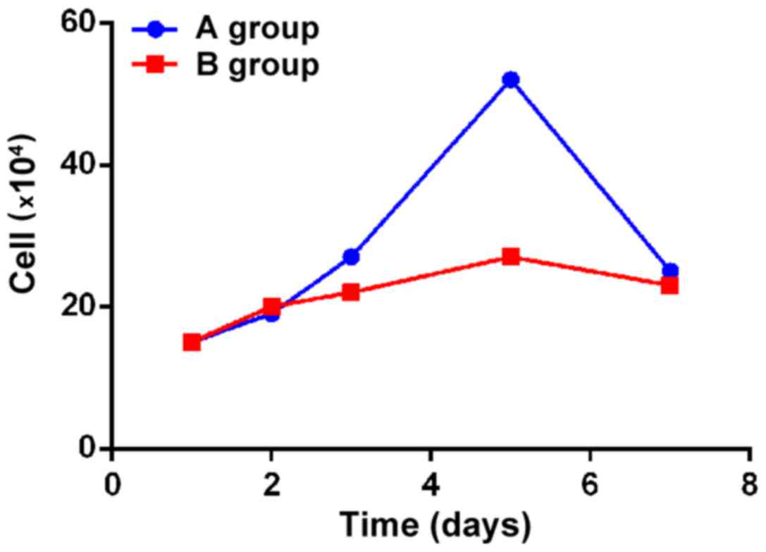
Analyses of cell growth curves in the
two groups
In this study, the growth curves were drawn in both
groups. It was manifested that the cell growth rate in group A
added with BMP-4 was significantly higher than that in group B
without treatment, and there was a statistically significant
difference between the two groups (p<0.05). The cell growth
rates in both groups were increased remarkably in an exponential
manner at day 3 after culture, the cell proliferation reached the
peak at day 5, and then slowed down (Fig. 1).
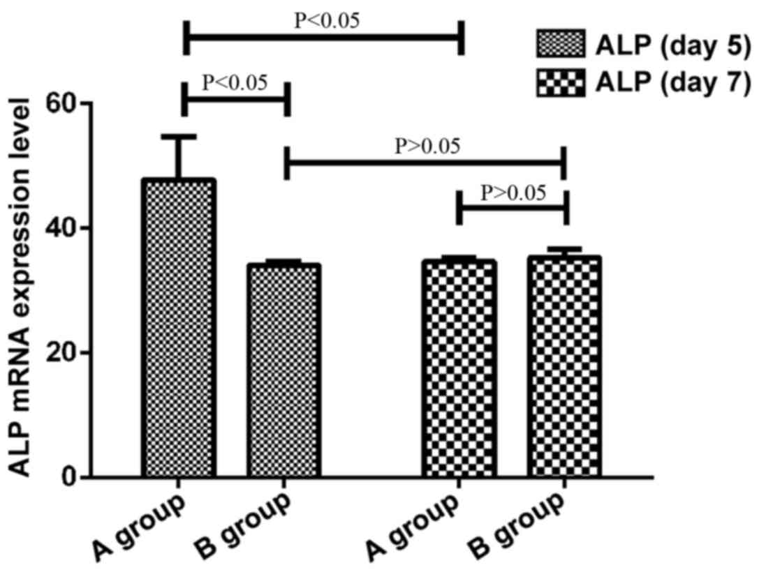
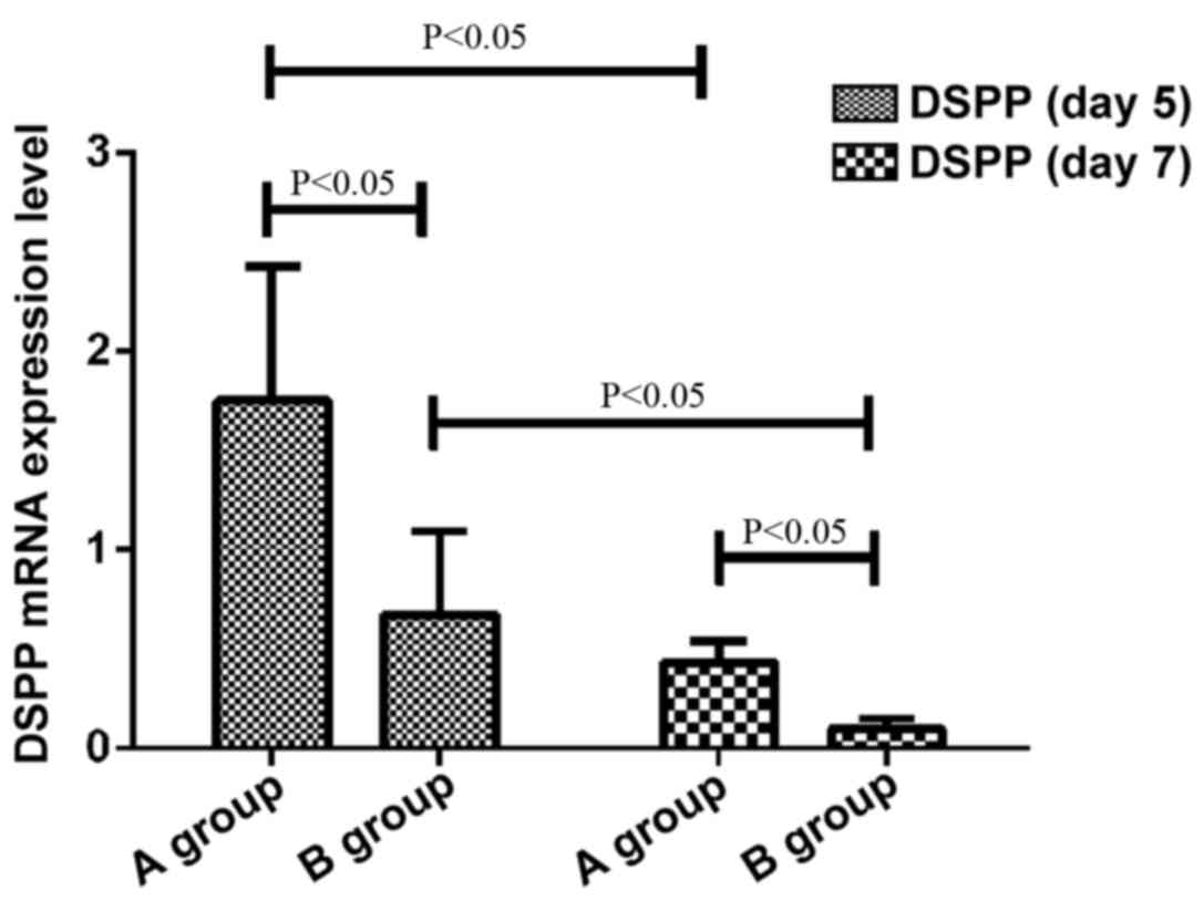
Detection of the expression of ALP,
DSPP and DMP-1 in cells by PCR
At day 5 and 7 after culture, cells in both groups
were taken to detect the expression of ALP, DSPP and DMP-1 via
RT-qPCR. The expression levels of ALP, DSPP and DMP-1 in group A at
day 5 after culture were significantly increased compared with
those in group B, and there were statistically significant
differences (p<0.05). Compared with those at day 5 after
culture, the expression levels of DSPP in both groups of cells were
significantly decreased at day 7 after culture (p<0.05), and the
expression levels of DMP-1 in both groups of cells were
significantly increased (p<0.05). Besides, compared with that at
day 5 after culture, the expression level of ALP in group A was
decreased (p<0.05), but there was no significant difference in
group B (p>0.05) (Figs.
2–4).
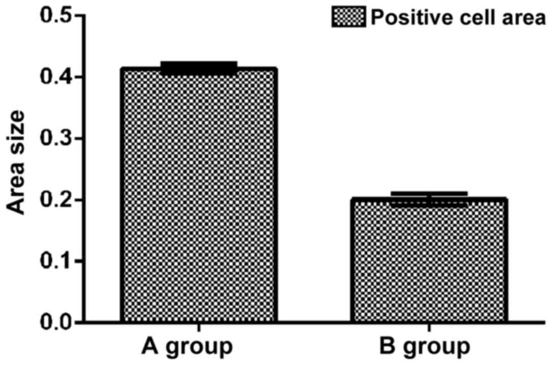
ALP staining results
At day 12 after culture, cells in both groups were
stained. Staining results displayed that cells in both groups were
stained successfully, and purple-black sediment could be observed
in both groups, but it was significantly darker in group A than
that in group B, showing strongly positive. Then, the proportion of
positive area was determined using the Image-Pro Plus image
software. Results showed that there was a significant difference in
the proportion of positive cells in group B (0.201±0.010) compared
with that in group A (0.414±0.008) (p<0.05) (Fig. 5).
Discussion
Dental pulp cells are predominantly distributed in
adult dental pulp cells that possess multiple differentiation and
self-renewal capacities, and the same characteristics as other
tissue stem cells in the body (8).
Studies have proved that (9)
differentiation and self-renewal capacities of dental pulp cells
are regulated by biological factors and signaling pathways.
Recently it was demonstrated in a large number of studies that BMP,
as an indispensable morphogenetic protein in tooth growth, can
promote the growth and development of human teeth through the
influence of multiple pathways (10–12).
As an extracellular signal molecule of transforming
growth factor-β family, BMP-4 plays an important role in the tooth
and bone growth and repair processes in the body, and also plays a
regulatory role in the growth, apoptosis and differentiation of
human dental pulp cells, which can maintain the renewal capacity of
dental pulp cells through blocking the mitogen-activated protein
kinase signaling pathway (13). In
addition to its effects on the growth and development of teeth and
bones, BMP-4 also exerts important effects on the embryonic
development and differentiation in digestive, reproductive and
nervous systems in the human body (14,15).
In this study, human dental pulp cells were cultured
in vitro to observe the effect of BMP-4 on dental pulp
cells. Through drawing the growth curves in both groups of cells,
it was found that the cell growth rate in group A pre-treated with
BMP-4 was significantly higher than that in group B, reaching the
peak at day 5 after culture, and the cell proliferation was
weakened after 5 days, possibly because the cell density was lower
in the early stage of growth, and cells contacted less with BMP-4.
With the cell growth, the secretion of BMP-4 was insufficient in
both groups, but the content of BMP-4 in cells in group A was
higher than that in group B due to the pre-treatment with BMP-4,
thus increasing the cell growth rate.
It has been reported that (16) the inactivation of BMP-4 in mouse
mandibular mesenchyme in animal model test leads to growth cease of
mandibular molar in mouse, but the growth of maxillary molar is
normal without significant differences from normal mouse. Moreover,
Zhang et al (17) implanted
the rhBMP4-attached agarose beads into the mouse tooth germ, and
they found after culture in vitro for 24 h that the cell
differentiation is significantly accelerated. It is visible through
the above studies that BMP-4 plays an important role in the growth
of human teeth, confirming the results of this experiment. ALP
activity is an important index of cell mineralization ability, and
the ALP expression level can directly reflect the degree of cell
differentiation. Therefore, ALP can serve as a sign of dental pulp
cell differentiation and formation (18). A number of scholars regard DSPP, one
of the important components of dentin non-collagen protein, as the
specific protein of dentin cells (19). Besides, DMP-1 can induce mesenchymal
cells to form dentin cells, and promote the formation of minera-
lization (20). In this experiment,
therefore, RT-qPCR was performed for both groups of cells cultured
for 5 and 7 days, and the expression of ALP, DSPP and DMP-1 was
detected. According to the results, the expression levels of ALP,
DSPP and DMP-1 in group A at day 5 after culture were obviously
increased with statistically significant differences compared with
those in group B (p<0.05), exactly illustrating that
differentiation markers of dental pulp cells pre-treated with BMP-4
are remarkably increased, and also well demonstrating that BMP-4
contributes to the growth of dental pulp cells. Finally, the
proportion of positive cell area was detected via ALP staining, and
results displayed that the positive area in group A was
significantly larger than that in group B (p<0.05), indicating
that pre-treatment with BMP-4 can increase the ALP activity and
promote cell matrix calcification.
There were also some defects in this study. Its
mechanism was not investigated deeply, and its regulatory mechanism
and pathways involved in regulation were not fully understood.
Therefore, its mechanism will be further studied in future
experiments.
In conclusion, pre-treatment with BMP-4 can
effectively promote the proliferation and differentiation of dental
pulp cells, providing a new method for the differentiation of
dental pulp cells.
Acknowledgements
Not applicable.
Funding
No funding was received.
Availability of data and materials
The datasets used and/or analyzed during the present
study are available from the corresponding author on reasonable
request.
Authors' contributions
NS designed this study, collected and analyzed the
data, as well as wrote this manuscript. TJ and CW contributed to
the extraction and passage of dental pulp cells. HS performed
immunocytochemical staining. QZ conducted RT-qPCR. LL recorded and
analyzed cell staining. All authors read and approved the final
manuscript.
Ethics approval and consent to
participate
The study was approved by the Ethics Committee of
School of Stomatology, China Medical University (Shenyang, China).
Before extraction, patients were informed and signed the informed
consent.
Patient consent for publication
Not applicable.
Competing interests
The authors declare that they have no competing
interests.
References
|
1
|
Lamont RJ, Hajishengallis GN and Jenkinson
HF: Oral Microbiology and Immunology. 2nd. ASM Press; Washington,
DC: 2014
|
|
2
|
Clokie CM, Yau DM and Chano L: Autogenous
tooth transplantation: An alternative to dental implant placement?
J Can Dent Assoc. 67:92–96. 2001.PubMed/NCBI
|
|
3
|
Lai WF, Lee JM and Jung HS: Molecular and
engineering approaches to regenerate and repair teeth in mammals.
Cell Mol Life Sci. 71:1691–1701. 2014. View Article : Google Scholar : PubMed/NCBI
|
|
4
|
Nakanishi T, Mukai K, Hosokawa Y, Takegawa
D and Matsuo T: Catechins inhibit vascular endothelial growth
factor production and cyclooxygenase-2 expression in human dental
pulp cells. Int Endod J. 48:277–282. 2015. View Article : Google Scholar : PubMed/NCBI
|
|
5
|
Antebi YE, Linton JM, Klumpe H, Bintu B,
Gong M, Su C, McCardell R and Elowitz MB: Combinatorial signal
perception in the BMP pathway. Cell. 170:1184–1196.e24. 2017.
View Article : Google Scholar : PubMed/NCBI
|
|
6
|
Jia S, Kwon HE, Lan Y, Zhou J, Liu H and
Jiang R: Bmp4-Msx1 signaling and Osr2 control tooth organogenesis
through antagonistic regulation of secreted Wnt antagonists. Dev
Biol. 420:110–119. 2016. View Article : Google Scholar : PubMed/NCBI
|
|
7
|
Nascimento MA, Nonaka CF, Barboza CA,
Freitas RA, Pereira Pinto L and Souza LB: Immunoexpression of BMP-2
and BMP-4 and their receptors, BMPR-IA and BMPR-II, in
ameloblastomas and adenomatoid odontogenic tumors. Arch Oral Biol.
73:223–229. 2017. View Article : Google Scholar : PubMed/NCBI
|
|
8
|
Hwang HI, Lee TH, Kang KJ, Ryu CJ and Jang
YJ: Immunomic screening of cell surface molecules on
undifferentiated human dental pulp stem cells. Stem Cells Dev.
24:1934–1945. 2015. View Article : Google Scholar : PubMed/NCBI
|
|
9
|
Chang MC, Chang HH, Lin PS, Huang YA, Chan
CP, Tsai YL, Lee SY, Jeng PY, Kuo HY, Yeung SY and Jeng JH: Effects
of TGF-β1 on plasminogen activation in human dental pulp cells:
Role of ALK5/Smad2, TAK1 and MEK/ERK signalling. J Tissue Eng Regen
Med. 12:854–863. 2018. View Article : Google Scholar : PubMed/NCBI
|
|
10
|
Yang J, Ye L, Hui TQ, Yang DM, Huang DM,
Zhou XD, Mao JJ and Wang CL: Bone morphogenetic protein 2-induced
human dental pulp cell differentiation involves p38
mitogen-activated protein kinase-activated canonical WNT pathway.
Int J Oral Sci. 7:95–102. 2015. View Article : Google Scholar : PubMed/NCBI
|
|
11
|
Luukko K and Kettunen P: Integration of
tooth morphogenesis and innervation by local tissue interactions,
signaling networks, and semaphorin 3A. Cell Adhes Migr. 10:618–626.
2016. View Article : Google Scholar
|
|
12
|
Zhang F, Song J, Zhang H, Huang E, Song D,
Tollemar V, Wang J, Wang J, Mohammed M, Wei Q, et al: Wnt and BMP
signaling crosstalk in regulating dental stem cells: Implications
in dental tissue engineering. Genes Dis. 3:263–276. 2016.
View Article : Google Scholar : PubMed/NCBI
|
|
13
|
Wang Y, He H, Cao Z, Fang Y, Du M and Liu
Z: Regulatory effects of bone morphogenetic protein-4 on tumour
necrosis factor-α-suppressed Runx2 and osteoprotegerin expression
in cementoblasts. Cell Prolif. 50:e123442017. View Article : Google Scholar
|
|
14
|
Xie FY, Xu WH, Yin C, Zhang GQ, Zhong YQ
and Gao J: Nanomedicine strategies for sustained, controlled, and
targeted treatment of cancer stem cells of the digestive system.
World J Gastrointest Oncol. 8:735–744. 2016. View Article : Google Scholar : PubMed/NCBI
|
|
15
|
Yuzaki M: Two classes of secreted synaptic
organizers in the central nervous system. Annu Rev Physiol.
80:243–262. 2018. View Article : Google Scholar : PubMed/NCBI
|
|
16
|
Jia S, Zhou J, Gao Y, Baek JA, Martin JF,
Lan Y and Jiang R: Roles of Bmp4 during tooth morphogenesis and
sequential tooth formation. Development. 140:423–432. 2013.
View Article : Google Scholar : PubMed/NCBI
|
|
17
|
Zhang J, Tian W, Liu L, Li S and Tang W:
Expression of bone morphogenetic protein (BMP)-2, 4 in mouse
embryonic tooth during the bud stage. Hua Xi Kou Qiang Yi Xue Za
Zhi = Huaxi kouqiang yixue zazhi. 19:392–393. 2001.(In Chinese).
PubMed/NCBI
|
|
18
|
Chang SW, Lee SY, Kum KY and Kim EC:
Effects of ProRoot MTA Bioaggregate, and Micromega MTA on
odontoblastic differentiation in human dental pulp cells. J Endod.
40:113–118. 2014. View Article : Google Scholar : PubMed/NCBI
|
|
19
|
Koli K, Saxena G and Ogbureke KU:
Expression of matrix metalloproteinase (MMP)-20 and potential
interaction with dentin sialophosphoprotein (DSPP) in human major
salivary glands. J Histochem Cytochem. 63:524–533. 2015. View Article : Google Scholar : PubMed/NCBI
|
|
20
|
Lourenço Neto N, Marques NC, Fernandes AP,
Rodini CO, Sakai VT, Abdo RC, Machado MA, Santos CF and Oliveira
TM: Immunolocalization of dentin matrix protein-1 in human primary
teeth treated with different pulp capping materials. J Biomed Mater
Res B Appl Biomater. 104:165–169. 2016. View Article : Google Scholar : PubMed/NCBI
|