Spandidos Publications Logo
  • About
    • About Spandidos
    • Aims and Scopes
    • Abstracting and Indexing
    • Editorial Policies
    • Reprints and Permissions
    • Job Opportunities
    • Terms and Conditions
    • Contact
  • Journals
    • All Journals
    • Oncology Letters
      • Oncology Letters
      • Information for Authors
      • Editorial Policies
      • Editorial Board
      • Aims and Scope
      • Abstracting and Indexing
      • Bibliographic Information
      • Archive
    • International Journal of Oncology
      • International Journal of Oncology
      • Information for Authors
      • Editorial Policies
      • Editorial Board
      • Aims and Scope
      • Abstracting and Indexing
      • Bibliographic Information
      • Archive
    • Molecular and Clinical Oncology
      • Molecular and Clinical Oncology
      • Information for Authors
      • Editorial Policies
      • Editorial Board
      • Aims and Scope
      • Abstracting and Indexing
      • Bibliographic Information
      • Archive
    • Experimental and Therapeutic Medicine
      • Experimental and Therapeutic Medicine
      • Information for Authors
      • Editorial Policies
      • Editorial Board
      • Aims and Scope
      • Abstracting and Indexing
      • Bibliographic Information
      • Archive
    • International Journal of Molecular Medicine
      • International Journal of Molecular Medicine
      • Information for Authors
      • Editorial Policies
      • Editorial Board
      • Aims and Scope
      • Abstracting and Indexing
      • Bibliographic Information
      • Archive
    • Biomedical Reports
      • Biomedical Reports
      • Information for Authors
      • Editorial Policies
      • Editorial Board
      • Aims and Scope
      • Abstracting and Indexing
      • Bibliographic Information
      • Archive
    • Oncology Reports
      • Oncology Reports
      • Information for Authors
      • Editorial Policies
      • Editorial Board
      • Aims and Scope
      • Abstracting and Indexing
      • Bibliographic Information
      • Archive
    • Molecular Medicine Reports
      • Molecular Medicine Reports
      • Information for Authors
      • Editorial Policies
      • Editorial Board
      • Aims and Scope
      • Abstracting and Indexing
      • Bibliographic Information
      • Archive
    • World Academy of Sciences Journal
      • World Academy of Sciences Journal
      • Information for Authors
      • Editorial Policies
      • Editorial Board
      • Aims and Scope
      • Abstracting and Indexing
      • Bibliographic Information
      • Archive
    • International Journal of Functional Nutrition
      • International Journal of Functional Nutrition
      • Information for Authors
      • Editorial Policies
      • Editorial Board
      • Aims and Scope
      • Abstracting and Indexing
      • Bibliographic Information
      • Archive
    • International Journal of Epigenetics
      • International Journal of Epigenetics
      • Information for Authors
      • Editorial Policies
      • Editorial Board
      • Aims and Scope
      • Abstracting and Indexing
      • Bibliographic Information
      • Archive
    • Medicine International
      • Medicine International
      • Information for Authors
      • Editorial Policies
      • Editorial Board
      • Aims and Scope
      • Abstracting and Indexing
      • Bibliographic Information
      • Archive
  • Articles
  • Information
    • Information for Authors
    • Information for Reviewers
    • Information for Librarians
    • Information for Advertisers
    • Conferences
  • Language Editing
Spandidos Publications Logo
  • About
    • About Spandidos
    • Aims and Scopes
    • Abstracting and Indexing
    • Editorial Policies
    • Reprints and Permissions
    • Job Opportunities
    • Terms and Conditions
    • Contact
  • Journals
    • All Journals
    • Biomedical Reports
      • Information for Authors
      • Editorial Policies
      • Editorial Board
      • Aims and Scope
      • Abstracting and Indexing
      • Bibliographic Information
      • Archive
    • Experimental and Therapeutic Medicine
      • Information for Authors
      • Editorial Policies
      • Editorial Board
      • Aims and Scope
      • Abstracting and Indexing
      • Bibliographic Information
      • Archive
    • International Journal of Epigenetics
      • Information for Authors
      • Editorial Policies
      • Editorial Board
      • Aims and Scope
      • Abstracting and Indexing
      • Bibliographic Information
      • Archive
    • International Journal of Functional Nutrition
      • Information for Authors
      • Editorial Policies
      • Editorial Board
      • Aims and Scope
      • Abstracting and Indexing
      • Bibliographic Information
      • Archive
    • International Journal of Molecular Medicine
      • Information for Authors
      • Editorial Policies
      • Editorial Board
      • Aims and Scope
      • Abstracting and Indexing
      • Bibliographic Information
      • Archive
    • International Journal of Oncology
      • Information for Authors
      • Editorial Policies
      • Editorial Board
      • Aims and Scope
      • Abstracting and Indexing
      • Bibliographic Information
      • Archive
    • Medicine International
      • Information for Authors
      • Editorial Policies
      • Editorial Board
      • Aims and Scope
      • Abstracting and Indexing
      • Bibliographic Information
      • Archive
    • Molecular and Clinical Oncology
      • Information for Authors
      • Editorial Policies
      • Editorial Board
      • Aims and Scope
      • Abstracting and Indexing
      • Bibliographic Information
      • Archive
    • Molecular Medicine Reports
      • Information for Authors
      • Editorial Policies
      • Editorial Board
      • Aims and Scope
      • Abstracting and Indexing
      • Bibliographic Information
      • Archive
    • Oncology Letters
      • Information for Authors
      • Editorial Policies
      • Editorial Board
      • Aims and Scope
      • Abstracting and Indexing
      • Bibliographic Information
      • Archive
    • Oncology Reports
      • Information for Authors
      • Editorial Policies
      • Editorial Board
      • Aims and Scope
      • Abstracting and Indexing
      • Bibliographic Information
      • Archive
    • World Academy of Sciences Journal
      • Information for Authors
      • Editorial Policies
      • Editorial Board
      • Aims and Scope
      • Abstracting and Indexing
      • Bibliographic Information
      • Archive
  • Articles
  • Information
    • For Authors
    • For Reviewers
    • For Librarians
    • For Advertisers
    • Conferences
  • Language Editing
Login Register Submit
  • This site uses cookies
  • You can change your cookie settings at any time by following the instructions in our Cookie Policy. To find out more, you may read our Privacy Policy.

    I agree
Search articles by DOI, keyword, author or affiliation
Search
Advanced Search
presentation
Experimental and Therapeutic Medicine
Join Editorial Board Propose a Special Issue
Print ISSN: 1792-0981 Online ISSN: 1792-1015
Journal Cover
December-2018 Volume 16 Issue 6

Full Size Image

Sign up for eToc alerts
Recommend to Library

Journals

International Journal of Molecular Medicine

International Journal of Molecular Medicine

International Journal of Molecular Medicine is an international journal devoted to molecular mechanisms of human disease.

International Journal of Oncology

International Journal of Oncology

International Journal of Oncology is an international journal devoted to oncology research and cancer treatment.

Molecular Medicine Reports

Molecular Medicine Reports

Covers molecular medicine topics such as pharmacology, pathology, genetics, neuroscience, infectious diseases, molecular cardiology, and molecular surgery.

Oncology Reports

Oncology Reports

Oncology Reports is an international journal devoted to fundamental and applied research in Oncology.

Experimental and Therapeutic Medicine

Experimental and Therapeutic Medicine

Experimental and Therapeutic Medicine is an international journal devoted to laboratory and clinical medicine.

Oncology Letters

Oncology Letters

Oncology Letters is an international journal devoted to Experimental and Clinical Oncology.

Biomedical Reports

Biomedical Reports

Explores a wide range of biological and medical fields, including pharmacology, genetics, microbiology, neuroscience, and molecular cardiology.

Molecular and Clinical Oncology

Molecular and Clinical Oncology

International journal addressing all aspects of oncology research, from tumorigenesis and oncogenes to chemotherapy and metastasis.

World Academy of Sciences Journal

World Academy of Sciences Journal

Multidisciplinary open-access journal spanning biochemistry, genetics, neuroscience, environmental health, and synthetic biology.

International Journal of Functional Nutrition

International Journal of Functional Nutrition

Open-access journal combining biochemistry, pharmacology, immunology, and genetics to advance health through functional nutrition.

International Journal of Epigenetics

International Journal of Epigenetics

Publishes open-access research on using epigenetics to advance understanding and treatment of human disease.

Medicine International

Medicine International

An International Open Access Journal Devoted to General Medicine.

Journal Cover
December-2018 Volume 16 Issue 6

Full Size Image

Sign up for eToc alerts
Recommend to Library

  • Article
  • Citations
    • Cite This Article
    • Download Citation
    • Create Citation Alert
    • Remove Citation Alert
    • Cited By
  • Similar Articles
    • Related Articles (in Spandidos Publications)
    • Similar Articles (Google Scholar)
    • Similar Articles (PubMed)
  • Download PDF
  • Download XML
  • View XML
Case Report Open Access

Rare case of idiopathic sclerosing cholangitis, which was difficult to distinguish from cholangiocarcinoma: A case report

  • Authors:
    • Shunsuke Shichi
    • Takahiro Einama
    • Mayu Suzuki
    • Hiroki Matsui
    • Ryo Kanazawa
    • Kazuaki Shibuya
    • Takashi Suzuki
    • Fumihiko Matsuzawa
    • Kohei Nakachi
    • Taku Hashimoto
    • Nobuo Kondo
    • Hironori Abe
    • Akinobu Taketomi
  • View Affiliations / Copyright

    Affiliations: Department of Surgery, Hokkaido Social Work Association Obihiro Hospital, Obihiro, Hokkaido 080‑0805, Japan, Department of Gastroenterology, Hokkaido Social Work Association Obihiro Hospital, Obihiro, Hokkaido 080‑0805, Japan, GeneticLab Co., Ltd., Sapporo, Hokkaido 060‑0009, Japan, Department of Gastroenterological Surgery I, Hokkaido University Graduate School of Medicine, Sapporo, Hokkaido 060‑8638, Japan
    Copyright: © Shichi et al. This is an open access article distributed under the terms of Creative Commons Attribution License.
  • Pages: 5224-5226
    |
    Published online on: October 5, 2018
       https://doi.org/10.3892/etm.2018.6832
  • Expand metrics +
Metrics: Total Views: 0 (Spandidos Publications: | PMC Statistics: )
Metrics: Total PDF Downloads: 0 (Spandidos Publications: | PMC Statistics: )
Cited By (CrossRef): 0 citations Loading Articles...

This article is mentioned in:


Abstract

It is often difficult to correctly diagnose patients who present with dilation of the bile duct. Cholangiocarcinoma, primary sclerosing cholangitis (PSC) and immunoglobulin (Ig)G4‑related sclerosing cholangitis must be considered as potential diagnoses for these cases. The current study presents a 73‑year‑old female patient who presented with a high fever and abdominal pain. Contrast‑enhanced computed tomography and magnetic resonance cholangiopancreatography revealed stenosis and dilation of the intrahepatic bile duct without solid components. It was suspected that the patient had intrahepatic cholangiocarcinoma. A left liver lobectomy, cholecystectomy and distal gastrectomy combined with a D2 lymph node dissection were performed. A pathological examination of the liver revealed increased fibrosis in the stroma, irregular bile duct dilation and clusters of inflamed lymph cells. No carcinoma or IgG4‑positive plasma cells were observed and the typical findings of PSC were not detected. Based on these clinical and pathological results, the diagnosis was idiopathic sclerosing cholangitis, which is particularly rare. It is often difficult to preoperatively differentiate between cholangiocarcinoma and benign bile duct stenosis.

Introduction

It is difficult to rule out cholangiocarcinoma in patients with dilated bile ducts. Therefore, clinicians must consider primary sclerosing cholangitis (PSC) and immunoglobulin (Ig)G4-related sclerosing cholangitis (IgG4-SC) as potential diagnoses in such cases. The incidence of benign stricture of the hilar bile duct was reported to be approximately 10% of surgically resected cases involving a preoperative diagnosis of cholangiocarcinoma (1). Most benign hilar strictures detected in surgically resected cases involved PSC, IgG4-SC, or secondary sclerosing cholangitis (SSC). Here, we report a rare case of idiopathic sclerosing cholangitis, which was preoperatively diagnosed as cholangiocarcinoma. This case had some pathological features of SC, but it could not be classified into either of the reported types of benign SCs. So it was diagnosed as an idiopathic sclerosing cholangitis. We report this case including the pathological features about SCs.

Case report

A 73-year-old female presented with high fever and abdominal pain during follow-up after endoscopic mucosal resection (EMR) for gastric cancer. On admission, her laboratory findings were as follows: White blood cell count and C reactive protein level were elevated to 14,960/µl (neutrophils=92.0%) and 23.55 mg/dl, respectively. In addition, her total bilirubin, aspartate aminotransferase, and alanine aminotransferase levels were elevated 0.62 mg/dl, 48, and 39 U/l, respectively. Carcinoembryonic antigen level was within the reference limits, whereas carbohydrate antigen level was slightly elevated, 68 U/ml. Unfortunately, preoperative serum IgG4 was not measured. Postoperative serum IgG4 was measured periodically, but it was within normal range at 10 to 18 mg/dl. 
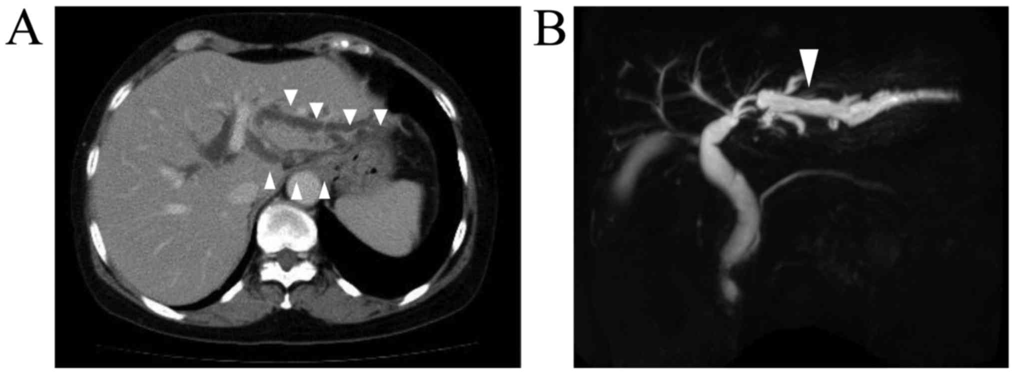
Contrast-enhanced computed tomography and magnetic resonance cholangiopancreatography showed stenosis and dilation of the intrahepatic bile duct, in the left hepatic lobe; i.e., within segments 2 and 3 of the liver (Fig. 1). During percutaneous transhepatic biliary drainage, the contrast medium that was injected into the drainage tube could not pass into the center of the bile duct. Endoscopic retrograde cholangiopancreatography revealed obstruction of the intrahepatic bile duct. Scrape cytology of this narrow segment of the bile duct revealed some atypical cells, which were suggestive of hilar cholangiocarcinoma. In addition, follow-up upper endoscopy was conducted, and a biopsy of a scar on the patient's stomach caused by the previous EMR was carried out, which revealed tubular adenocarcinoma. According to these results, we suspected intrahepatic cholangiocarcinoma and diagnosed the patient with locally recurrent gastric cancer. Surgical resection of the liver and stomach were recommended. A lobectomy of the left side of the liver, cholecystectomy, and a distal gastrectomy combined with a D2 lymph node dissection were performed. The operation time was 10 h and 28 min, and 840 ml of intraoperative blood loss occurred. An examination of the surgical specimens showed that the bile duct was thickened and extended and the length of stenosis was 2 cm, but there was no tumor. The patient did not suffer any complications 
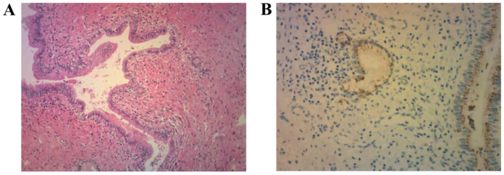
during the postoperative period. A pathological examination of her liver showed marked fibrosis in the stroma and clusters of lymphoid cells. The bile duct exhibited irregular dilation and stenotic compression from the outside, but no thickening of the bile duct wall was noted. No carcinoma or IgG4-positive plasma cells were observed, and we did not detect the typical findings of PSC. The patient had no history of SSC (Fig. 2). Based on these clinical and pathological results, we made a diagnosis of idiopathic sclerosing cholangitis. Incidentally, the pathological findings from the patient's stomach showed early gastric carcinoma, but there was no evidence of a pathological relationship between the bile duct stenosis and the gastric cancer.

Figure 1.

(A) Contrast-enhanced computed tomography and (B) magnetic resonance cholangiopancreatography of the abdomen revealed stenosis and dilation of the intrahepatic bile duct in the left hepatic lobe (segments 2 and 3 of the liver) as indicated by the arrowheads.

Figure 2.

(A) Histopathological examination (hematoxylin and eosin staining) of the liver revealing marked fibrosis in the stroma, irregular bile duct dilation and clusters of lymph cells. The bile duct demonstrated irregular dilation or stenosis compressed from the outside but the walls of the bile duct were not thickened. No carcinoma was observed and the typical findings of PSC were not detected. Magnification ×100. (B) IgG4 immunostaining of the specimen for IgG4 did not detect any IgG4-positive plasma cells. Magnification, ×400. Ig, Immunoglobulin G; PSC, primary sclerosing cholangitis.

Discussion

It is difficult to differentiate between cholangiocarcinoma and benign bile duct stenosis preoperatively. In this case, we did not detect any obvious malignant findings; however, based on the progression of the intrahepatic bile duct stenosis and dilation, we suspected that the patient might have cholangiocarcinoma and decided to perform a lobectomy of the left side of the liver.

It remains difficult to distinguish bile duct malignancy from PSC or IgG4-SC based on imaging features alone. The presence of pancreatic abnormalities, including a peripancreatic rind, atrophication, abnormal enhancement, or high T2 signal intensity, strongly favors a diagnosis of IgG4-SC (2). However, benign bile duct stenosis is detected at a relatively high frequency during liver surgery (8–17%) (1). Thus, additional diagnostic strategies are likely to be vital for distinguishing between these conditions (3).

PSC, IgG4-SC, and SSC are the most common causes of benign bile duct stenosis. PSC and IgG4-SC exhibit similar pathological features, but do differ in some aspects. For example, the pathological features of PSC include onion-skin fibrosis of the bile duct, whereas those of IgG4-SC include infiltration by IgG4-positive plasma cells, obliterative phlebitis, and storiform fibrosis (4). SSC displays suppurative inflammation and erosion of the bile duct mucosa during infections and ischemia. Some representative diseases, such as PSC, IgG4-SC, and SSC, cause benign bile duct stenosis and dilation. This case did not display the typical findings of PSC or IgG4-SC, and the patient did not have a history that was suggestive of SSC.

Fujita et al reported the pathological features of 5 cases of benign sclerosing cholangitis of unknown origin (1). Pathologically, fibroinflammatory changes and lymphoplasmacytic infiltration were observed in all 5 cases. Furthermore, these cases were classified into two types. One type exhibited a thickened bile duct wall and dense fibrosis as well as the formation of numerous lymphoid follicles with germinal centers under the mucosal layer. The other type involved a single focal stricture of the bile duct and subtle lymphoplasmacytic infiltration. The pathological features of our case included fibrosis and lymphoplasmacytic infiltration, which were seen in the previously reported cases, and lymphoid follicle formation, whereas thickening of the bile duct wall was absent. So, this case cannot be classified into either of the reported types of benign sclerosing cholangitis.

Therefore, it cannot be classified into typical benign bile duct stenosis, and as far as we know, no similar cases have been reported. So, we consider that this case was a rare case of idiopathic sclerosing cholangitis involving atypical pathological findings, such as benign bile duct stenosis.

In conclusion a rare case of idiopathic sclerosing cholangitis was determined. It is often difficult to differentiate between cholangiocarcinoma and benign bile duct stenosis preoperatively. Additional diagnostic strategies are likely to be vital for distinguishing between these diseases.

Acknowledgements

Not applicable.

Funding

No funding was received.

Availability of data and materials

All data generated or analyzed during this study are included in this published article.

Authors' contributions

SS, TE, MS, HM, RK, KS, TS, FM, TH and HA performed the operation and perioperative medical treatment. KN performed preoperative examination and diagnosis. NK performed the histopathological examination of the liver. AT analyzed the pathological findings, assisted with patient diagnosis and was a major contributor in writing the manuscript. All authors read and approved the final manuscript.

Ethics approval and consent to participate

Written informed consent was obtained from the participating patient.

Patient consent for publication

Written informed consent for publication was obtained from the patient.

Competing interests

The authors declare that they have no competing interests.

References

1 

Fujita T, Kojima M, Gotohda N, Takahashi S, Nakagohri T, Konishi M, Ochiai A and Kinoshita T: Incidence, clinical presentation and pathological features of benign sclerosing cholangitis of unknown origin masquerading as biliary carcinoma. J Hepatobiliary Pancreat Sci. 17:139–146. 2010. View Article : Google Scholar : PubMed/NCBI

2 

Gardner CS, Bashir MR, Marin D, Nelson RC, Choudhury KR and Ho LM: Diagnostic performance of imaging criteria for distinguishing autoimmune cholangiopathy from primary sclerosing cholangitis and bile duct malignancy. Abdom Imaging. 40:3052–3061. 2015. View Article : Google Scholar : PubMed/NCBI

3 

Kalaitzakis E, Levy M, Kamisawa T, Johnson GJ, Baron TH, Topazian MD, Takahashi N, Kanno A, Okazaki K, Egawa N, et al: Endoscopic retrograde cholangiography does not reliably distinguish IgG4-associated cholangitis from primary sclerosing cholangitis or cholangiocarcinoma. Clin Gastroenterol Hepatol. 9:800–803.e2. 2011. View Article : Google Scholar : PubMed/NCBI

4 

Nakazawa T, Naitoh I, Hayashi K, Miyabe K, Simizu S and Joh T: Diagnosis of IgG4-related sclerosing cholangitis. World J Gastroenterol. 19:7661–7670. 2013. View Article : Google Scholar : PubMed/NCBI

Related Articles

  • Abstract
  • View
  • Download
  • Twitter
Copy and paste a formatted citation
Spandidos Publications style
Shichi S, Einama T, Suzuki M, Matsui H, Kanazawa R, Shibuya K, Suzuki T, Matsuzawa F, Nakachi K, Hashimoto T, Hashimoto T, et al: Rare case of idiopathic sclerosing cholangitis, which was difficult to distinguish from cholangiocarcinoma: A case report. Exp Ther Med 16: 5224-5226, 2018.
APA
Shichi, S., Einama, T., Suzuki, M., Matsui, H., Kanazawa, R., Shibuya, K. ... Taketomi, A. (2018). Rare case of idiopathic sclerosing cholangitis, which was difficult to distinguish from cholangiocarcinoma: A case report. Experimental and Therapeutic Medicine, 16, 5224-5226. https://doi.org/10.3892/etm.2018.6832
MLA
Shichi, S., Einama, T., Suzuki, M., Matsui, H., Kanazawa, R., Shibuya, K., Suzuki, T., Matsuzawa, F., Nakachi, K., Hashimoto, T., Kondo, N., Abe, H., Taketomi, A."Rare case of idiopathic sclerosing cholangitis, which was difficult to distinguish from cholangiocarcinoma: A case report". Experimental and Therapeutic Medicine 16.6 (2018): 5224-5226.
Chicago
Shichi, S., Einama, T., Suzuki, M., Matsui, H., Kanazawa, R., Shibuya, K., Suzuki, T., Matsuzawa, F., Nakachi, K., Hashimoto, T., Kondo, N., Abe, H., Taketomi, A."Rare case of idiopathic sclerosing cholangitis, which was difficult to distinguish from cholangiocarcinoma: A case report". Experimental and Therapeutic Medicine 16, no. 6 (2018): 5224-5226. https://doi.org/10.3892/etm.2018.6832
Copy and paste a formatted citation
x
Spandidos Publications style
Shichi S, Einama T, Suzuki M, Matsui H, Kanazawa R, Shibuya K, Suzuki T, Matsuzawa F, Nakachi K, Hashimoto T, Hashimoto T, et al: Rare case of idiopathic sclerosing cholangitis, which was difficult to distinguish from cholangiocarcinoma: A case report. Exp Ther Med 16: 5224-5226, 2018.
APA
Shichi, S., Einama, T., Suzuki, M., Matsui, H., Kanazawa, R., Shibuya, K. ... Taketomi, A. (2018). Rare case of idiopathic sclerosing cholangitis, which was difficult to distinguish from cholangiocarcinoma: A case report. Experimental and Therapeutic Medicine, 16, 5224-5226. https://doi.org/10.3892/etm.2018.6832
MLA
Shichi, S., Einama, T., Suzuki, M., Matsui, H., Kanazawa, R., Shibuya, K., Suzuki, T., Matsuzawa, F., Nakachi, K., Hashimoto, T., Kondo, N., Abe, H., Taketomi, A."Rare case of idiopathic sclerosing cholangitis, which was difficult to distinguish from cholangiocarcinoma: A case report". Experimental and Therapeutic Medicine 16.6 (2018): 5224-5226.
Chicago
Shichi, S., Einama, T., Suzuki, M., Matsui, H., Kanazawa, R., Shibuya, K., Suzuki, T., Matsuzawa, F., Nakachi, K., Hashimoto, T., Kondo, N., Abe, H., Taketomi, A."Rare case of idiopathic sclerosing cholangitis, which was difficult to distinguish from cholangiocarcinoma: A case report". Experimental and Therapeutic Medicine 16, no. 6 (2018): 5224-5226. https://doi.org/10.3892/etm.2018.6832
Follow us
  • Twitter
  • LinkedIn
  • Facebook
About
  • Spandidos Publications
  • Careers
  • Cookie Policy
  • Privacy Policy
How can we help?
  • Help
  • Live Chat
  • Contact
  • Email to our Support Team