Introduction
Alzheimer's disease (AD) is a neurological type of
neurodegenerative disease. It is mainly caused by aging and
apoptosis of nerve cells affected by internal and external
environmental factors, and a large amount of degeneration and loss
of dopaminergic neurons. AD mainly occurs in the elderly at the age
of >65 years and is manifested as memory loss and cognitive
problems resulting in serious inconvenience of their daily lives
(1,2). In Europe, the United States and other
countries, AD is a major neurological disease that causes the death
of the elderly. The number of deaths from AD in China also ranks in
the top 10 countries (3). At
present, popular drugs used in the treatment of AD on the market do
not substantially improve the loss of dopaminergic neurons,
especially in patients with advanced AD (4). However, AD patients are often
accompanied by an increase of inflammatory cytokines in the body
and brain, and inflammation is considered by many researchers as
one of the key causes of neurodegenerative diseases that trigger AD
and dementia. Therefore, many traditional Chinese and Western
anti-inflammatory drugs also appear in various studies on treating
AD (5,6). Stem cells can be renewable and
differentiable, and thus considered to be a promising material for
the treatment of AD. Bone marrow mesenchymal stem cells (BMSCs)
have been characterized as having a wide range of sources, easiness
to draw materials, simple to culture and difficult in tumor
formation in vivo, thereby differentiating them into
dopaminergic neurons in brain tissues to make up for the loss of
cells caused by AD, thus fundamentally treating AD. (7–9). In this
study, the improvement of AD symptoms by BMSCs and the effects of
BMSCs on in vivo inflammatory cytokines were observed and
investigated through studying AD mice.
Materials and methods
Experimental animals
A total of 20 amyloid precursor protein
(APP)/presenilin-1 (PS1) double transgenic mice (Beijing Biocytogen
Co., Ltd., Beijing, China) were randomly divided into two groups:
the AD control group (n=10) and the stem cell treatment group
(n=10). Ten C57 black 6 bred in the Jackson Laboratory (C57BL/6J)
non-transgenic mice were included in the normal control group. The
mice were kept in cages with controlled temperature and light
cycles (24°C and 12/12 light cycles) and had free access to food
and water. The humidity was 60±10%. There were no statistically
significant differences in age, sex and weight among the three
groups of mice (P>0.05), and the data were comparable (Table I). The study was approved by the
Ethics Committee of the Second Hospital of Shandong University
(Jinan, China).
 | Table I.Comparisons of general data among the
three groups of mice (mean ± SD). |
Table I.
Comparisons of general data among the
three groups of mice (mean ± SD).
| Group | n | Age (weeks) | Weight (g) | Sex
(male/female) |
|---|
| Normal control | 10 | 7.2±0.3 | 24.2±4.2 | 5/5 |
| AD control | 10 | 7.1±0.2 | 23.4±3.9 | 5/5 |
| Treatment | 10 | 6.9±0.2 | 23.7±3.8 | 5/5 |
| P-value | – | >0.05 | >0.05 | >0.05 |
Reagents
Cell culture Dulbecco's modified Eagle's medium
(DMEM), fetal bovine serum (FBS) and trypsin powder were purchased
from Gibco (Gibco; Thermo Fisher Scientific, Inc., Waltham, MA,
USA). RNAiso Plus, PrimeScript® Real-Time (RT) Reagent
kit with genomic deoxyribonucleic acid (gDNA) Eraser (Perfect
Real-time) and SYBR® Premix Ex Taq™ II [Tli ribonuclease
H (RNase H) Plus] were purchased from Takara Biotechnology Co.,
Ltd. (Dalian, China). Enzyme-linked immunosorbent assay (ELISA)
kits were purchased from Zhongshan Golden Bridge Biotechnology Co.,
Ltd. (Zhongshan Golden Bridge Biotechnology Co., Ltd.; OriGene
Technologies, Beijing, China).
Separation and culture of BMSCs
Operations were conducted under aseptic environment.
A 200-mesh sieve was used for cell screening. The parameters of
cell and tissue centrifugation for the separation process were 800
× g/min at 4°C for 3 min. Phosphate-buffered saline (PBS) was used
for thorough washing, as the clearer and cleaner the solution was
the better the results would be. The washing was conducted each
time for approximately ≥2 times. In addition, the number of living
cells was determined using trypan blue staining, and counted using
a cell counter. Cell culture reagents, consumable items and
environmental conditions (temperature and humidity) were strictly
adjusted in accordance with the characteristics of the cell growth.
After the cell growth area reached ~80% of the total area of the
flat bottom, the passage could be performed, and the passage ratio
would be determined according to the amount of cells required for
the next experiment.
Determination by flow cytometry
Flow cytometry detection (10): i) the test objects were the
fourth-generation BMSCs. ii) The centrifugation parameters for the
preparation of the flow cytometry samples were 800 × g/min at 4°C
for 3 min. iii) The cells were mixed and suspended in PBS, and the
number of cells was 1–2×10⁶. ⅳ) Surface antibody (4 µl)
was added in each tube and incubated at 25°C for 30 min avoiding
light. v) PBS (500 µl) was used to wash the cells. Then the cells
were collected and centrifuged at 800 × g for 5 min. The
supernatant was discarded. vi) PBS (500 µl) was added for
resuspension. Then the cells were added to the BD FACSCalibur flow
cytometer (BD Biosciences, Franklin Lakes, NJ, USA). ⅶ) The cell
flow rate was moderate, and the cell population conforming to the
characteristics was selected according to forward scatters (FSCs)
and side scatters (SSCs). ⅷ) The voltage was adjusted according to
the graphics to make the graphics easy to observe and for better
display results.
Tail intravenous injection of
mice
BMSC suspension was prepared. Stem cells for
intravenous injection were digested from the adherent state to the
suspended state, and washed with normal saline, followed by
centrifugation at 800 × g/min at 4°C for 3 min twice and filtering
by a cell sieve filter once. Then the filtered stem cell suspension
was immediately placed on ice and transferred to the intravenous
injection site. The mice aged 7 weeks were fixed in a rat fixator,
and 1 ml suspension containing 2×10⁶ stem cells was
injected using a 2.5 ml syringe each time. The normal control group
and the AD control group were injected with the same volume of
normal saline once a day.
Morris water maze test (11)
The experiment began on the 22nd day after gavage
treatment and was divided into two stages. In the first stage, mice
received 3-day continuous training twice a day, and the
experimental time was not recorded. In the second phase, mice
underwent the fixed-position cruise for 5 consecutive days, once
daily: the mice were placed facing the wall of the pool and put in
pools from different quadrants to record the time they searched for
the underwater surface hidden in the third quadrant (escape
latency). If the mice could not find the platform within 90 sec,
they were guided to the platform for a rest for 30 sec, and the
latency was recorded as 90 sec. At the end of fixed-position
cruise, the space exploration experiment was conducted for 1 day:
the platform was withdrawn, the time occupied by the mice in the
third quadrant within the 90 sec and the frequency of crossing the
original platform were recorded. Morris water maze image automatic
monitoring and processing system was applied to collect and process
data.
Indicator detection
ELISA kits were used to detect the contents of
interleukin (IL)-1, IL-2, IL-10, tumor necrosis factor-α (TNF-α),
and interferon-γ (IFN-γ) in plasma as well as amyloid β (Aβ)1–42
content in brain tissues. The relative expression levels of β-site
APP cleaving enzyme 1 (BACE1) and α-2-macroglobulin (A2M) genes
were detected by fluorescence quantitative polymerase chain
reaction (qPCR), and the primer sequences are shown in Table II. mRNA levels were determined by
Bio-Rad CFX96 fluorescence qPCR detection system (Bio-Rad
Laboratories, Inc., Hercules, CA, USA). The thermocycling
conditions were as follows: 5°C for 5 min; 94°C for 10 sec, 50°C
for 30 sec, 40 cycles; 72°C for another 10 min. The image was
analyzed with GraphPad Prism statistical software (GraphPad
Software, Inc., La Jolla, CA, USA).
| Table II.qPCR primer sequences. |
Table II.
qPCR primer sequences.
| Gene name | Primer sequence |
|---|
| BACE1 | F:
5′-3′CAAGTTCATTACCTCCCTA |
|
| R:
5′-3′CGACTGACCCTTGTGG |
| A2M | F:
5′-3′AAAGGAAATCGCATCG |
|
| R:
5′-3′CATGTTCATTGTCACGGTTT |
| β-actin | F:
5′-3′TCAGGTCATCACTATCGGCAAT |
|
| R:
5′-3′ACGCAAATGTGGGAAAGAAA |
Statistical methods
All data were processed using Statistical Product
and Service Solutions (SPSS) 16.0 software (Cabit Information
Technology Co., Ltd., Shanghai, China). Measurement data were
expressed as mean ± SD, and the comparison of mean was conducted
using t-test and one-way analysis of variance. The post hoc test
used was LSD test. P<0.05 was considered to indicate a
statistically significant difference.
Results
Separation and identification of
BMSCs
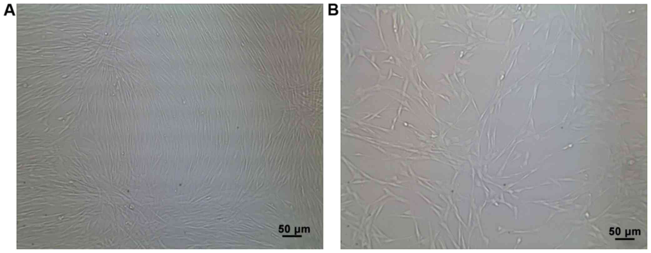
BMSCs were successfully isolated. They were
spindle-shaped and spirally arranged, and grew adhering to the
culture dishes (Fig. 1). The
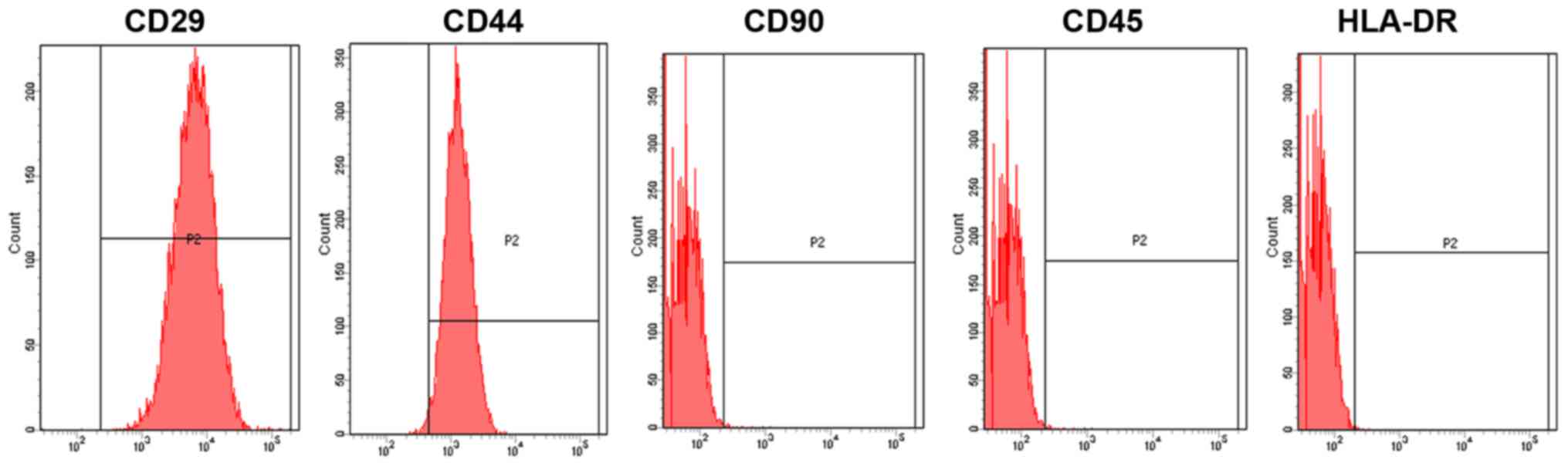
obtained BMSCs were identified using a flow cytometer, which
revealed that cluster of differentiation (CD)29, CD44 and CD90 were
positive, whereas CD45 and human leukocyte antigen
antigen-D-related (HLA-DR) were negative (Fig. 2).
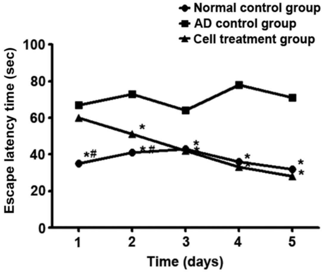
Comparison of memory function using
the water maze test
From day 2, the escape latency in the water maze in
the stem cell treatment group was shortened compared with that in
the AD control group (P<0.05). From day 3, there was no
significant difference in the escape latency between the stem cell
treatment group and the normal control group (P>0.05) (Fig. 3). The space exploration experiment
showed that the time of crossing the ring for the first time in the
stem cell treatment group was shorter than that in the AD control
group (P<0.05), but the frequency was higher than that in the AD
control group (P<0.05) (Table
III).
| Table III.The time of crossing the ring for the
first time and its frequency (mean ± SD). |
Table III.
The time of crossing the ring for the
first time and its frequency (mean ± SD).
| Group | Time of crossing the
ring for the first time (sec) | Frequency of crossing
the ring (times) |
|---|
| Normal control | 16.32±9.32 | 4.8±1.3 |
| AD control | 27.32±12.43 | 1.5±0.4 |
| Stem cell
treatment |
22.38±11.54a | 4.9±0.9a |
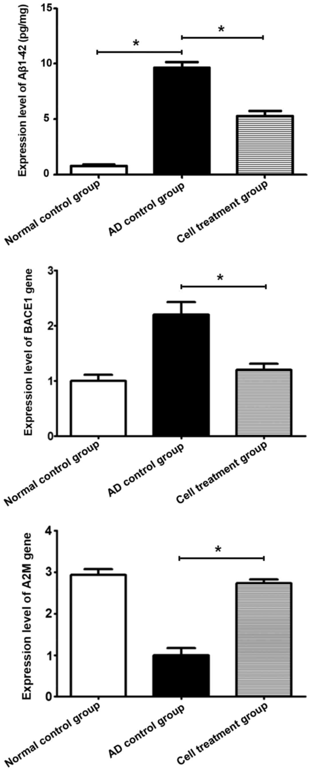
Aβ1–42 content and the relative
expression levels of BACE1 and A2M genes in brain tissues
Aβ1–42 content in the AD control group was higher
than that in the stem cell treatment group and the normal control
group (P<0.05). The expression level of BACE1 in the stem cell
treatment group was lower than that in the AD control group
(P<0.05), while the expression level of A2M gene was increased
in the stem cell treatment group compared to that in the AD control
group (P<0.05) (Fig. 4).
Comparison of inflammatory cytokines
between the three groups
At 14 days after treatment, the contents of IL-1,
IL-2, TNF-α and IFN-γ in blood in the stem cell treatment group
were lower than those in the AD control group (P<0.05) (Table IV).
| Table IV.Comparison of several inflammatory
cytokines in blood (mean ± SD). |
Table IV.
Comparison of several inflammatory
cytokines in blood (mean ± SD).
| Group | IL-1 (pg/ml) | IL-2 (pg/ml) | TNF-α (pg/ml) | IFN-γ (pg/ml) | IL-10 (pg/ml) |
|---|
| Normal control | 43.36±12.32 | 59.33±21.32 | 49.55±19.34 | 39.44±15.21 | 54.22±24.54 |
| AD control | 143.21±34.21 | 121.32±12.43 | 109.22±22.12 | 108.23±35.21 | 41.21±19.32 |
| Stem cell
treatment |
53.21±19.43a |
67.35±21.11a |
55.21±20.32a |
44.43±19.54a | 47.43±17.43 |
Discussion
AD has been characterized as a refractory disease
worldwide and has long been a hotspot in the field of
neuromedicine. Research on AD mainly focuses on the pathogenesis
and treatment of the disease. The pathogenesis of AD is very
complex, and the two currently-recognized theories are the
reduction of cholinergic and the increase of Aβ protein (12,13). AD
is treated mainly through drug actions on different
neurotransmitter systems, so as to enhance the high-level
activities of the central nervous system, reduce the symptoms
occurring in the disease process, and delay the further development
of dementia. However, most of the oral drugs exert certain effects
in the treatment of AD in the early stage, but in the late stage,
due to the large-scale decline of nerve cells in the hippocampus,
drugs cannot fully pass the blood-brain barrier and exert few
effects (14). In addition, AD is
often accompanied by an increase of inflammatory cytokines, causing
other complications, so it is also treated by a combination of
multiple drugs (15). As a bioactive
material, stem cells can be transformed into nerve cells by
environmental stimuli to make up for the lack of original nerve
cells, and they can be expanded in vivo to some extent so
that the therapeutic effects endure longer. Besides, MSCs can also
secrete factors that promote cell growth such as epidermal growth
factor (EGF), vascular endothelial growth factor (VEGF) and
insulin-like growth factor (IGF) under the action of paracrine and
reduce the complications such as inflammation caused by AD
(16,17). Compared with embryonic stem cells,
BMSCs, as a type of stem cells, are easier to obtain and to be
isolated and cultured in vitro, and it is not easy for them
to form tumors. Therefore, BMSCs have the advantages of low cost
and high safety.
Han and He (18)
treated Aβ modeling Sprague-Dawley (SD) rats with MSCs in the
umbilical cord. At 14 days after injection, the water maze test
demonstrated that the frequency of mice in stem cell treatment
group crossing the target quadrant was significantly increased.
Some studies have pointed out that if stem cells are induced into
neural stem cells in vitro, the direct injection of neural
stem cells into the hippocampus of the brain will be more effective
in the treatment of AD, but the difficulty lies in how to inject
neural stem cells into a designated place and avoid trauma to the
brain (19). It was found in the
study of Farina et al (20)
that BMSCs can attenuate the memory impairment of Aβ deposits and
stimulate the signal pathways of the primary tumor, change the
expression levels of relevant genes, and inhibit apoptosis of nerve
cells. Other studies have revealed that the occurrence process of
AD is accompanied by inflammation, which can cause inflammation
responses, resulting in changes in the in vivo contents of
some factors associated with the verification. This study showed
that the escape latency in the water maze in the stem cell
treatment group was shortened, the time of mice crossing the ring
for the first time was reduced, and the frequency of crossing the
ring was increased. The relative expression level of BACE1 gene in
the stem cell treatment group was decreased compared with that in
the AD control group, while that of A2M gene was increased. The
increased level of BACE1 gene would produce more encephaloclastic
Aβ proteins, while A2M gene encoded inhibitors that eliminated Aβ
proteins, and helped eliminate Aβ proteins. However, at 14 days
after treatment, Aβ1–42 content in brain tissues in the stem cell
treatment group was lower than that in the AD control group, and
the expression levels of IL-1, IL-2, TNF-α and IFN-γ in blood and
hippocampus tissues in the stem cell treatment group were lower
than those in the AD control group.
In conclusion, human BMSCs can ameliorate the
symptoms of AD by decreasing the level of inflammatory cytokines
and regulating the expression levels of Aβ-related genes.
Acknowledgements
Not applicable.
Funding
The study was supported by the Fundamental Research
Funds of Shandong University (2016JC022) and the Foundation of the
Second Hospital of Shandong University (S2014010007).
Availability of data and materials
The datasets used and/or analyzed during the current
study are available from the corresponding author on reasonable
request.
Authors' contributions
YW wrote the manuscript and was responsible for the
separation and culture of BMSCs. ZX prepared the mice and was also
involved in the conception of the study. JB assisted with flow
cytometry. ZZ performed Morris water maze test. All authors read
and approved the final manuscript.
Ethics approval and consent to
participate
The study was approved by the Ethics Committee of
the Second Hospital of Shandong University (Jinan, China).
Patient consent for publication
Not applicable.
Competing interests
The authors declare that they have no competing
interests.
References
|
1
|
Bahaeddin Z, Yans A, Khodagholi F and
Sahranavard S: Dietary supplementation with Allium
hirtifolium and/or Astragalus hamosus improved memory
and reduced neuro-inflammation in the rat model of Alzheimer's
disease. Appl Physiol Nutr Metab. 43:558–564. 2018. View Article : Google Scholar : PubMed/NCBI
|
|
2
|
Elnagar MR, Walls AB, Helal GK, Hamada FM,
Thomsen MS and Jensen AA: Probing the putative α7 nAChR/NMDAR
complex in human and murine cortex and hippocampus: Different
degrees of complex formation in healthy and Alzheimer brain tissue.
PLoS One. 12:e01895132017. View Article : Google Scholar : PubMed/NCBI
|
|
3
|
Swaminathan A and Jicha GA: Nutrition and
prevention of Alzheimer's dementia. Front Aging Neurosci.
6:2822014. View Article : Google Scholar : PubMed/NCBI
|
|
4
|
Mohamad Nasir NF, Zainuddin A and
Shamsuddin S: Emerging roles of sirtuin 6 in Alzheimer's disease. J
Mol Neurosci. 64:157–161. 2018. View Article : Google Scholar : PubMed/NCBI
|
|
5
|
Momtaz S, Hassani S, Khan F, Ziaee M and
Abdollahi M: Cinnamon, a promising prospect towards Alzheimer's
disease. Pharmacol Res. 130:241–258. 2018. View Article : Google Scholar : PubMed/NCBI
|
|
6
|
Vinceti M, Chiari A, Eichmüller M, Rothman
KJ, Filippini T, Malagoli C, Weuve J, Tondelli M, Zamboni G,
Nichelli PF, et al: A selenium species in cerebrospinal fluid
predicts conversion to Alzheimer's dementia in persons with mild
cognitive impairment. Alzheimers Res Ther. 9:1002017. View Article : Google Scholar : PubMed/NCBI
|
|
7
|
Hagl S, Heinrich M, Kocher A, Schiborr C,
Frank J and Eckert GP: Curcumin micelles improve mitochondrial
function in a mouse model of Alzheimer's disease. J Prev Alzheimers
Dis. 1:80–83. 2014.PubMed/NCBI
|
|
8
|
Grundman M: Alzheimer's disease drug
development: Trial-ready cohorts should help. J Prev Alzheimers
Dis. 1:69–70. 2014.PubMed/NCBI
|
|
9
|
Bateman RJ and Morris JC: A new era:
Disease modifying Alzheimer disease prevention trials. J Prev
Alzheimers Dis. 1:682014.PubMed/NCBI
|
|
10
|
Hu X, Qu Y, Chu Q, Li W and He J:
Investigation of the neuroprotective effects of Lycium
barbarum water extract in apoptotic cells and Alzheimer's
disease mice. Mol Med Rep. 17:3599–3606. 2018.PubMed/NCBI
|
|
11
|
Wang P, Guo Q, Zhou Y, Chen K, Xu Y, Ding
D, Hong Z and Zhao Q: Lack of association between triggering
receptor expressed on myeloid cells 2 polymorphism rs75932628 and
late-onset Alzheimer's disease in a Chinese Han population.
Psychiatr Genet. 28:16–18. 2018.PubMed/NCBI
|
|
12
|
Flanagan KJ, Copland DA, Chenery HJ, Byrne
GJ and Angwin AJ: Semantic feature disturbance in Alzheimer
disease: Evidence from an object decision task. Cogn Behav Neurol.
30:159–171. 2017. View Article : Google Scholar : PubMed/NCBI
|
|
13
|
Roland KP and Chappell NL: Caregiver
experiences across three neurodegenerative diseases: Alzheimer's,
Parkinson's, and Parkinson's with dementia. J Aging Health. Oct
1–2017.(Epub ahead of print). View Article : Google Scholar : PubMed/NCBI
|
|
14
|
Botchway BOA, Moore MK, Akinleye FO, Iyer
IC and Fang M: Nutrition: Review on the possible treatment for
Alzheimer's disease. J Alzheimers Dis. 61:867–883. 2018. View Article : Google Scholar : PubMed/NCBI
|
|
15
|
Wang SS, Jia J and Wang Z: Mesenchymal
stem cell-derived extracellular vesicles suppresses iNOS expression
and ameliorates neural impairment in Alzheimer's disease mice. J
Alzheimers Dis. 61:1005–1013. 2018. View Article : Google Scholar : PubMed/NCBI
|
|
16
|
Saksida T, Koprivica I, Vujičić M,
Stošić-Grujičić S, Perović M, Kanazir S and Stojanović I: Impaired
IL-17 production in gut-residing immune cells of 5×FAD mice with
Alzheimer's disease pathology. J Alzheimers Dis. 61:619–630. 2018.
View Article : Google Scholar : PubMed/NCBI
|
|
17
|
Eldholm RS, Barca ML, Persson K, Knapskog
AB, Kersten H, Engedal K, Selbaek G, Braekhus A, Skovlund E and
Saltvedt I: Progression of Alzheimer's disease: A longitudinal
study in Norwegian Memory Clinics. J Alzheimers Dis. 61:1221–1232.
2018. View Article : Google Scholar : PubMed/NCBI
|
|
18
|
Han X and He G: Toward a rational design
to regulate β-amyloid fibrillation for Alzheimer's disease
treatment. ACS Chem Neurosci. 9:198–210. 2018. View Article : Google Scholar : PubMed/NCBI
|
|
19
|
Harding A, Gonder U, Robinson SJ, Crean S
and Singhrao SK: Exploring the association between Alzheimer's
disease, oral health, microbial endocrinology and nutrition. Front
Aging Neurosci. 9:3982017. View Article : Google Scholar : PubMed/NCBI
|
|
20
|
Farina E, Baglio F, Pomati S, D'Amico A,
Campini IC, Di Tella S, Belloni G and Pozzo T: The mirror neurons
network in aging, mild cognitive impairment, and Alzheimer disease:
A functional MRI study. Front Aging Neurosci. 9:3712017. View Article : Google Scholar : PubMed/NCBI
|