Introduction
There is a high incidence rate of deep venous
thrombosis (DVT) of lower extremity in China, and the pulmonary
thromboembolism formed by detachment of thrombus is the leading
cause for death of people (1).
Studies have suggested that inflammatory reaction plays a key role
in the occurrence and development of DVT, and that inflammatory
cytokine is a bioactive peptide that not only acts as a signal
transduction factor, but also performs as an effector molecule
(2). Inflammatory factors can
directly cause injuries of endothelial cells, and they can also
promote the release of inflammatory factors by the blood
coagulation system and further accelerate the inflammatory reaction
and blood coagulation, thus having very close relationships with
the blood coagulation, anticoagulation and fibrinolysis processes
(3).
Matrix metalloproteinases (MMPs) is a category of
enzymes with zinc ion as the prosthetic group, which can degrade
extracellular matrix proteins (4).
Under the normal physiological status of tissues, the expression of
MMPs is at a low level, but changes in the expression proportion of
the MMPs to their inhibitory factors may lead to pathological
responses such as inflammation, neovascularization and neoplasm
metastasis (5). MMP-1 and MMP-2
belong to gelatinases, and their activation plays a vital role in
the process of thromboembolism (4,6).
Increasing the MMP-2 gene expression levels or activating enzyme
activities may involve the venous thromboembolism (7). MMP-1, MMP-2 and activator of
plasminogen are regarded as key factors for the occurrence and
development of venous diseases of the lower extremities (8). The relative expression levels of MMP
and its inhibitor can adjust inflammation and thrombosis (6).
Some scholars believe that MMP-1, MMP-2 and
inflammatory factors may play key roles in the occurrence and
development of DVT (9). To
investigate the effects of MMP-1, MMP-2 and inflammation-associated
factors on DVT, a total of 50 patients with DVT of lower extremity
admitted and treated in the Department of Vascular Surgery of
People's Hospital of Jiyang (Jinan, China) and another 50
volunteers receiving health examination were selected. The
concentrations of MMP-1, MMP-2, interleukin-6 (IL-6), IL-8 and
tumor necrosis factor-α (TNF-α) in the serum were tested,
respectively, and the expression levels of MMP-1 and MMP-2 proteins
as well as IL-6, IL-8 and TNF-α messenger ribonucleic acids (mRNAs)
in peripheral blood mononuclear cells (PBMCs) of DVT patients were
determined. In the meantime, the circumference at 15 cm above the
knee and 10 cm below the knee of both extremities were measured
before treatment and at 7 days after treatment, and the difference
between the circumferences of unaffected and affected extremities
was calculated. In addition, correlation analysis was conducted
respectively for the levels of MMP-1, MMP-2, IL-6, IL-8 and TNF-α
in the serum of patients in the DVT group, aiming to explore the
functions of these factors in DVT.
Materials and methods
General data
Primer synthesis, reverse transcription kit and
real-time fluorescent quantitative polymerase chain reaction (PCR)
kit (Takara Biotechnology Co., Ltd., Dalian, China), TRIzol kit
(Ambion; Thermo Fisher Scientific, Dallas, TX, USA). Rabbit
anti-human MMP-1, MMP-2, glyceraldehyde-3-phosphate dehy-drogenase
(gAPDh) primary polyclonal antibodies and goat anti-rabbit
horseradish peroxidase (hRP)-labeled secondary polyclonal antibody
(cat nos. 10371-2-AP, 10373-2-AP, 10494-1-AP, SA00001-2;
(Proteintech Group, Inc., Wuhan, China), bicinchoninic acid (BCA)
protein assay kit and cell lysis buffer (Beyotime Institute of
Biotechnology, Jiangsu, China).
A total of 50 DVT patients diagnosed in the
Department of Vascular Surgery of People's Hospital of Jiyang from
February 2016 to February 2017 were selected as the DVT group,
including 27 males and 23 females aged 25–57 years, with an average
age of 55.72±11.46 years. Inclusion criteria: i) patients who had
DVT onset within 7 days, ii) patients who had similar disease
conditions and iii) patients who had venous flow obstruction of the
lower extremity which was confirmed by the anterograde venography
of deep vein of lower extremity. Patients with the following
conditions were excluded: pregnancy, infection, tumor, inherited
anticoagulation factor deficiency, vascular abnormality or diabetes
mellitus. Before treatment, all the patients had such clinical
manifestations as high muscle tension and swelling in their
affected extremities, and the DVT of unilateral lower extremity was
verified via color Doppler ultrasound or venography examinations
after admission to hospital. Another 50 healthy individuals
receiving health examination in People's Hospital of Jiyang were
enrolled as the normal control group which included 28 men and 22
women aged 26–58 years, with an average age of 52.35±10.38 years.
They were all healthy adults without cardiac, hepatic, cerebral,
renal and infectious diseases or diabetes mellitus. There were no
statistically significant differences in the age, gender and weight
between the two groups (P>0.05) (Table I).
 | Table I.Comparison of general data. |
Table I.
Comparison of general data.
| Characteristics | DVT group (n=50) | Normal control group
(n=50) | P-value |
|---|
| Age (years) |
55.72±11.46 | 52.35±10.38 | >0.05 |
| Sex [n
(male/female)] | 27/23 | 28/22 | >0.05 |
| Weight (kg, mean ±
SD) | 61.56±9.05 | 64.84±10.53 | >0.05 |
The study was approved by the Ethics Committee of
People's Hospital of Jiyang. Signed informed consents were obtained
from the patients or guardians.
Clinical treatment
The patients with DVT of lower extremity were
treated with continuous intravenous infusion of 200,000 units of
urokinases for 5 days from the first day after the diagnosis was
confirmed. Moreover, 4,000 units of low molecular weight heparin
was injected subcutaneously twice daily, and 100 mg aspirin, an
anti-platelet aggregation drug, was orally administered once
daily.
Acquisition and detection of serum
specimens
A total of 5 ml fasting venous blood was collected
in the morning from each patient in the DVT group before treatment
and at 7 days after treatment as well as each healthy person in the
normal control group, respectively. The blood was added into a
vacuum blood collection tube without any anticoagulant and
centrifuged at 4,000 × g 4°C for 15 min. After that, the
supernatant was absorbed carefully and then stored in a
refrigerator at −80°C for standby use.
Enzyme-linked immunosorbent assay (ELISA) was
performed to measure the levels of MMP-1, MMP-2, IL-6, IL-8 and
TNF-α in the serum in accordance with the steps recommended in the
kit instructions.
Isolation and culture of patient
PBMCs
Before treatment and at 7 days after treatment, 10
ml fasting venous blood was drawn in the morning from each patient
in the DVT group, and Ficoll-Paque PLUS was utilized to isolate the
PBMCs. After the cells were cultured with Roswell Park Memorial
Institute (RPMI)-1640 medium containing 10% serum in an incubator
with 5% CO2 at 37°C for 2 h, the suspended cells were
washed out, and the adherent cells obtained were the PBMCs, which
were adopted for subsequent experiments.
Detection of MMP-1 and MMP-2 protein expression
levels in patient PBMCs via western blotting. Patient PBMCs were
digested with trypsin and then collected, which were lysed using
the lysis buffer and centrifuged at 3,000 × g for 10 min at 4°C to
collect the supernatant protein. The protein concentration was
determined by virtue of the BCA protein assay kit, and 30 µg
protein loading in each specimen was taken for sodium dodecyl
sulfate-polyacrylamide gel electrophoresis (SDS-PAGE), followed by
transfer to a polyvinylidene fluoride (PVDF) membrane, sealing with
blocking buffer for 2 h and addition of MMP-1, MMP-2 and GAPDH
primary antibodies (dilution, 1:1,000) for incubation in the
refrigerator at 4°C overnight. Next, the membrane was washed with
Tris-buffered saline Tween-20 (TBST) 3 times, and then the
secondary antibodies (dilution, 1:1,200) were added for incubation
at room temperature for 2 h, followed by membrane washing with TBST
3 times. After that, the membrane was placed in enhanced
chemiluminescence (ECL) liquid (MilliporeSigma, Burlington, MA,
USA) for development in the dark, which was scanned and recorded by
means of a gel imager for gray analysis. The densitometry was
analyzed by Image J professional image analysis software (NIH,
Bethesda, MD, USA).
Detection of mRNA expression levels of
IL-6, IL-8 and TNF-α in patient PBMCs via reverse
transcription-quantitative polymerase chain reaction (RT-qPCR)
The PBMCs of the patients collected after trypsin
digestion were applied to extract the total RNA of the cells via
the TRIzol kit. Then the samples with an absorbance ratio
(A260/A280) of 1.8–2.0 were selected for
reverse transcription, followed by PCR with complementary
deoxyribonucleic acid (cDNA) as the template. The primer sequences
are shown in Table II, and the
reaction conditions are as follows: pre-denaturation at 94°C for 3
min and then at 95°C for 1 min, annealing at 50°C for 50 sec,
extension at 72°C for 1 min, a total of 40 cycles of amplification
and extension for 10 min. With GAPDH mRNA as the control, the
experimental results were analyzed using the 2−∆∆Cq
method (10).
| Table II.Primer sequences of RT-qPCR. |
Table II.
Primer sequences of RT-qPCR.
| Genes | Primer sequences |
|---|
| IL-6 | F: 5′-
CTGTTGTTGCTGTGGCTGAT-3′ |
|
| R:
5′-TCCGTCCACAAGCAATGAGT-3′ |
| IL-8 | F:
5′-ATGACTTCCAAGCTGGCCGTGGCT-3′ |
|
| R:
5′-TCTCAGCCCTCTTCAAAAACTTCTC-3′ |
| TNF-α | F:
5′-TACTGAACTTCGGGGTGATTGGTCC-3′ |
|
| R:
5′-CAGCCTTGTCCCTTGAAGAGAACC-3′ |
| GAPDH | F:
5′-ATGGCACCGTCAAGGCTGAG-3′ |
|
| R:
5′-GCAGTGATGGCATGGACTGT-3′ |
Measurement of circumference of the
lower extremity
The circumferences at 15 cm above the knee and 10 cm
below the knee of both extremities were measured while the blood
samples of the DVT patients were collected. The difference between
the circumference of affected extremity and unaffected extremity
was calculated, and the circumference difference of the affected
and unaffected extremities was compared before and after
treatment.
Statistical analysis
Statistical Product and Service Solutions (SPSS)
17.0 software (IBM Corp., Armonk, NY, USA) was adopted for data
processing. Measurement data were presented as mean ± standard
deviation, and t-test was used for intergroup comparison. Linear
(Pearson's) correlation analysis was utilized to analyze
correlations, and P≤0.05 was considered to indicate a statistically
significant difference.
Results
Comparison of levels of serum MMP-1,
MMP-2 and inflammatory factors
The ELISA results indicated that the levels of
MMP-1, MMP-2, IL-6, IL-8 and TNF-α in the serum of the DVT group
were remarkably higher than those of the normal control group
before treatment, and the differences were statistically
significant (P<0.01) (Table
III).
| Table III.Concentration of serum MMP-1, MMP-2
and inflammatory factors in the DVT and normal control groups
before treatment. |
Table III.
Concentration of serum MMP-1, MMP-2
and inflammatory factors in the DVT and normal control groups
before treatment.
| Groups | MMP-1 (µmol/l) | MMP-2 (µmol/l) | IL-6 (µg/l) | IL-8 (µg/l) | TNF-α (µg/l) |
|---|
| DVT | 3.255±0.572 | 4.073±0.546 | 0.152±0.038 | 0.797±0.155 | 54.37±11.48 |
| Control group | 0.642±0.127 | 0.926±0.283 | 0.094±0.012 | 0.344±0.087 | 16.21±3.772 |
| P-value | <0.01 | <0.01 | <0.01 | <0.01 | <0.01 |
After 7 days of treatment, the levels of serum
MMP-1, MMP-2, IL-6, IL-8 and TNF-α in the DVT group were markedly
lower compared with those before treatment, showing statistically
significant differences (P<0.01) (Table IV).
| Table IV.Concentration of serum MMP-1, MMP-2
and inflammatory factors in patients of the DVT group before and
after treatment. |
Table IV.
Concentration of serum MMP-1, MMP-2
and inflammatory factors in patients of the DVT group before and
after treatment.
| Groups | MMP-1 (µmol/l) | MMP-2 (µmol/l) | IL-6 (µg/l) | IL-8 (µg/l) | TNF-α (µg/l) |
|---|
| Before treatment | 3.255±0.572 | 4.073±0.546 | 0.152±0.038 | 0.797±0.155 | 54.37±11.48 |
| After treatment | 1.591±0.382 | 1.952±0.457 | 0.094±0.012 | 0.459±0.146 | 34.52±8.971 |
| P-value | <0.01 | <0.01 | <0.01 | <0.01 | <0.01 |
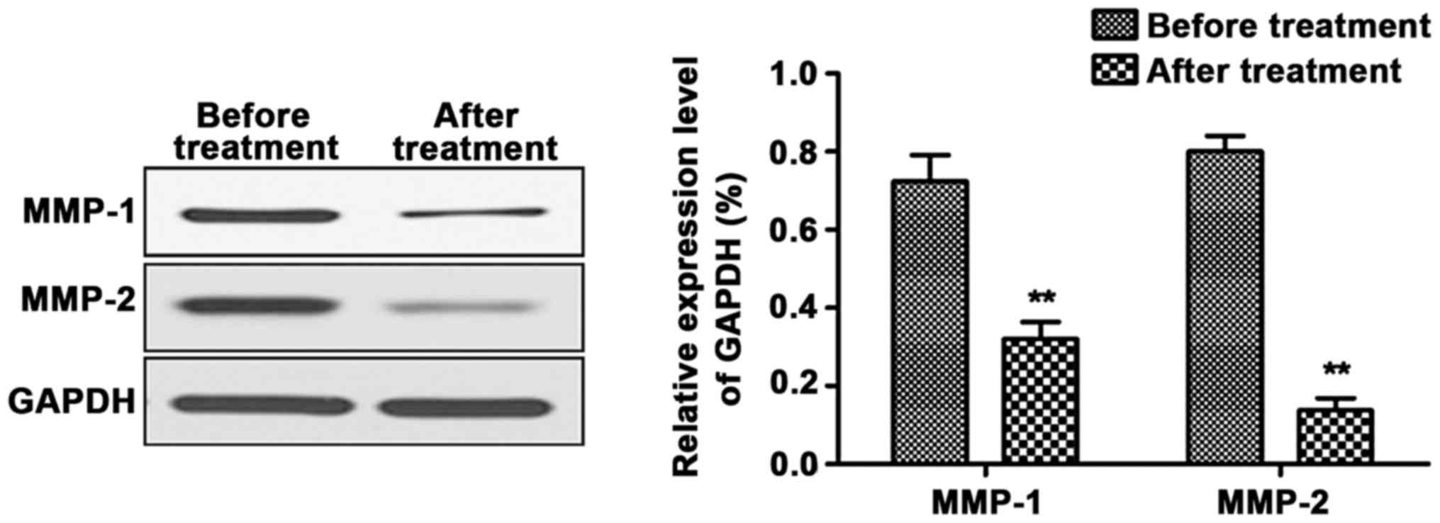
Expression levels of MMP-1 and MMP-2
proteins in PBMCs of the patients before and after treatment
The results of western blotting revealed that
compared with those before treatment, the expression levels of
MMP-1 and MMP-2 proteins in the PBMCs were decreased notably after
treatment, and the differences were statistically significant
(P<0.01) (Fig. 1).
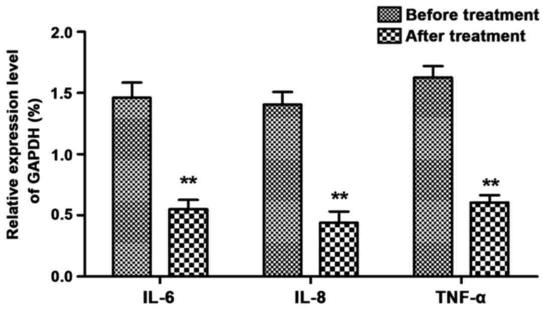
Expression levels of IL-6, IL-8 and
TNF-α mRNAs in PBMCs of the patients before and after
treatment
As shown in Fig. 2,
the RT-qPCR results manifested that the expression levels of IL-6,
IL-8 and TNF-α mRNAs in the PBMCs declined compared with those
before treatment, with statistically significant difference
(P<0.01).
Comparison of circumference of the
affected and unaffected extremities of patients in the DVT group
before and after treatment
The degree of limb swelling of the DVT patients was
alleviated obviously after treatment, and the difference between
the circumferences of unaffected extremity and affected extremity
of the patients was decreased remarkably at 7 days after treatment
in comparison with that before treatment, displaying statistically
significant difference (P<0.01) (Table V).
| Table V.Comparison of circumference of the
affected and unaffected extremities of patients in the DVT group
before and after treatment. |
Table V.
Comparison of circumference of the
affected and unaffected extremities of patients in the DVT group
before and after treatment.
| Groups | 15 cm above the
knee | 10 cm below the
knee |
|---|
| Before treatment | 6.22±1.74 | 3.03±0.68 |
| After treatment | 2.84±0.76 | 3.16±0.92 |
| P-value | <0.01 | <0.01 |
Correlation analysis of MMP-1 and
MMP-2 with inflammatory factors in the serum of patients in the DVT
group
The levels of IL-6, IL-8, TNF-α, MMP-1 and MMP-2 in
the serum of DVT patients were recorded for Pearson's correlation
analysis. It was indicated that the serum IL-6, IL-8 and TNF-α
levels were positively correlated with MMP-1 and MMP-2 levels of
patients in the DVT group, respectively (P<0.05 or P<0.01)
(Table VI).
| Table VI.Correlation of serum IL-6, IL-8 and
TNF-α with MMP-1 and MMP-2 of patients in the DVT group. |
Table VI.
Correlation of serum IL-6, IL-8 and
TNF-α with MMP-1 and MMP-2 of patients in the DVT group.
| Correlation
coefficient | MMP-1 | MMP-2 |
|---|
| IL-6 | 0.551b | 0.638b |
| IL-8 | 0.493a | 0.674b |
| TNF-α | 0.533b | 0.573b |
Discussion
Color Doppler ultrasound is a reliable and effective
method for diagnosing DVT, with the advantages of noninvasive and
repeatable examinations. However, imaging examinations still cannot
determine whether fibrinolysis reaction exists in the human body.
Therefore, studying thromboembolism at the molecular level and
monitoring the expression levels of relevant molecules in the DVT
progression have become new research hotspots (11).
In recent years, studies worldwide have demonstrated
that inflammation participates in the venous thromboembolism.
Inflammation can cause venous wall injury and induce thrombosis
which can further stimulate apparent inflammatory reactions on the
venous wall (12). Inflammation is
stimulated and regulated by cytokines to some extent, and
relatively high levels of inflammatory cytokines in the blood are
important risk factors for the venous thromboembolism (13). It is discovered in research that such
inflammatory cytokines as IL-6, IL-8 and TNF-α are involved in the
process of venous thromboembolism (14).
Studies on thrombus have revealed that IL-6 can
significantly increase the expression levels of cell adhesion
molecules [cluster of differentiation molecule 11b (CD11b)/CD18]
and directly decrease the transcription and translation of
L-selectin (CD62L) (15). IL-8 is a
key factor for venous thromboembolism, and research has manifested
that IL-8 is directly implicated in the thrombosis, accelerates
thrombolysis and ultimately induces neovascularization (16). TNF-α is a type of inflammatory
cytokine secreted by mononuclear macrophages and eosinophils.
Studies have discovered that TNF-α is able to affect the adhesion
and migration of leukocytes and influence the amount of thrombus
generated at the same time (17).
The content of serum TNF-α can indicate the degree of inflammation
and tissue injuries in the body and regulate the growth and
differentiation of multiple cells. Moreover, it can control the
life activities of the cells by virtue of their autocrine (18).
Studies have suggested that inflammatory cytokines
are capable of regulating the expression levels of MMP genes, whose
major process is that they activate the activating transcription
factor 2 and C-transcriptase via ceramide signaling pathway and
then activate the activator protein-1 (AP-1), followed by binding
to the AP-1 sites on the MMP genes, thus elevating the
transcription levels of the MMP genes (19). It is found through research that
blood stasis and venous hypertension can induce increase in MMP-2
expression, which is able to trigger degradation of the
extracellular matrix, thus damaging the venous wall (20). In addition, platelets adhered to
exposed collagens may lead to activation of MMP-1, thus directly
lysing the protease-activated receptor type 1 (PAR1) of the
platelets (21).
In order to investigate the roles of MMP-1, MMP-2
and inflammation-associated cytokines in DVT, a total of 50
patients with DVT of the lower extremity admitted and treated in
the Department of Vascular Surgery of People's Hospital of Jiyang
and 50 volunteers undergoing health examinations were selected to
detect the concentrations of MMP-1, MMP-2, IL-6, IL-8 and TNF-α in
their serum. The results indicated that before treatment, the
levels of serum MMP-1, MMP-2, IL-6, IL-8 and TNF-α of the DVT group
were remarkably higher than those of the normal control group, and
that after treatment, the levels of serum MMP-1, MMP-2, IL-6, IL-8
and TNF-α in the DVT group were markedly lower compared with those
before treatment. Meanwhile, the expression levels of MMP-1 and
MMP-2 proteins as well as IL-6, IL-8 and TNF-α mRNAs in the PBMCs
of the DVT patients were detected before and after treatment. It
was manifested that the expression levels of MMP-1 and MMP-2
proteins as well as IL-6, IL-8 and TNF-α mRNAs in the PBMCs were
decreased notably after treatment. Moreover, the circumference at
15 cm above the knee and 10 cm below the knee of both extremities
of the patients were measured before treatment and at 7 days after
treatment, and the results showed that the difference between the
circumferences of unaffected extremity and affected extremity of
the patients was decreased remarkably after treatment in comparison
with that before treatment. Pearson's correlation analysis
demonstrated that the serum IL-6, IL-8 and TNF-α had positive
correlation with MMP-1 and MMP-2 levels of patients in the DVT
group, respectively.
In conclusion, MMP-1, MMP-2 and inflammatory factors
play important roles in the occurrence and development of DVT, of
which the levels of IL-6, IL-8 and TNF-α are positively correlated
with the levels of MMP-1 and MMP-2, respectively. Therefore,
determining the concentrations of MMP-1, MMP-2, IL-6, IL-8 and
TNF-α in the peripheral blood is of significant value in clinical
practice for the diagnosis, progression and treatment effect
judgement of DVT.
Acknowledgements
Not applicable.
Funding
No funding was received.
Availability of data and materials
The datasets used and/or analyzed during the present
study are available from the corresponding author on reasonable
request.
Authors' contributions
TZ drafted the manuscript. TZ and QL were mainly
devoted to ELISA. TZ and LW performed RT-qPCR. LW and GL were
responsible for measurement of circumference of the lower
extremity. All authors read and approved the final manuscript.
Ethics approval and consent to
participate
The study was approved by the Ethics Committee of
People's Hospital of Jiyang (Jinan, China). Signed informed
consents were obtained from the patients or guardians.
Patient consent for publication
Not applicable.
Competing interests
The authors declare that they have no competing
interests.
References
|
1
|
Crop MJ, Siemes C, Berendes P, van der
Straaten F, Willemsen S and Levin MD: Influence of C-reactive
protein levels and age on the value of D-dimer in diagnosing
pulmonary embolism. Eur J Haematol. 92:147–155. 2014. View Article : Google Scholar : PubMed/NCBI
|
|
2
|
Rodrigues CA, Ferrarotto R, Kalil Filho R,
Novis YA and Hoff PM: Venous thromboembolism and cancer: A
systematic review. J Thromb Thrombolysis. 30:67–78. 2010.
View Article : Google Scholar : PubMed/NCBI
|
|
3
|
Tichelaar YI, Kluin-Nelemans HJ and Meijer
K: Infections and inflammatory diseases as risk factors for venous
thrombosis. A systematic review. Thromb Haemost. 107:827–837. 2012.
View Article : Google Scholar : PubMed/NCBI
|
|
4
|
Galis ZS and Khatri JJ: Matrix
metalloproteinases in vascular remodeling and atherogenesis: The
good, the bad, and the ugly. Circ Res. 90:251–262. 2002. View Article : Google Scholar : PubMed/NCBI
|
|
5
|
Liu YE, Wang M, Greene J, Su J, Ullrich S,
Li H, Sheng S, Alexander P, Sang QA and Shi YE: Preparation and
characterization of recombinant tissue inhibitor of
metalloproteinase 4 (TIMP-4). J Biol Chem. 272:20479–20483. 1997.
View Article : Google Scholar : PubMed/NCBI
|
|
6
|
Visse R and Nagase H: Matrix
metalloproteinases and tissue inhibitors of metalloproteinases:
Structure, function, and biochemistry. Circ Res. 92:827–839. 2003.
View Article : Google Scholar : PubMed/NCBI
|
|
7
|
Yadav L, Puri N, Rastogi V, Satpute P,
Ahmad R and Kaur G: Matrix metalloproteinases and cancer - roles in
threat and therapy. Asian Pac J Cancer Prev. 15:1085–1091. 2014.
View Article : Google Scholar : PubMed/NCBI
|
|
8
|
Raffetto JD and Khalil RA: Matrix
metalloproteinases and their inhibitors in vascular remodeling and
vascular disease. Biochem Pharmacol. 75:346–359. 2008. View Article : Google Scholar : PubMed/NCBI
|
|
9
|
Becattini C and Agnelli G: Pathogenesis of
venous thromboembolism. Curr Opin Pulm Med. 8:360–364. 2002.
View Article : Google Scholar : PubMed/NCBI
|
|
10
|
Livak KJ and Schmittgen TD: Analysis of
relative gene expression data using real-time quantitative PCR and
the 2(-Delta Delta C(T)) method. Methods. 25:402–408. 2001.
View Article : Google Scholar : PubMed/NCBI
|
|
11
|
Squizzato A and Ageno W: D-dimer testing
in ischemic stroke and cerebral sinus and venous thrombosis. Semin
Vasc Med. 5:379–386. 2005. View Article : Google Scholar : PubMed/NCBI
|
|
12
|
Sullivan VV, Hawley AE, Farris DM, Knipp
BS, Varga AJ, Wrobleski SK, Thanapron P, Eagleton MJ, Myers DD Jr,
Fowlkes JB, et al: Decrease in fibrin content of venous thrombi in
selectin-deficient mice. J Surg Res. 109:1–7. 2003. View Article : Google Scholar : PubMed/NCBI
|
|
13
|
Tsai AW, Cushman M, Rosamond WD, Heckbert
SR, Tracy RP, Aleksic N and Folsom AR: Coagulation factors,
inflammation markers, and venous thromboembolism: The longitudinal
investigation of thromboembolism etiology (LITE). Am J Med.
113:636–642. 2002. View Article : Google Scholar : PubMed/NCBI
|
|
14
|
Fichtlscherer S, Breuer S, Heeschen C,
Dimmeler S and Zeiher AM: Interleukin-10 serum levels and systemic
endothelial vasoreactivity in patients with coronary artery
disease. J Am Coll Cardiol. 44:44–49. 2004. View Article : Google Scholar : PubMed/NCBI
|
|
15
|
Suwa T, Hogg JC, Quinlan KB and Van Eeden
SF: The effect of interleukin-6 on L-selectin levels on
polymorphonuclear leukocytes. Am J Physiol Heart Circ Physiol.
283:H879–H884. 2002. View Article : Google Scholar : PubMed/NCBI
|
|
16
|
Henke PK, Wakefield TW, Kadell AM, Linn
MJ, Varma MR, Sarkar M, Hawley A, Fowlkes JB and Strieter RM:
Interleukin-8 administration enhances venous thrombosis resolution
in a rat model. J Surg Res. 99:84–91. 2001. View Article : Google Scholar : PubMed/NCBI
|
|
17
|
Eppihimer MJ and Schaub RG:
P-Selectin-dependent inhibition of thrombosis during venous stasis.
Arterioscler Thromb Vasc Biol. 20:2483–2488. 2000. View Article : Google Scholar : PubMed/NCBI
|
|
18
|
Wisithphrom K and Windsor LJ: The effects
of tumor necrosis factor-alpha, interleukin-1beta, interleukin-6,
and transforming growth factor-beta1 on pulp fibroblast mediated
collagen degradation. J Endod. 32:853–861. 2006. View Article : Google Scholar : PubMed/NCBI
|
|
19
|
Matrisian LM: Metalloproteinases and their
inhibitors in matrix remodeling. Trends Genet. 6:121–125. 1990.
View Article : Google Scholar : PubMed/NCBI
|
|
20
|
Deatrick KB, Elfline M, Baker N, Luke CE,
Blackburn S, Stabler C, Wakefield TW and Henke PK: Postthrombotic
vein wall remodeling: preliminary observations. J Vasc Surg.
53:139–146. 2011. View Article : Google Scholar : PubMed/NCBI
|
|
21
|
Chesler NC, Ku DN and Galis ZS: Transmural
pressure induces matrix-degrading activity in porcine arteries ex
vivo. Am J Physiol. 277:H2002–H2009. 1999.PubMed/NCBI
|