Introduction
Skin inflammation has a key role in both
physiological and pathological conditions. Its evaluation is of
great potential to provide essential information regarding the
association between inflammatory processes and systemic or
cutaneous diseases. However, existing classical methods can
themselves induce inflammation and are not suitable for studying
the dynamic, in vivo components of skin inflammation
(1).
Noninvasive, in vivo imaging tools have
gained popularity in dermatology to overcome the burden and
limitations of histopathological examination. In vivo
confocal laser scanning microscopy (CLSM) is a novel imaging
technique that provides the noninvasive, morphological and dynamic
characterization of skin structures with a resolution that comes
close to that of light microscopy, therefore performing a skin
‘optical biopsy’ (2). As it allows
repeated imaging of the same skin area at different time-points, it
is an excellent method for monitoring disease course, response to
treatment or specific stimuli and a path to study dynamic phenomena
in real-time (1–6).
To date, two different variants of in vivo
CLSM have been authorized in dermatological field, namely the
reflectance confocal microscopy (RCM) predominantly for clinical
diagnosis use and the fluorescence confocal microscopy mainly for
studying skin penetration of various substances (7). In vivo RCM achieves contrast
from backscattered light of various components of the skin, in
their native state and it uses a laser with near-infrared
wavelengths, enabling a maximum penetration depth of 200–300 µm
that corresponds to the epidermis and upper dermis (8). The restricted depth of examination is
the most recognized limitation of the currently commercially
available confocal microscopes, but there are attempts to develop
new devices that could overcome this drawback (9).
Skin components with high refractive index like
melanin and keratin provide high contrast and strongly backscatter
light. Therefore, cells containing melanin or keratin appear bright
in RCM images (3). The usefulness of
this novel technology has been recognized for the noninvasive
investigation of melanocytic (10–12) and
non-melanocytic lesions (13–16), of
various inflammatory dermatologic conditions (17–25) and
of various skin inflammatory processes (6). RCM can rapidly identify the
pathological features of dermatoses with atypical clinical
presentation with no associated pain or trauma for the patient
(26).
As it allows repeated imaging of the same skin area
at different time intervals, it has the advantage of monitoring
disease progression, as well as treatment efficacy and side effects
(27–29). In recent years, attention was turned
to the study of dynamic processes such as wound healing (30,31),
skin aging (32), ultraviolet
radiation (UVR)-induced alterations (33–35), in
real-time assessment of blood flow in response to various topical
stimuli (6,36,37) or
leucocyte migration (1,4).
This study describes the role of in vivo RCM
technique in the diagnosis and monitoring of inflammatory skin
diseases, as well as some promising research directions to study
the dynamics of skin inflammation using this method.
In vivo confocal laser scanning microscopy
imaging of inflammatory skin diseases
Plaque psoriaris
Plaque psoriasis is a common, chronic inflammatory
skin disorder (38), usually with a
typical clinical presentation. Sometimes, to exclude other similar
erythematous-squamous diseases and to confirm the clinical
suspicion, a skin biopsy is needed, despite its invasiveness
(39,40). In early stages of the disease, the
histopathological result may be equivocal and often cannot be
differentiated from spongiotic dermatitis.
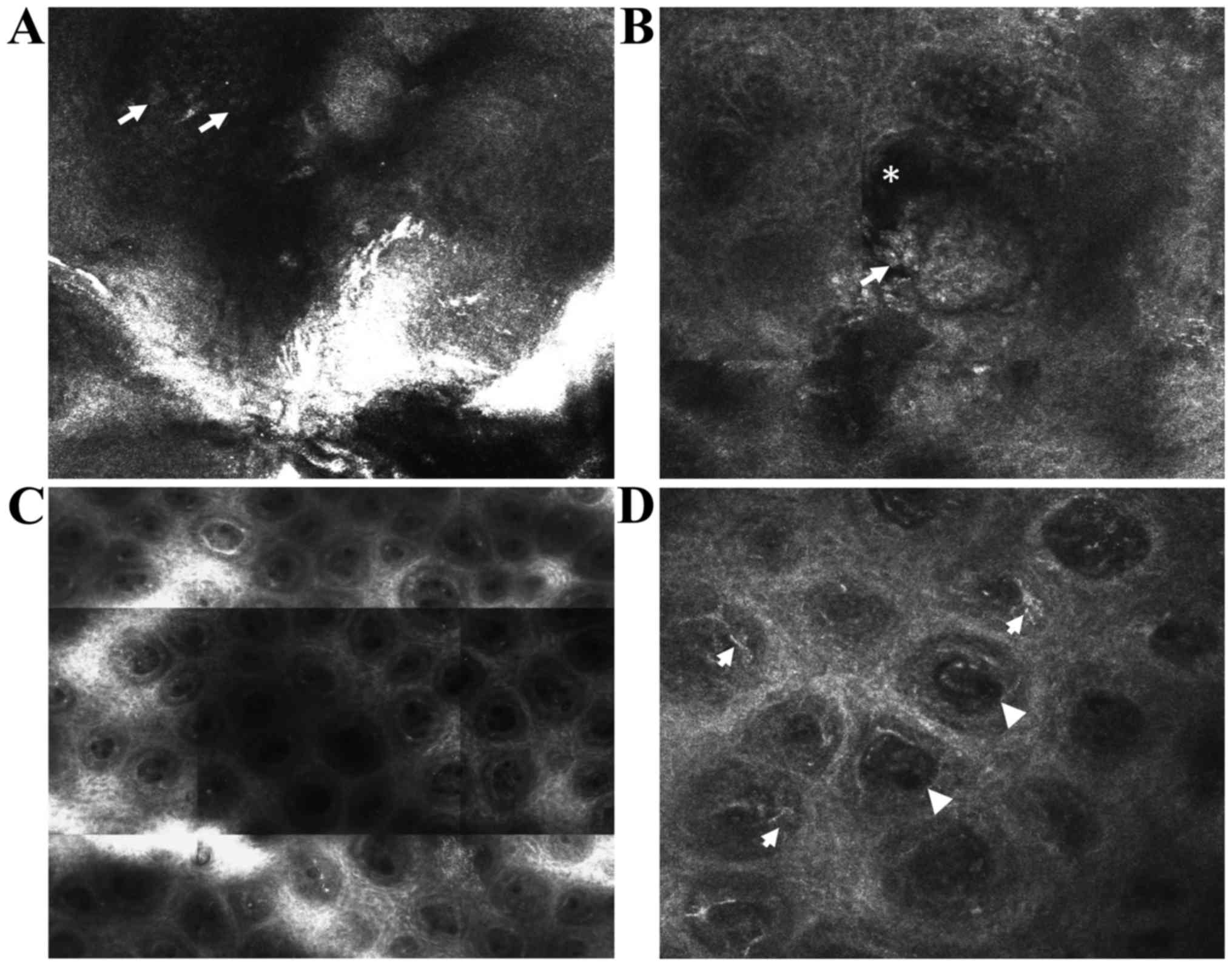
Characteristic micromorphological features of plaque
psoriasis lesions can be easily identified with in vivo RCM,
showing a high correlation with histology sections (19,20,41). In
the majority of cases, the stratum corneum is thickened
(hyperkeratosis) and associates small dark nuclei within its bright
cells (parakeratosis). Sometimes, clusters of highly refractile
round to polygonal cells can be seen between the corneocytes and
correspond to the diagnostic Munro's collections of neutrophils
(42). Going deeper into the
epidermis, a reduced or even absent granular layer (hypogranulosis)
is observed, whereas stratum spinosum has an increased thickness
(acanthosis). The horizontal RCM optical sections show an increase
in the diameter (>100 µm) and density of dermal papillae
(papillomatosis), as well as dilated blood vessels surrounded by
moderately refractile inflammatory cells in the superficial dermis
(Fig. 1) (17–20).
Moreover, in vivo RCM can be used for the objective
assessment of the response to various treatments at microscopic
level by performing serial determinations of the same skin area
(28,29,32).
Recently, our research group identified a method
(43) for the objective assessment
of psoriasis vulgaris lesions using in vivo RCM technique
that is potentially applicable to clinical studies and monitoring
the evolution of lesions under treatment.
Lichen planus
Lichen planus is an inflammatory mucocutaneous
disease with still unclear etiopathogenesis. In vivo RCM has
been used for the noninvasive evaluation of cutaneous lichen planus
and enabled the identification of its distinctive features, with a
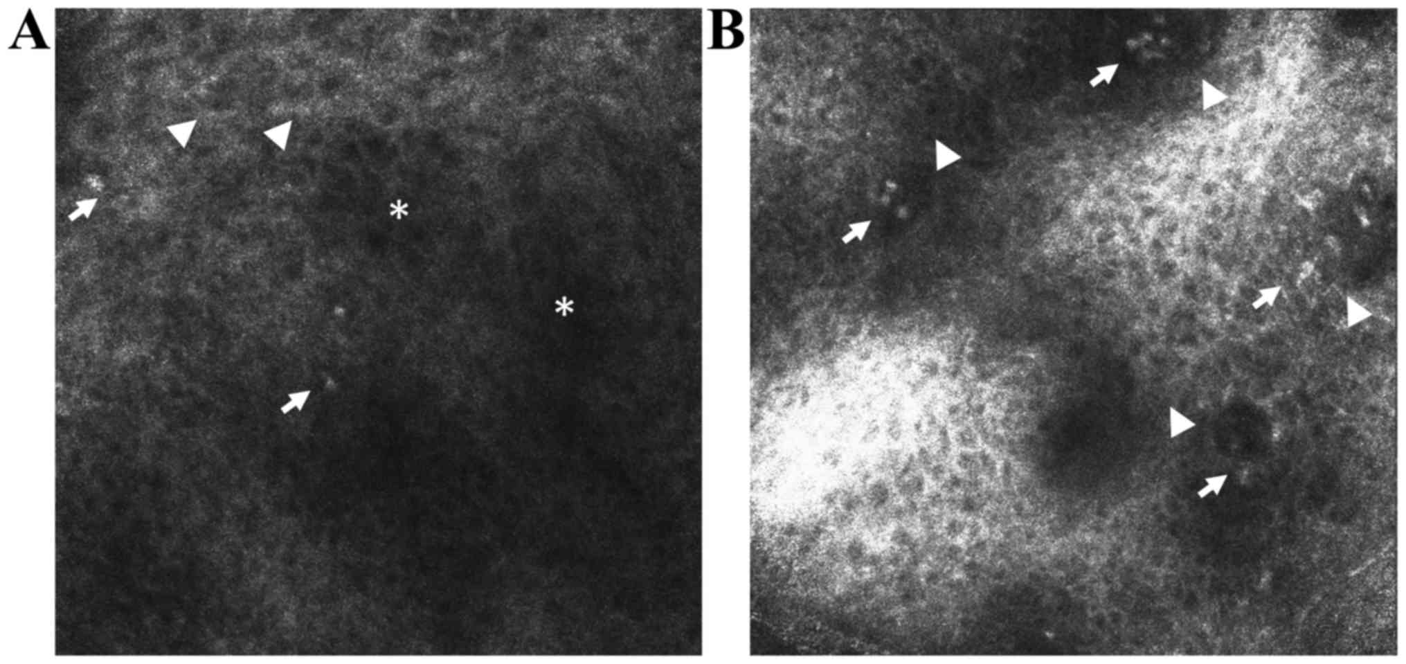
good correlation with histological findings (Fig. 2) (21). Confocal features of the epidermal
layers include increased intercellular spaces (spongiosis), large,
polygonal cells (hypergranulosis) disposed in a thickened
wedge-shaped granular cell layer (corresponding to Wickham's
striae) and the presence of grouped round-to-polygonal bright cells
(inflammatory infiltrates).
Due to the extensive inflammatory cell infiltrate at
the epidermal-dermal junction level, the replacement of the bright
ring-like structures around dermal papillae with smeared refractile
rings can be observed (vacuolar degeneration of the basal layer).
Large bright, plump, oval to stellate cells (melanophages) and
dilated blood vessels can be seen in the superficial dermis
(21,44). In vivo RCM represents a useful
tool for the noninvasive diagnosis of lichen planus, but further
studies are necessary in order to distinguish between different
subtypes of interface dermatitis (21).
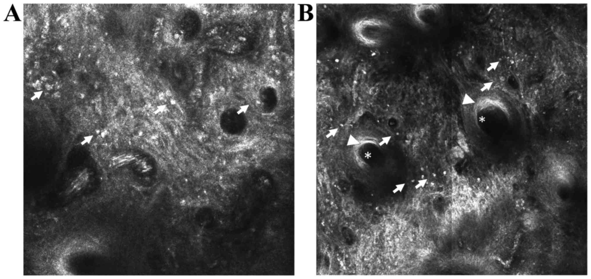
Discoid lupus erythematosus (DLE)
DLE is an inflammatory condition that can be
difficult to distinguish clinically from other erythematosquamous
skin diseases. In vivo RCM evaluation of DLE lesions enables
the identification of key diagnostic features seen in
histopathological samples including epidermal atrophy, interface
changes, as well as epidermal, dermal and periadnexial inflammatory
cell infiltration (Fig. 3) (22). The maximum depth of imaging limited
to the upper dermis and the inability to distinguish lymphocytes
from other inflammatory cells are disadvantages of RCM examination
in DLE. However, combining dermoscopy with RCM has an important
role in choosing the apropriate biopsy site for more histologic
diagnostic criteria (22,45).
In vivo RCM proved to be useful also in the
diagnosis of inflammatory dermatoses with special localizations
like the scalp, including alopecia areata (23,24),
lichen planopilaris and DLE secondary scarring alopecia (45). RCM criteria for these inflammatory
skin conditions strongly correlate with histologic features and are
sensitive enough to differentiate between entities with similar
clinical presentation (25). Also,
during follow-up of lesions, some micromorphological features
helped in choosing the appropriate therapeutic option.
In vivo confocal laser scanning microscopy
imaging for skin conditions associated with inflammation
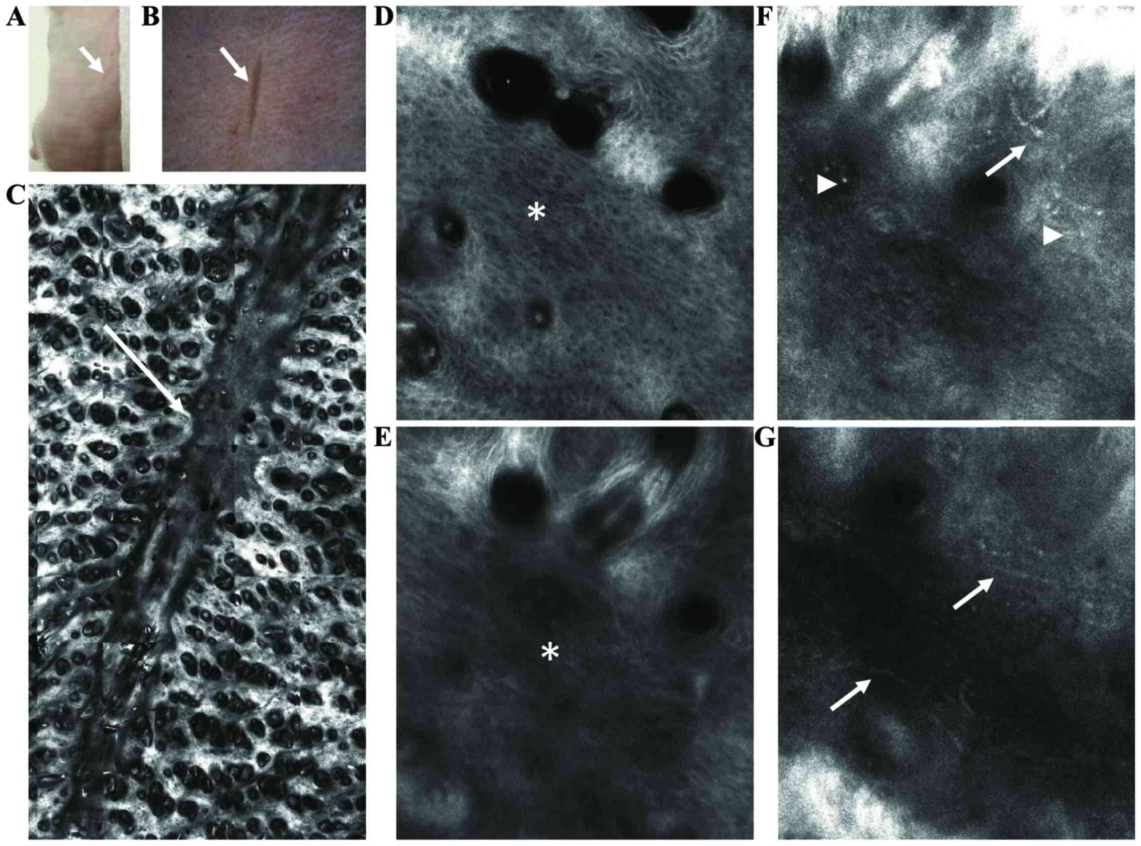
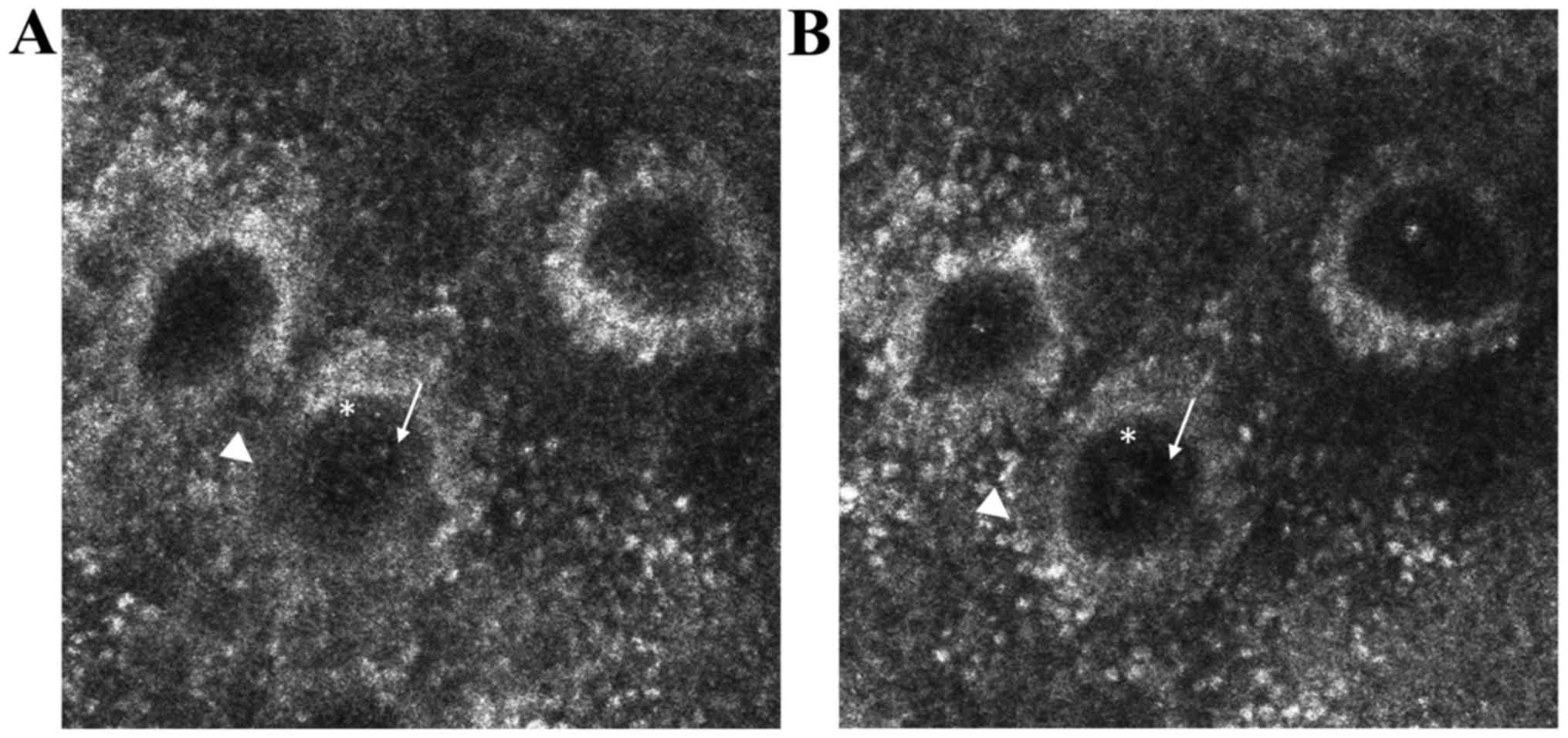
Cutaneous wound healing
Cutaneous wound healing is a continuously expanding
research area and various noninvasive imaging techniques have been
evaluated for their applicability in monitoring healing of skin
wounds (46). Among these
techniques, in vivo RCM was able to visualize the
inflammatory, vascular and tissue remodeling features associated
with cutaneous wound repair in real-time, at different time
intervals (30). A recent study
performed by our research group on BALB/c mice showed that in
vivo RCM is able to assess the extent of wound dehiscence,
restoration of the normal honeycombed pattern of epidermis and of
dermal fibro-elastic structures, as well as the aspect of dermal
blood vessels and the presence of inflammatory cells (unpublished
data) (Fig. 4).
Moreover, it can provide an objective and
noninvasive assessment of burn wounds gravity either alone, with
the advantage of its near cellular resolution (47), or combined with optical coherence
tomography for a better depth penetration (48). Moreover, in vivo RCM can
predict the healing course of burn wounds of indeterminate depth
based on serial determinations of microcirculation, morphology and
inflammatory cell traffic (49).
This noninvasive high-resolution technique was also able to assess
the effects of changes in cutaneous microcirculation on tissue
morphology during burn wound healing (31).
Skin aging
Skin aging is subject of increased research in
dermatology and cosmetology reflected in the continuously
development of products and methods that could prevent or reverse
this complex inevitable process. In vivo CLSM proved to be a
reliable method for assessment of skin aging and revealed the
presence of keratinocyte alterations, irregular pigmentation and
increased compactness of collagen fibers that became more
pronounced with aging (32). This
noninvasive method offers potential applications in choosing the
appropriate antiaging method and testing cosmetic treatment
efficacy.
UVR-induced alterations
UVR-induced alterations are responsible for
extrinsic aging and play an important role in skin cancer
development (33). In vivo
RCM was able to identify and monitor UVR produced skin alterations
beginning with skin inflammation, followed by the appearance of
microvesicles, apoptotic keratinocytes and activated melanocytes
and finally, loss of the epidermal structure (34). Specifically, ultraviolet A (UVA)
radiation effects upon the skin were analyzed using this
noninvasive technique, confirming that UVA-induced accelerated
blood flow is a prerequisite for the development of immediate and
delayed tanning (50). Another study
showed the negative effects of sun exposure upon epidermal
architecture, dermal collagen and overall skin thickness (35).
Moreover, a recent study demonstrated the utility of
RCM for an objective quantitative definition of sensitive skin and
showed that disarranged as well as reduced honeycomb pattern depth
and the presence of spongiform edema were predominant confocal
features for this type of skin (51).
In vivo confocal laser scanning microscopy
imaging for investigation of skin inflammation components
Leukocyte migration
Leukocyte migration or relocation is a synchronized
active process through which both innate and adaptive immune system
work to resolve a distally located inflammation or injury (52). Although intensively studied as
important immune defenders, the complex voyage of leukocytes,
especially neutrophils, alongside the blood vessel claims new
imaging methods such as CLSM, or even a three-dimensional (3D)
approach, in order to reveal their in-depth spatiotemporal scene in
vessels and surrounding tissues. Thus, acknowledged to confocal
fluorescent microscopy it becomes possible to envisage
intravascular migration of neutrophils, extravasation as well as
interstitial passage thorough peripheral vessels as a prompt
response to physically damaged tissue, bacteria and virus invaders
in order to reduce the injury and preserve the body homeostasis
(53).
Why is a 3D imaging approach so worthy in monitoring
a fine cellular process like leukocyte transmigration cascade?
Firstly, it catches cellular aspects which, although important,
cannot be detected with other in vitro investigation
methods. For instance, besides endothelial cells pericytes are also
involved which are rarely included and examined by in vitro
methods, although they express key adhesion molecules (e.g.,
VCAM-1), are an integrant part of the vessel wall and perform an
active role in transmigration (54).
Secondly, a 3D approach would clasp in real-time the timing
process; for example, in vitro detection of neutrophils
passage was reported within 2 min while in vivo methods
reported 15–45 min for neutrophil migration through endothelial
cells (55).
These aspects become very important when, for
instance the effect of a stimulus or a therapeutic agent on
leukocyte functions is tested, and also when investigator would
directly track the whole picture of leukocyte behavior in a
physiologically milieu including all junctions and intercellular
interactions. The final step of leukocyte/neutrophil migration
(extravasation) was evidenced and reported quite recently; thus,
the uropod elongation as final step of extravasation was imaged by
multiphoton intravital microscopy in cremaster muscle leukocytes of
CD18-mCFP KI mice following neutrophil stimulation (CXCL2, fMLP and
TNF) and Texas Red-dextran staining of blood vessels. Images reveal
that during extravasation the CD18+ neutrophils marked
with Alexa Fluor 488-anti-Gr1 antibody migrated from cremaster
venules are about 4 times elongated compared with rolling and
crawling leukocytes (56). Thus,
migration process imaging provides valuable data regarding accurate
function and regulation of leukocyte recruitment to injury site
with potential impact in therapeutic purposes (4).
Capillary blood flow
Capillary blood flow within dermal vascularization
is a parameter that can be easily monitored in real-time, in
vivo with RCM imaging due to brightly reflecting of blood
cells. Several factors are usually recorded, thus, the quantitative
blood cell flow per minute is measured, taking into account the
number of capillary loop and multiple fields of view digitally
measured for a fixed time-point (e.g., every 30 sec). Further, the
capillary loop diameter and the density of dermal capillaries per
area are registered in the dermal papillae of the epidermal-dermal
junction, where multiple fields of view are captured in real-time
images (36). In addition, in
real-time investigation of skin blood flow changes induced by
topical capsaicin using RCM was proposed as a research model to
test neurovascular reactivity (5).
An alternative to RCM is two-photon excitation
microscopy that provides advantages for 3D and deep tissue imaging.
A widespread application of this tool is to evaluate the blood flow
via blood cell velocity and brightness, these measurements being
very useful in setting different experimental models such as
stroke, embryo development (57) or
different skin cancers (basal cell carcinoma, squamous cell
carcinoma) where morphological and functional assessments of skin
layers are primarily made by combining techniques as laser Doppler
flowmetry and RCM (58).
Skin reactivity to topical
stimuli
Skin reactivity to certain local stimuli could be an
excellent model for revising various skin conditions through
evaluation of the inflammatory process at the skin level (6,37).
Neurogenic inflammation was experimentally locally induced by
capsaicin and further CLSM in reflectance mode enabled the
assessment of the cutaneous micro-vascularization (Fig. 5). This could represent an important
research model for studying the link between cutaneous diseases and
the nervous system (6).
As an innovative non-invasive method for in
vivo imaging of skin structure, CLSM could be successfully
applied in dermatological fields less studied until now in terms of
imaging, such as experimental contact dermatitis. Such attempts
have been made for almost one decade when in situ imaging of
skin reactions produced by sodium lauryl sulphate and pelargonic
acid as experimental irritants was reported; the group describes
extended cell boundaries, keratinocyte swelling (pelargonic acid)
and induction of parakeratosis within the stratum corneum (sodium
lauryl sulphate) (59).
CLSM was applied also in a recent investigation in
helping the patch test interpretations, a test known as the gold
standard for contact dermatitis validation, and thus to
differentiate very precisely between allergic, irritant, and
equivocal patch test reactions; in this study, in vivo CLSM
assessment revealed that 40% from equivocal reactions displayed
confocal patterns in line with the positive allergic reactions
patterns (60).
Conclusions
In vivo CLSM is a novel imaging technique
that provides the morphological and dynamic characterization of
skin structures with a high, quasi-microscopic resolution. The
non-invasive character of the examination and the possibility to
evaluate the same skin area at different time-points make in
vivo CLSM a useful tool in the diagnosis and monitoring of
inflammatory skin diseases. Moreover, it is an excellent method to
study in real-time the dynamic components of skin inflammation,
response to treatment or specific stimuli with broad applications
ranging from clinical to experimental, functional studies involving
the skin. Studying the components of skin inflammation is also of
great potential to unravel pathways in the pathogenesis of diseases
associated with skin inflammation and might contribute to the
development of new treatment strategies.
Acknowledgements
Not applicable.
Funding
This study was partially supported by a grant of the
Romanian Ministry of Research and Innovation, CCCDI-UEFISCDI
(project nos. 61PCCDI⁄2018 PN-III-P1-1.2-PCCDI-2017-034 and
PN-III-P2-2.1-BG-2016-0443), within PNCDI-III.
Availability of data and materials
Not applicable.
Authors' contributions
MAI, CCa, DL, MT, SRG, MMC, CCo, MN, SAZ and DB
contributed equally to acquisition, analysis and systematization of
data, manuscript writing and critical revision of it for important
intellectual content. All authors read and approved the final
version of the manuscript.
Ethics approval and consent to
participate
Not applicable.
Patient consent for publication
Not applicable.
Competing interests
The authors declare that they have no competing
interests.
References
|
1
|
Peppelman M, Wolberink EA, Gerritsen MJ,
van de Kerkhof PC and van Erp PE: Application of leukotriene B4 and
reflectance confocal microscopy as a noninvasive in vivo model to
study the dynamics of skin inflammation. Skin Res Technol.
21:232–240. 2015. View Article : Google Scholar : PubMed/NCBI
|
|
2
|
Diaconeasa A, Boda D, Neagu M, Constantin
C, Căruntu C, Vlădău L and Guţu D: The role of confocal microscopy
in the dermato-oncology practice. J Med Life. 4:63–74.
2011.PubMed/NCBI
|
|
3
|
Rajadhyaksha M, Grossman M, Esterowitz D,
Webb RH and Anderson RR: In vivo confocal scanning laser microscopy
of human skin: Melanin provides strong contrast. J Invest Dermatol.
104:946–952. 1995. View Article : Google Scholar : PubMed/NCBI
|
|
4
|
González S, Sackstein R, Anderson RR and
Rajadhyaksha M: Real-time evidence of in vivo leukocyte trafficking
in human skin by reflectance confocal microscopy. J Invest
Dermatol. 117:384–386. 2001. View Article : Google Scholar : PubMed/NCBI
|
|
5
|
Ghiţă MA, Căruntu C, Rosca AE, Căruntu A,
Moraru L, Constantin C, Neagu M and Boda D: Real-time investigation
of skin blood flow changes induced by topical capsaicin. Acta
Dermatovenerol Croat. 25:223–227. 2017.PubMed/NCBI
|
|
6
|
Căruntu C and Boda D: Evaluation through
in vivo reflectance confocal microscopy of the cutaneous neurogenic
inflammatory reaction induced by capsaicin in human subjects. J
Biomed Opt. 17:0850032012. View Article : Google Scholar : PubMed/NCBI
|
|
7
|
Meyer LE, Otberg N, Sterry W and Lademann
J: In vivo confocal scanning laser microscopy: Comparison of the
reflectance and fluorescence mode by imaging human skin. J Biomed
Opt. 11:0440122006. View Article : Google Scholar : PubMed/NCBI
|
|
8
|
Skvara H, Plut U, Schmid JA and Jonak C:
Combining in vivo reflectance with fluorescence confocal microscopy
provides additive information on skin morphology. Dermatol Pract
Concept. 2:3–12. 2012. View Article : Google Scholar : PubMed/NCBI
|
|
9
|
Izatt JA, Kulkarni MD, Hsing-Wen W,
Kobayashi K and Sivak MV: Optical coherence tomography and
microscopy in gastrointestinal tissues. IEEE J Sel Top Quantum
Electron. 2:1017–1028. 1996. View Article : Google Scholar
|
|
10
|
Pellacani G, Guitera P, Longo C, Avramidis
M, Seidenari S and Menzies S: The impact of in vivo reflectance
confocal microscopy for the diagnostic accuracy of melanoma and
equivocal melanocytic lesions. J Invest Dermatol. 127:2759–2765.
2007. View Article : Google Scholar : PubMed/NCBI
|
|
11
|
Guida S, Longo C, Casari A, Ciardo S,
Manfredini M, Reggiani C, Pellacani G and Farnetani F: Update on
the use of confocal microscopy in melanoma and non-melanoma skin
cancer. G Ital Dermatol Venereol. 150:547–563. 2015.(In Italian).
PubMed/NCBI
|
|
12
|
Guida S, Longo C, Casari A, Ciardo S,
Manfredini M, Reggiani C, Pellacani G and Farnetani F: Distinct
melanoma types based on reflectance confocal microscopy. Exp
Dermatol. 23:414–418. 2014. View Article : Google Scholar : PubMed/NCBI
|
|
13
|
Ghita MA, Caruntu C, Rosca AE, Kaleshi H,
Caruntu A, Moraru L, Docea AO, Zurac S, Boda D, Neagu M, et al:
Reflectance confocal microscopy and dermoscopy for in vivo,
non-invasive skin imaging of superficial basal cell carcinoma.
Oncol Lett. 11:3019–3024. 2016. View Article : Google Scholar : PubMed/NCBI
|
|
14
|
Căruntu C, Boda D, Guţu DE and Căruntu A:
In vivo reflectance confocal microscopy of basal cell carcinoma
with cystic degeneration. Rom J Morphol Embryol. 55:1437–1441.
2014.PubMed/NCBI
|
|
15
|
Lupu M, Caruntu C, Solomon I, Popa A,
Lisievici C, Draghici C, Papagheorghe L, Voiculescu V and
Giurcaneanu C: The use of in vivo reflectance confocal microscopy
and dermoscopy in the preoperative determination of basal cell
carcinoma histopathological subtypes. Dermatovenerologia.
62:265–275. 2017.
|
|
16
|
Lupu M, Caruntu A, Caruntu C, Boda D,
Moraru L, Voiculescu V and Bastian A: Non-invasive imaging of
actinic cheilitis and squamous cell carcinoma of the lip. Mol Clin
Oncol. 8:640–646. 2018.PubMed/NCBI
|
|
17
|
Białek-Galas K, Wielowieyska-Szybińska D,
Dyduch G and Wojas-Pelc A: The use of reflectance confocal
microscopy in selected inflammatory skin diseases. Pol J Pathol.
66:103–108. 2015. View Article : Google Scholar : PubMed/NCBI
|
|
18
|
Wolberink EA, van Erp PE, Teussink MM, van
de Kerkhof PC and Gerritsen MJ: Cellular features of psoriatic
skin: Imaging and quantification using in vivo reflectance confocal
microscopy. Cytometry B Clin Cytom. 80:141–149. 2011. View Article : Google Scholar : PubMed/NCBI
|
|
19
|
Ardigo M, Cota C, Berardesca E and
González S: Concordance between in vivo reflectance confocal
microscopy and histology in the evaluation of plaque psoriasis. J
Eur Acad Dermatol Venereol. 23:660–667. 2009. View Article : Google Scholar : PubMed/NCBI
|
|
20
|
González S, Rajadhyaksha M, Rubinstein G
and Anderson RR: Characterization of psoriasis in vivo by
reflectance confocal microscopy. J Med. 30:337–356. 1999.PubMed/NCBI
|
|
21
|
Moscarella E, González S, Agozzino M,
Sánchez-Mateos JL, Panetta C, Contaldo M and Ardigò M: Pilot study
on reflectance confocal microscopy imaging of lichen planus: A
real-time, non-invasive aid for clinical diagnosis. J Eur Acad
Dermatol Venereol. 26:1258–1265. 2012. View Article : Google Scholar : PubMed/NCBI
|
|
22
|
Ardigò M, Maliszewski I, Cota C, Scope A,
Sacerdoti G, Gonzalez S and Berardesca E: Preliminary evaluation of
in vivo reflectance confocal microscopy features of Discoid lupus
erythematosus. Br J Dermatol. 156:1196–1203. 2007. View Article : Google Scholar : PubMed/NCBI
|
|
23
|
Ardigò M, Tosti A, Cameli N, Vincenzi C,
Misciali C and Berardesca E: Reflectance confocal microscopy of the
yellow dot pattern in alopecia areata. Arch Dermatol. 147:61–64.
2011. View Article : Google Scholar : PubMed/NCBI
|
|
24
|
Rudnicka L, Olszewska M and Rakowska A: In
vivo reflectance confocal microscopy: Usefulness for diagnosing
hair diseases. J Dermatol Case Rep. 2:55–59. 2008. View Article : Google Scholar : PubMed/NCBI
|
|
25
|
Koller S, Gerger A, Ahlgrimm-Siess V,
Weger W, Smolle J and Hofmann-Wellenhof R: In vivo reflectance
confocal microscopy of erythematosquamous skin diseases. Exp
Dermatol. 18:536–540. 2009. View Article : Google Scholar : PubMed/NCBI
|
|
26
|
Ma J, Zhang X, Lv Y, Zhao C, Li Q, Yang X
and Zhao J: Clinical application of confocal laser scanning
microscopy for atypical dermatoses. Cell Biochem Biophys.
73:199–204. 2015. View Article : Google Scholar : PubMed/NCBI
|
|
27
|
González S, Sánchez V, González-Rodríguez
A, Parrado C and Ullrich M: Confocal microscopy patterns in
nonmelanoma skin cancer and clinical applications. Actas
Dermosifiliogr. 105:446–458. 2014. View Article : Google Scholar : PubMed/NCBI
|
|
28
|
Wolberink EA, van Erp PE, de Boer-van
Huizen RT, van de Kerkhof PC and Gerritsen MJ: Reflectance confocal
microscopy: An effective tool for monitoring ultraviolet B
phototherapy in psoriasis. Br J Dermatol. 167:396–403. 2012.
View Article : Google Scholar : PubMed/NCBI
|
|
29
|
Ardigò M, Agozzino M, Longo C, Conti A, Di
Lernia V, Berardesca E and Pellacani G: Psoriasis plaque test with
confocal microscopy: Evaluation of different microscopic response
pathways in NSAID and steroid treated lesions. Skin Res Technol.
19:417–423. 2013.PubMed/NCBI
|
|
30
|
Lange-Asschenfeldt S, Bob A, Terhorst D,
Ulrich M, Fluhr J, Mendez G, Roewert-Huber HJ, Stockfleth E and
Lange-Asschenfeldt B: Applicability of confocal laser scanning
microscopy for evaluation and monitoring of cutaneous wound
healing. J Biomed Opt. 17:0760162012. View Article : Google Scholar : PubMed/NCBI
|
|
31
|
Altintas AA, Altintas MA, Ipaktchi K,
Guggenheim M, Theodorou P, Amini P and Spilker G: Assessment of
microcirculatory influence on cellular morphology in human burn
wound healing using reflectance-mode-confocal microscopy. Wound
Repair Regen. 17:498–504. 2009. View Article : Google Scholar : PubMed/NCBI
|
|
32
|
Longo C, Casari A, Beretti F, Cesinaro AM
and Pellacani G: Skin aging: In vivo microscopic assessment of
epidermal and dermal changes by means of confocal microscopy. J Am
Acad Dermatol. 68:e73–e82. 2013. View Article : Google Scholar : PubMed/NCBI
|
|
33
|
Solovastru LG, Vâta D, Statescu L,
Constantin MM and Andrese E: Skin cancer between myth and reality,
yet ethically constrained. Rev Rom Bioet. 12:47–52. 2014.
|
|
34
|
Koller S, Inzinger M, Rothmund M,
Ahlgrimm-Siess V, Massone C, Arzberger E, Wolf P and
Hofmann-Wellenhof R: UV-induced alterations of the skin evaluated
over time by reflectance confocal microscopy. J Eur Acad Dermatol
Venereol. 28:1061–1068. 2014. View Article : Google Scholar : PubMed/NCBI
|
|
35
|
Haytoglu NS, Gurel MS, Erdemir A, Falay T,
Dolgun A and Haytoglu TG: Assessment of skin photoaging with
reflectance confocal microscopy. Skin Res Technol. 20:363–372.
2014. View Article : Google Scholar : PubMed/NCBI
|
|
36
|
Altintas MA, Altintas AA, Guggenheim M,
Steiert AE, Aust MC, Niederbichler AD, Herold C and Vogt PM:
Insight in human skin microcirculation using in vivo
reflectance-mode confocal laser scanning microscopy. J Digit
Imaging. 23:475–481. 2010. View Article : Google Scholar : PubMed/NCBI
|
|
37
|
Altintas AA, Guggenheim M, Oezcelik A,
Gehl B, Aust MC and Altintas MA: Local burn versus local cold
induced acute effects on in vivo microcirculation and
histomorphology of the human skin. Microsc Res Tech. 74:963–969.
2011. View Article : Google Scholar : PubMed/NCBI
|
|
38
|
Caruntu C, Boda D, Dumitrascu G,
Constantin C and Neagu M: Proteomics focusing on immune markers in
psoriatic arthritis. Biomarkers Med. 9:513–528. 2015. View Article : Google Scholar
|
|
39
|
Schön MP and Boehncke WH: Psoriasis. N
Engl J Med. 352:1899–1912. 2005. View Article : Google Scholar : PubMed/NCBI
|
|
40
|
Wu H, Shapiro B and Harrist TJ:
Noninfectious erythematous, popular, and squamous diseases.
Psoriasis. Lever's Histopathology of the Skin. 9th. Elder DE,
Elenitzas R, Johnson BL and Murphy GF: Lippincott Williams &
Wilkins; Philadelphia, PA: pp. 59–60. 2005
|
|
41
|
Căruntu C, Boda D, Căruntu A, Rotaru M,
Baderca F and Zurac S: In vivo imaging techniques for psoriatic
lesions. Rom J Morphol Embryol. 55:1191–1196. 2014.PubMed/NCBI
|
|
42
|
Zhong LS, Wei ZP and Liu YQ: Sensitivity
and specificity of Munro microabscess detected by reflectance
confocal microscopy in the diagnosis of psoriasis vulgaris. J
Dermatol. 39:282–283. 2012. View Article : Google Scholar : PubMed/NCBI
|
|
43
|
Batani A, Brănișteanu DE, Ilie MA, Boda D,
Ianosi S, Ianosi G and Caruntu C: Assessment of dermal papillary
and microvascular parameters in psoriasis vulgaris using in vivo
reflectance confocal microscopy. Exp Ther Med. 15:1241–1246.
2018.PubMed/NCBI
|
|
44
|
González S: Lichen planus. Reflectance
Confocal Microscopy in Dermatology: Fundamentals and Clinical
Applications. Grupo Aula Medica; Madrid: pp. 25–26. 2012
|
|
45
|
Agozzino M, Tosti A, Barbieri L,
Moscarella E, Cota C, Berardesca E and Ardigò M: Confocal
microscopic features of scarring alopecia: Preliminary report. Br J
Dermatol. 165:534–540. 2011.PubMed/NCBI
|
|
46
|
Mani R: Science of measurements in wound
healing. Wound Repair Regen. 7:330–334. 1999. View Article : Google Scholar : PubMed/NCBI
|
|
47
|
Altintas MA, Altintas AA, Knobloch K,
Guggenheim M, Zweifel CJ and Vogt PM: Differentiation of
superficial-partial vs. deep-partial thickness burn injuries in
vivo by confocal-laser-scanning microscopy. Burns. 35:80–86. 2009.
View Article : Google Scholar : PubMed/NCBI
|
|
48
|
Iftimia N, Ferguson RD, Mujat M, Patel AH,
Zhang EZ, Fox W and Rajadhyaksha M: Combined reflectance confocal
microscopy/optical coherence tomography imaging for skin burn
assessment. Biomed Opt Express. 4:680–695. 2013. View Article : Google Scholar : PubMed/NCBI
|
|
49
|
Altintas AA, Guggenheim M, Altintas MA,
Amini P, Stasch T and Spilker G: To heal or not to heal: Predictive
value of in vivo reflectance-mode confocal microscopy in assessing
healing course of human burn wounds. J Burn Care Res. 30:1007–1012.
2009.PubMed/NCBI
|
|
50
|
Yamashita T, Akita H, Astner S, Miyakawa
M, Lerner EA and González S: In vivo assessment of pigmentary and
vascular compartments changes in UVA exposed skin by
reflectance-mode confocal microscopy. Exp Dermatol. 16:905–911.
2007. View Article : Google Scholar : PubMed/NCBI
|
|
51
|
Ma YF, Yuan C, Jiang WC, Wang XL and
Humbert P: Reflectance confocal microscopy for the evaluation of
sensitive skin. Skin Res Technol. 23:227–234. 2017. View Article : Google Scholar : PubMed/NCBI
|
|
52
|
Yadav R, Larbi KY, Young RE and Nourshargh
S: Migration of leukocytes through the vessel wall and beyond.
Thromb Haemost. 90:598–606. 2003. View Article : Google Scholar : PubMed/NCBI
|
|
53
|
Park SA and Hyun YM: Neutrophil
extravasation cascade: What can we learn from two-photon intravital
imaging? Immune Netw. 16:317–321. 2016. View Article : Google Scholar : PubMed/NCBI
|
|
54
|
Proebstl D, Voisin M-B, Woodfin A,
Whiteford J, D'Acquisto F, Jones GE, Rowe D and Nourshargh S:
Pericytes support neutrophil subendothelial cell crawling and
breaching of venular walls in vivo. J Exp Med. 209:1219–1234. 2012.
View Article : Google Scholar : PubMed/NCBI
|
|
55
|
Stein B, Khew-Goodall Y, Gamble J and
Vadas MA: Transmigration of leukocytes. Endothelium in Clinical
Practice: Source and Target of Novel Therapies. Rubanyi GM and Dzau
VJ: Marcel Dekker, Inc.; New York, NY: pp. 149–202. 1997
|
|
56
|
Hyun YM, Sumagin R, Sarangi PP, Lomakina
E, Overstreet MG, Baker CM, Fowell DJ, Waugh RE, Sarelius IH and
Kim M: Uropod elongation is a common final step in leukocyte
extravasation through inflamed vessels. J Exp Med. 209:1349–1362.
2012. View Article : Google Scholar : PubMed/NCBI
|
|
57
|
Benninger RK and Piston DW: Two-photon
excitation microscopy for the study of living cells and tissues.
Curr Protoc Cell Biol Chapter. 4:1–24. 2013.doi:
10.1002/0471143030.cb0411s59.
|
|
58
|
Mowla A, Taimre T, Lim YL, Bertling K,
Wilson SJ, Prow TW, Soyer H and Rakić A: Concurrent reflectance
confocal microscopy and laser doppler flowmetry to improve skin
cancer imaging: A Monte Carlo model and experimental validation.
Sensors (Basel). 16:14112016. View Article : Google Scholar
|
|
59
|
Suihko C and Serup J: Fluorescence
confocal laser scanning microscopy for in vivo imaging of epidermal
reactions to two experimental irritants. Skin Res Technol.
14:498–503. 2008. View Article : Google Scholar : PubMed/NCBI
|
|
60
|
Slodownik D, Levi A, Lapidoth M, Ingber A,
Horev L and Enk CD: Noninvasive in vivo confocal laser scanning
microscopy is effective in differentiating allergic from
nonallergic equivocal patch test reactions. Lasers Med Sci.
30:1081–1087. 2015. View Article : Google Scholar : PubMed/NCBI
|