Introduction
Stress has become a major component of modern
lifestyle, the human body being frequently faced to the need for
adaptation. Through its impact on the mechanisms of homeostasis,
stress can alter health, stress-related illnesses being an alarming
ongoing problem in developed countries, both through social costs
and healthcare spending, but also economic losses from productivity
decline (1–4).
Numerous studies have suggested that stress can be
involved in triggering or aggravating various dermatological
conditions, from skin cancer to inflammatory skin diseases such as
psoriasis, atopic dermatitis, seborrheic eczema, acne, prurigo
nodularis, lichen planus, chronic urticaria, rosacea and alopecia
areata (5–15). The complexity of interconnections
between the nervous system and the skin are surprising at the first
glance; the action of stress on skin can be exerted both directly
through the peripheral nervous system and indirectly through
endocrine and immune systems.
Neurogenic inflammation is a particular type of
inflammatory response, highlighting the multitude of neurocutaneous
connections. The neurogenic inflammatory process developed in the
skin is a potential factor involved in the onset and progression of
some of the inflammatory dermatological diseases (9,16).
Hence, the study of mechanisms of activation and modulation of
neurogenic inflammation has both clinical and fundamental
importance.
Previous studies have highlighted the effect of
stress on the mechanisms involved in the cutaneous neurogenic
reaction, such as modulating the density and activity of
nociceptive nerve fibers sensitive to capsaicin, modulating the
activity of their neurons, increasing SP release from the
unmyelinated nerve endings of the skin and the degranulation of
cutaneous mast cells (17–21).
In this study, we aimed to assess the influence of
stress on the capsaicin-induced skin inflammatory reaction, one of
the most commonly used neurogenic skin inflammation models in
scientific research (22–24). To achieve this goal we proposed a
good control of unwanted external stressors. In order to induce
psychological stress, we proposed the development and
implementation of an easy-to-use protocol that contains various
tasks capable of generating stress differently, thus allowing
stress to be induced to as many categories of subjects as possible
and avoiding the installation of habituation. We also sought to
evaluate the capsaicin-induced inflammatory response by methods in
which the subjective factor had the least possible involvement.
Subjects and methods
Subjects
In this study, 31 healthy subjects of both sexes
(M=13, F=18) were enrolled, aged between 20 and 35 years (mean age
25.096 years).
The study was carried out with the approval of the
Ethics Committee of Carol Davila University of Medicine and
Pharmacy (Bucharest, Romania). At the time of the study, all
participants attended university studies and enrollment in the
study was done on a voluntary basis. The participation of the
subjects in the study was conditioned by a written consent.
Subjects with cardiovascular or respiratory
diseases, autoimmune diseases, neoplasms, organ transplants,
allergic reactions to the substances used in the study, as well as
subjects treated with drugs that may influence the physiological
parameters studied were excluded from the study. Other exclusion
criteria were major psychiatric diseases and major stress or
infectious diseases in the previous month. Moreover, pregnant or
lactating women were excluded from the study.
Study participants were asked to avoid drinking
psychoactive substances, alcohol, coffee, tea, energy drinks or
containing caffeine, smoking, medication, and intense physical
effort 24 h before the test.
Subjects included in the study were randomized into
two groups: stress group of 17 subjects (F=10, M=7, mean age
25.12±4.96 years) undergoing a stress-inducing protocol; and
control group of 14 subjects (F=8, M=6, mean age 25.07±4.32 years)
who were exposed to indifferent conditions.
Procedures
Experiments were performed during the afternoon
(12:00 p.m. - 18:00 p.m.) and lasted approximately 2 h per subject.
During the experiments, a temperature of 22±2°C and a humidity of
50±5% were maintained in the laboratory.
Upon arrival at the place of the experiment, the
subjects were given a 15-min period of accommodation with the
laboratory conditions, completing the inclusion forms in the
study.
In order to quantify the impact of stressors in
everyday life prior to participating in the study, we assessed the
level of perceived stress for each subject using the Perceived
Stress Scale (PSS) questionnaire (25).
Subsequently, each of the subjects participated in a
30-min computerized stage in which the subjects in the stress group
underwent a stress-inducing protocol, and those in the control
group were exposed to indifferent conditions. The stress-inducing
protocol contained three phases of similar duration: a phase of
viewing images with negative emotional load; an arithmetic
counter-clock test phase; and a counter-clock intelligence test
phase. Exposure to indifferent conditions was accomplished by
viewing images with no negative emotional load.
Both during the accommodation and during the
experiments the subjects were placed in a sitting position on a
comfortable chair.
For 1 min before and during the computerized stage,
the skin conductance was recorded in each subject. At the end of
this stage, each of the subjects quantified on a scale from 0 to 10
the level of stress experienced during exposure to the stress
induction protocol and indifferent conditions, respectively.
At the end of the computerized stage, the subjects
in both groups participated in the capsaicin exposure stage in
which, at the middle third of the non-dominant anterior forearm,
topical capsaicin cream was administered at 0.1%. The cream was
kept in contact with the skin of the subjects for 10 min, after
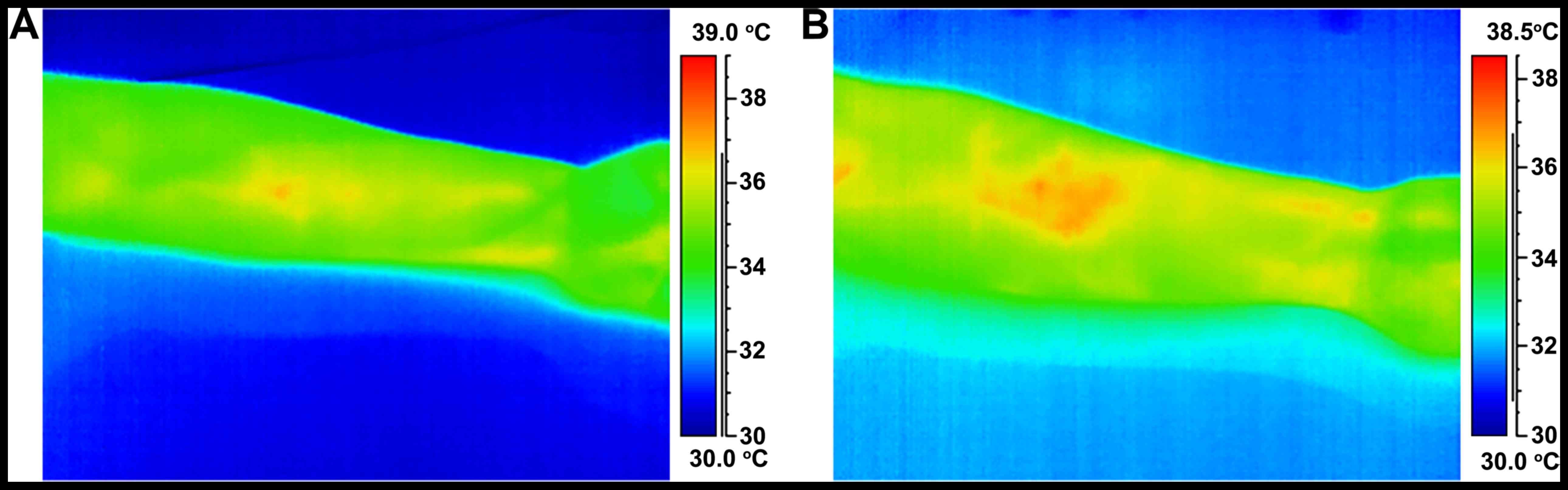
which it was removed. The assessment of the local inflammatory
reaction induced by capsaicin was performed by cutaneous
thermography, and the thermographic images taken at 25 and 40 min
after the onset of capsaicin administration were analyzed.
Viewing images with/without negative
emotional load
The phase of viewing images with negative emotional
load in the stress-inducing protocol and exposure to indifferent
conditions by viewing images without unpleasant connotations were
performed using the IAPS image set (26). The images used in the experiment were
chosen on the basis of the individual emotional, activation and
dominance values indicated in the IAPS technical report and were
divided into two sets: a set of images without negative emotional
load that contained neutral, pleasant or intense pleasant images;
and a set of negative emotional images that contained neutral,
unpleasant or intense unpleasant images.
The images were displayed on the screen of a monitor
using a software developed by our group (27). The image display interval was 7 sec
and was chosen experimentally so that the subjects were exposed for
long enough to the visual stimulus in order to achieve a reaction
and at the same time to avoid habituation.
Arithmetic counter-clock test
The arithmetic test phase of the stress induction
protocol was based on a similar MIST stress test concept (28). It was accomplished through a program
that displays on a monitor screen arithmetical operations that the
subject is trained to solve mentally within a limited
time-frame.
Counter-clock intelligence test
For the phase of the stress-inducing intelligence
test, we used a computerized version of the Raven's Progressive
Matrices test (29), which is a
non-verbal evaluation of the level of general intelligence
(30).
The Raven test was used in the experiment because
the notion of psychological test and assessment of intelligence
level, accompanied by the investigator's assertion that test
results will also be important in the experiment, is an important
stress factor. In addition, the time frame available to solve the
problems was limited to 10 min.
Evaluation of cutaneous
conductance
The skin conductance was recorded via a Varioport-B
portable acquisition system (Becker Meditec, Karlsruhe, Germany) in
a 8-bit digital system (spectrum = 0–70 µsec) at a frequency of 250
Hz. The recording was performed in a bipolar system using two
circular-shaped Ag/AgCl electrodes with a 10 mm diameter contact
surface (Becker Meditec), with a constant 0.5 V. The applied
current had a low voltage so it could not be perceived by the
subjects.
Electrode placement was performed with caution, and
subjects were instructed to avoid changing position during the test
to reduce the risk of electrode detachment due to movement and
sweat secretion. The electrodes were positioned at the palm of the
non-dominant hand, on the thenar and hypothenar eminences, ensuring
a constant contact area and good contact between the electrodes and
the skin surface (27). An isotonic
paste with a composition close to that of the eccrine sweat
secretion (31) was applied to the
surface of the electrodes and they were applied to the skin using
double-sided adhesive rings.
Prior to applying the electrodes, the skin was
cleaned with water and soap, while avoiding skin abrasion maneuvers
or contact with solvent agents. The level of skin conductance was
determined for 1 min under baseline (reference) conditions. Then,
recording of skin conductance continued throughout the computerized
stage.
In 4 subjects, two in each lot, recording of skin
conductance could not be completed due to technical reasons
(recording artifacts or discontinuation of equipment activity).
For each subject, the average skin conductance level
was calculated for the reference period and for the computerized
stage. The average level of skin conductance for the computerized
stage (stress induction protocol or exposure to indifferent
conditions) was expressed as a percentage of basal level.
Administration of capsaicin
Capsaicin was topically applied as a 0.1% cream (CVS
Pharmacy, Woonsocket, RI, USA). For each subject, 75 mg of 0.1%
capsaicin cream on a 3.142 cm2 cutaneous area was
administered via a polycarbonate adhesive disc (Lucid Inc.,
Rochester, NY, USA). This capsaicin application method allowed
isolation of the investigated region from the adjacent skin, an
uniform administration of the same amount of active substance on a
similar skin surface to all subjects and a better penetration of
the active substance into the skin, generated by the occlusive
effect induced by the plastic adhesive disc.
Evaluation of the inflammatory
reaction by cutaneous thermography
Thermographic evaluation was performed with a FLIR
ThermoVision A320 camera (Flir Systems, Danderyd, Sweden). The
images were taken at a frequency of 30 Hz with a resolution of
320×240 pixels. The thermographic camera was fixed at a determined
distance by the non-dominant forearm of each subject, and during
the thermographic recording, subjects were asked to avoid altering
the position of the forearm.
The thermographic images taken at 25 and 40 min
after the onset of capsaicin application were analyzed.
Inflammatory area measurement was performed using the ImageJ 1.45s
image analysis program (http://rsbweb.nih.gov/ij/; National Institutes of
Health, Bethesda, MD, USA). Two measurements were made for each
subject and each experimental stage, while the value of the
inflammatory area was expressed as the mean of the two
measurements.
Statistical analysis
We used SPSS 15.0 software (SPSS, Inc., Chicago, IL,
USA) and GraphPad Prism (Graphpad Software, Inc., San Diego, CA,
USA) for statistical analysis of data. The confidence level between
the two measurements of the capsaicin-induced inflammatory area,
performed for each subject in the two groups and for each time
interval, was estimated by the intraclass correlation coefficient
(ICC) tests in a two-way mixed effects model using a definition of
absolute agreement. To test the normality and homogeneity of the
data distribution, Bartlett's test was used. The differences
between the two groups were evaluated using two-tailed independent
samples Student's t-test for normal and homogeneous distribution
and Mann-Whitney U test when the data did not express a normal and
homogeneous distribution. Within each of the two groups, an
evaluation of the inflammatory area evolution was performed using
the Wilcoxon signed-rank test. The results were presented as mean ±
standard deviation (SD). P<0.05 values were considered to
indicate statistically significant differences.
Results
Initially, a comparative analysis was performed in
the two groups of the global level of perceived stress, using the
PSS score. Control group subjects had a PSS score of
23.2857±5.2540, and those in the stress group had a PSS score of
22.5882±4.9882. There were no statistically significant differences
between groups (P=0.7080).
Further, a comparative analysis of the self-assessed
level of stress experienced during the computerized stage by the
subjects of the two groups was carried out. Moreover, the effects
of exposure to the stress induction protocol and indifferent
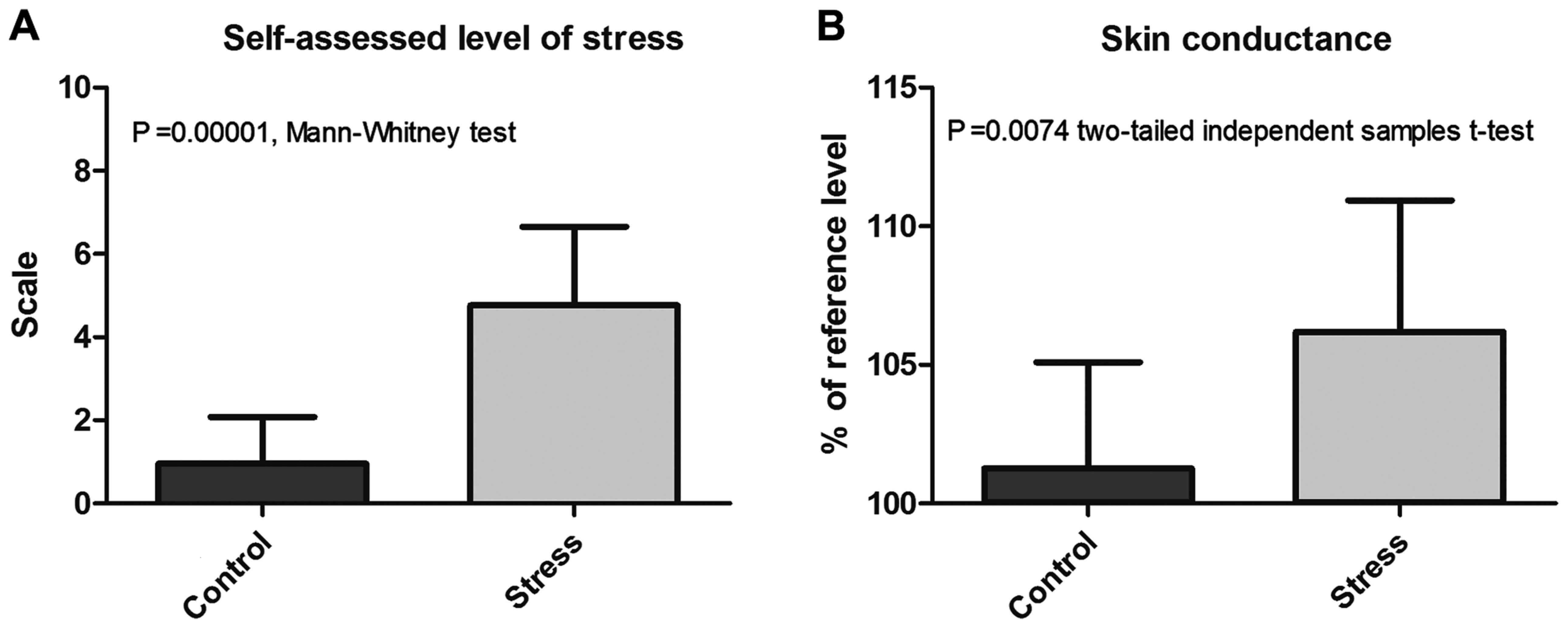
conditions on skin conductance were also compared (Table I and Fig.
1).
 | Table I.Comparative analysis of cutaneous
conductance and self-assessed stress at the end of the computerized
stage. |
Table I.
Comparative analysis of cutaneous
conductance and self-assessed stress at the end of the computerized
stage.
|
| Stress | Control |
|
|---|
|
|
|
|
|
|---|
| Parameters | Mean | SD | Range | Mean | SD | Range | Level of
significance |
|---|
| Skin conductance (%
reference level) | 106.1919 | 4.7425 | 19.5609 | 101.2734 | 3.8161 | 13.1926 | 0.0074a |
| Self-assessed
stress | 4.7647 | 1.888 | 5.5 | 0.9643 | 1.117 | 3 |
0.00001b |
The stress experienced during the computerized stage
had a score of 0.9643±1.117 in control group subjects and
4.7647±1.888 in subjects in the stress group, the difference
between the two groups being statistically significant
(P=0.00001).
Regarding the skin conductance reference level,
there were no statistically significant differences between the two
groups (P=0.5125). In contrast, exposure to the stress induction
protocol resulted in a statistically significant increase in the
level of skin conductance. Thus, the average level of skin
conductance during the computerized stage, expressed as a
percentage of the reference level, was 106.1919±4.7425% for the
stress group vs. 101.2734±3.8161% for control group (P=0.0074
two-tailed independent samples t-test).
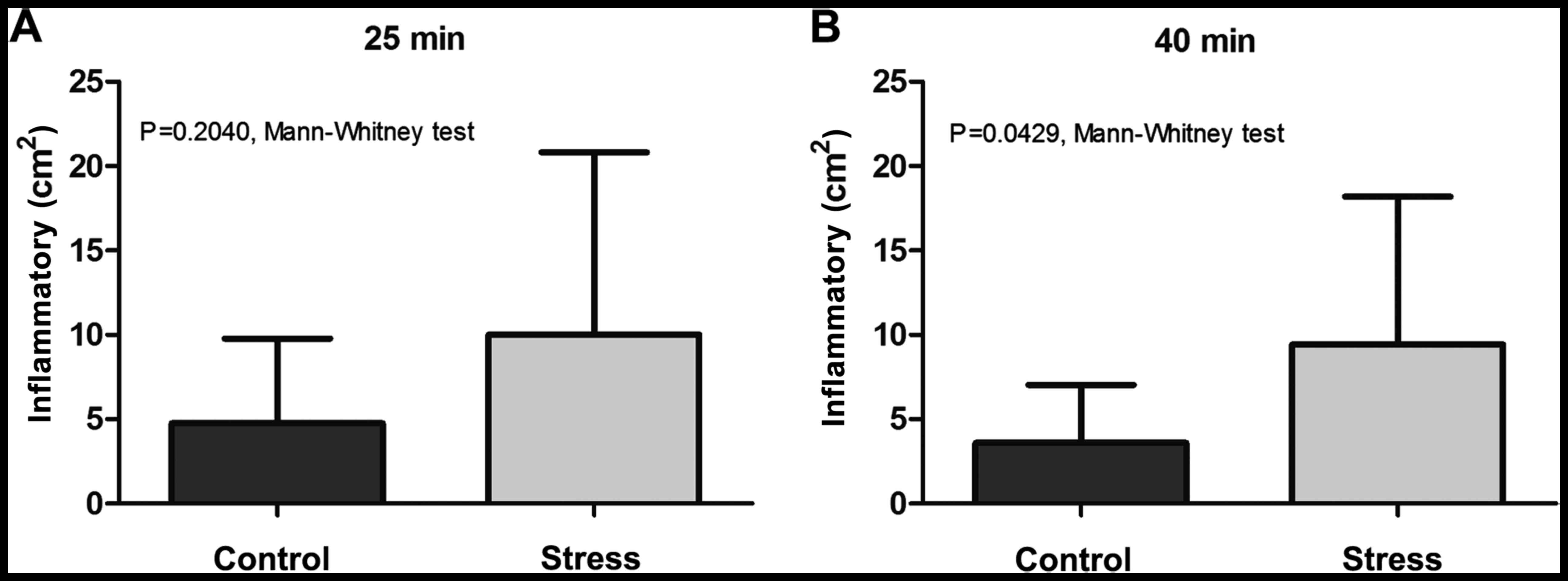
The evaluation of capsaicin-induced skin reaction
was performed by analyzing in each group the evolution of the
inflammatory area assessed by thermography at 25 and 40 min after
capsaicin application, followed by quantification of the
differences between the two groups for each of the above mentioned
time intervals. The ICC of the capsaicin-induced inflammatory area
measurements was 0.989.
Within the control group, the inflammatory area
evaluated 25 min after capsaicin administration was 4.7610±5.0133
cm2, while at 40 min post-application it appeared to
slightly decrease to 3.5842±3.4560 cm2, without the
difference between the two values reaching the level of statistical
significance (P=0.064039, two-tailed Wilcoxon signed ranks
test).
In stress group, 25 min after application of
capsaicin the inflammatory area had a value of 10.0063±10.8107
cm2 and at 40 min post-application was 9.4286±8.7749
cm2, the difference between the two values being not
statistically significant (P=0.90579).
Comparative analysis between groups (Table II and Figs. 2 and 3) showed that even if at 25 min after
capsaicin administration there were no statistically significant
differences (P=0.2040, Mann-Whitney test), at 40 min
post-treatment, the inflammatory area was significantly higher in
the stress group, compared to control group (P=0.0429).
| Table II.Comparative analysis of the
inflammatory area at 25 and 40 min after capsaicin application. |
Table II.
Comparative analysis of the
inflammatory area at 25 and 40 min after capsaicin application.
|
| Stress | Control |
|
|---|
|
|
|
|
|
|---|
| Parameters | Mean | SD | Range | Mean | SD | Range | Level of
significancea |
|---|
| Inflammatory area
at 25 min (cm2) | 10.0063 | 10.8107 | 34.3709 | 4.7610 | 5.0133 | 17.8211 | 0.2040 |
| Inflammatory area
at 40 min (cm2) | 9.4286 | 8.7749 | 28.9463 | 3.5842 | 3.4560 | 11.8757 | 0.0429 |
Discussion
Despite the great impact of neuroendocrine factors
in dermatological conditions and the extensive research in animal
or in vitro models, until now there are only a limited
number of studies in human subjects regarding the role of stress in
modulation of capsaicin-induced skin reactions. However, a recent
study on healthy women showed that increased numbers of stressful
life events are associated with a larger area of secondary
hyperalgesia after topical application of capsaicin (32). Another investigation by Lutgendorf
et al (33) on 50 healthy
subjects of both sexes showed a decreased capsaicin-induced skin
inflammation associated with stress reduction by relaxation
techniques but could not reveal a direct impact of stress on this
reaction. Another study of the same research group assessed the
impact of stress and relaxation on the pain sensation induced by
cutaneous administration of capsaicin, indicating that women
undergoing relaxation experienced a significantly lower level of
pain than those exposed to stress (34). Research has shown that stress
reduction techniques can lessen the neurogenic inflammatory
response induced by topical administration of capsaicin, the impact
being different depending on the technique used (35,36).
In our study regarding the stress influence on the
capsaicin-induced inflammatory neurogenic reaction we highlighted
that exposure to the stress induction protocol that we have
implemented determined a significantly higher level of stress
during the computerized stage. It also induced a significant
increase of skin conductance, indicating an important sympathetic
activation (37,38). Within the subjects in both groups,
topical application of capsaicin caused an inflammatory reaction
that was evidenced by thermograpy at 25 min and was maintained at
40 min post-application. The hyperthermal area induced by topical
application of capsaicin was higher at 40 min post-application in
subjects in the stress group, suggesting that acute stress exposure
is associated with an extension of the area and duration of action
of the mechanisms involved in capsaicin-induced skin neurogenic
inflammation.
Previous studies have highlighted the important role
played by dorsal root reflexes in the extension of the skin
inflammation induced by capsaicin (39,40).
Stress may increase the capsaicin-induced cutaneous inflammatory
response by facilitation of this type of reflexes by the modulator
actions of catecholamines on the activity of nerve fibers sensitive
to capsaicin and their neurons of origin (17,18). The
skin inflammatory response determined by capsaicin is dependent on
the integrity of the sympathetic nervous system, its modulation
being probably achieved through α1-adrenergic receptors (41,42).
Moreover, the increased inflammatory response induced by capsaicin
correlates with the high serum level of norepinephrine (33).
Another way of acting through which stress could
induce the amplification of the inflammatory area is to stimulate
mast cell degranulation. The important role of mast cells in the
capsaicin inflammatory response was highlighted in previous studies
(43), and stress exposure
determines a significant and rapid increase in the percentage of
degranulated mast cells (19,20).
In conclusion, our results show that acute stress
exposure induces an amplification of the capsaicin-induced skin
inflammatory response. It is likely that the influence of
sympathetic activation and possibly other mechanisms, such as
direct modulatory effects on mast cells, nociceptive nerve endings
and their neurons of origin, would have a strong impact on the
amplitude of capsaicin-induced inflammatory reaction in the
skin.
Acknowledgements
Authors wish to thank Ioana Safta and Valentin
Velican for their constant support during the course of this
work.
Funding
This study was partially supported by a grant of
Romanian Ministery of Research and Innovation, CCCDI-UEFISCDI
(project no. 61PCCDI⁄2018 PN-III-P1-1.2-PCCDI-2017-0341) within
PNCDI-III.
Availability of data and materials
The datasets used and/or analyzed during the current
study are available from the corresponding author on reasonable
request.
Authors' contributions
OG, AIM, IS, DB and CC were responsible for
acquisition, analysis and interpretation of the data and
contributed to drafting the manuscript and revising it critically
for important intellectual content. All authors read and approved
the final version of manuscript.
Ethics approval and consent to
participate
This study was approved by the Ethics Committee of
‘Carol Davila’ University of Medicine and Pharmacy (Bucharest,
Romania) and all participants provided written informed consent for
the publication of data in this study.
Patient consent for publication
Not applicable.
Competing interests
The authors declare that they have no competing
interests.
References
|
1
|
Lander F, Friche C, Tornemand H, Andersen
JH and Kirkeskov L: Can we enhance the ability to return to work
among workers with stress-related disorders? BMC Public Health.
9:3722009. View Article : Google Scholar : PubMed/NCBI
|
|
2
|
Dhabhar FS and McEwen BS: Enhancing versus
suppressive effects of stress hormones on skin immune function.
Proc Natl Acad Sci USA. 96:1059–1064. 1999. View Article : Google Scholar : PubMed/NCBI
|
|
3
|
Maddock C and Pariante CM: How does stress
affect you? An overview of stress, immunity, depression and
disease. Epidemiol Psichiatr Soc. 10:153–162. 2001. View Article : Google Scholar : PubMed/NCBI
|
|
4
|
Van Rhenen W, Blonk RW, van der Klink JJ,
van Dijk FJ and Schaufeli WB: The effect of a cognitive and a
physical stress-reducing programme on psychological complaints. Int
Arch Occup Environ Health. 78:139–148. 2005. View Article : Google Scholar : PubMed/NCBI
|
|
5
|
Koo J and Lebwohl A: Psycho dermatology:
The mind and skin connection. Am Fam Physician. 64:1873–1878.
2001.PubMed/NCBI
|
|
6
|
Arck P and Paus R: From the brain-skin
connection: The neuroendocrine-immune misalliance of stress and
itch. Neuroimmunomodulation. 13:347–356. 2006. View Article : Google Scholar : PubMed/NCBI
|
|
7
|
Tausk F, Elenkov I and Moynihan J:
Psychoneuroimmunology. Dermatol Ther. 21:22–31. 2008. View Article : Google Scholar : PubMed/NCBI
|
|
8
|
Gupta MA and Gupta AK: Psychiatric and
psychological co-morbidity in patients with dermatologic disorders:
Epidemiology and management. Am J Clin Dermatol. 4:833–842. 2003.
View Article : Google Scholar : PubMed/NCBI
|
|
9
|
Căruntu C, Grigore C, Căruntu A,
Diaconeasa A and Boda D: The role of stress in skin diseases.
Medicina Internă: Internal Medicine. 8:73–84. 2011.(In
Romanian).
|
|
10
|
Căruntu C, Ghiță MA, Căruntu A and Boda D:
The role of stress in the multifactorial etiopathogenesis of acne.
Ro Med J. 58:98–101. 2011.
|
|
11
|
Surcel M, Constantin C, Caruntu C, Zurac S
and Neagu M: Inflammatory cytokine pattern is sex-dependent in
mouse cutaneous melanoma experimental model. J Immunol Res.
2017:92121342017. View Article : Google Scholar : PubMed/NCBI
|
|
12
|
Caruntu C, Mirica A, Rosca AE, Mirica R,
Caruntu A, Tampa M, Matei C, Constantin C, Neagu M, Badarau AI, et
al: The role of estrogens and estrogen receptors in melanoma
development and progression. Acta Endo. 12:234–241. 2016.
View Article : Google Scholar
|
|
13
|
Caruntu C, Boda D, Constantin C, Caruntu A
and Neagu M: Catecholamines increase in vitro proliferation of
murine B16F10 melanoma cells. Acta Endocrinologica (Bucharest).
10:545–558. 2014.
|
|
14
|
Lupu M, Caruntu A, Caruntu C, Papagheorghe
LML, Ilie MA, Voiculescu V, Boda D, Constantin C, Tanase C, Sifaki
M, et al: Neuroendocrine factors: The missing link in non-melanoma
skin cancer (Review). Oncol Rep. 38:1327–1340. 2017. View Article : Google Scholar : PubMed/NCBI
|
|
15
|
Solomon I, Voiculescu VM, Caruntu C, Lupu
M, Popa A, Ilie MA, Albulescu R, Caruntu A, Tanase C, Constantin C,
et al: Neuroendocrine factors and head and neck squamous cell
carcinoma: An affair to remember. Dis Markers. 2018:97878312018.
View Article : Google Scholar : PubMed/NCBI
|
|
16
|
Zegarska B, Lelińska A and Tyrakowski T:
Clinical and experimental aspects of cutaneous neurogenic
inflammation. Pharmacol Rep. 58:13–21. 2006.PubMed/NCBI
|
|
17
|
Căruntu C, Boda D, Musat S, Căruntu A,
Poenaru E, Calenic B, Savulescu-Fiedler I, Draghia A, Rotaru M and
Badarau AI: Stress effects on cutaneous nociceptive nerve fibers
and their neurons of origin in rats. Rom Biotechnol Lett.
19:9525–9538. 2014.
|
|
18
|
Filippi A, Caruntu C, Gheorghe RO, Deftu
A, Amuzescu B and Ristoiu V: Catecholamines reduce transient
receptor potential vanilloid type 1 desensitization in cultured
dorsal root ganglia neurons. J Physiol Pharmacol. 67:843–850.
2016.PubMed/NCBI
|
|
19
|
Singh LK, Pang X, Alexacos N, Letourneau R
and Theoharides TC: Acute immobilization stress triggers skin mast
cell degranulation via corticotropin releasing hormone,
neurotensin, and substance P: A link to neurogenic skin disorders.
Brain Behav Immun. 13:225–239. 1999. View Article : Google Scholar : PubMed/NCBI
|
|
20
|
Căruntu C, Boda D, Musat S, Căruntu A and
Mandache E: Stress-induced mast cell activation in glabrous and
hairy skin. Mediators Inflamm. 2014:1059502014. View Article : Google Scholar : PubMed/NCBI
|
|
21
|
Pavlovic S, Daniltchenko M, Tobin DJ,
Hagen E, Hunt SP, Klapp BF, Arck PC and Peters EM: Further
exploring the brain-skin connection: Stress worsens dermatitis via
substance P-dependent neurogenic inflammation in mice. J Invest
Dermatol. 128:434–446. 2008. View Article : Google Scholar : PubMed/NCBI
|
|
22
|
Căruntu C, Negrei C, Ghiţă MA, Căruntu A,
Bădărău AI, Buraga I, Boda D, Albu A and Branisteanu D: Capsaicin,
a hot topic in skin pharmacology and physiology. Farmacia.
63:487–491. 2015.
|
|
23
|
Căruntu C and Boda D: Evaluation through
in vivo reflectance confocal microscopy of the cutaneous neurogenic
inflammatory reaction induced by capsaicin in human subjects. J
Biomed Opt. 17:0850032012. View Article : Google Scholar : PubMed/NCBI
|
|
24
|
Ghiţă MA, Căruntu C, Rosca AE, Căruntu A,
Moraru L, Constantin C, Neagu M and Boda D: Real-time investigation
of skin blood flow changes induced by topical capsaicin. Acta
Dermatovenerol Croat. 25:223–227. 2017.PubMed/NCBI
|
|
25
|
Cohen S, Kamarck T and Mermelstein R: A
global measure of perceived stress. J Health Soc Behav. 24:385–396.
1983. View
Article : Google Scholar : PubMed/NCBI
|
|
26
|
Lang PJ, Bradley MM and Cuthbert BN:
International affective picture system (IAPS): Affective ratings of
pictures and instruction manual. Technical Report A-8. University
of Florida; Gainesville, FL: 2008
|
|
27
|
Safta I, Grigore O and Căruntu C: Emotion
detection using psycho-physiological signal processing. 2011 7th
International Symposium on Advanced Topics in Electrical
Engineering (ATEE). May 12-14–2011.https://ieeexplore.ieee.org/document/5952203
|
|
28
|
Dedovic K, Renwick R, Mahani NK, Engert V,
Lupien SJ and Pruessner JC: The Montreal imaging stress task: Using
functional imaging to investigate the effects of perceiving and
processing psychosocial stress in the human brain. J Psychiatry
Neurosci. 30:319–325. 2005.PubMed/NCBI
|
|
29
|
Raven JC: Standardization of progressive
matrices, 1938. Br J Med Psychol. 19:137–150. 1941. View Article : Google Scholar
|
|
30
|
Raven J: The Raven's progressive matrices:
Change and stability over culture and time. Cognit Psychol.
41:1–48. 2000. View Article : Google Scholar : PubMed/NCBI
|
|
31
|
Schmidt S and Walach H: Electrodermal
activity (EDA): State of the art measurement and techniques for
parapsychological purposes. J Parapsychol. 64:139–163. 2000.
|
|
32
|
You DS, Creech SK and Meagher MW: Enhanced
area of secondary hyperalgesia in women with multiple stressful
life events: A pilot study. Pain Med. 17:1859–1864. 2016.
View Article : Google Scholar : PubMed/NCBI
|
|
33
|
Lutgendorf S, Logan H, Kirchner HL,
Rothrock N, Svengalis S, Iverson K and Lubaroff D: Effects of
relaxation and stress on the capsaicin-induced local inflammatory
response. Psychosom Med. 62:524–534. 2000. View Article : Google Scholar : PubMed/NCBI
|
|
34
|
Logan H, Lutgendorf S, Rainville P,
Sheffield D, Iverson K and Lubaroff D: Effects of stress and
relaxation on capsaicin-induced pain. J Pain. 2:160–170. 2001.
View Article : Google Scholar : PubMed/NCBI
|
|
35
|
Rosenkranz MA, Davidson RJ, Maccoon DG,
Sheridan JF, Kalin NH and Lutz A: A comparison of mindfulness-based
stress reduction and an active control in modulation of neurogenic
inflammation. Brain Behav Immun. 27:174–184. 2013. View Article : Google Scholar : PubMed/NCBI
|
|
36
|
Rosenkranz MA, Lutz A, Perlman DM,
Bachhuber DR, Schuyler BS, MacCoon DG and Davidson RJ: Reduced
stress and inflammatory responsiveness in experienced meditators
compared to a matched healthy control group.
Psychoneuroendocrinology. 68:117–125. 2016. View Article : Google Scholar : PubMed/NCBI
|
|
37
|
Storm H, Myre K, Rostrup M, Stokland O,
Lien MD and Raeder JC: Skin conductance correlates with
perioperative stress. Acta Anaesthesiol Scand. 46:887–895. 2002.
View Article : Google Scholar : PubMed/NCBI
|
|
38
|
Lidberg L and Wallin BG: Sympathetic skin
nerve discharges in relation to amplitude of skin resistance
responses. Psychophysiology. 18:268–270. 1981. View Article : Google Scholar : PubMed/NCBI
|
|
39
|
Lin Q, Li D, Xu X, Zou X and Fang L: Roles
of TRPV1 and neuropeptidergic receptors in dorsal root
reflex-mediated neurogenic inflammation induced by intradermal
injection of capsaicin. Mol Pain. 3:302007. View Article : Google Scholar : PubMed/NCBI
|
|
40
|
Lin Q, Wu J and Willis WD: Dorsal root
reflexes and cutaneous neurogenic inflammation after intradermal
injection of capsaicin in rats. J Neurophysiol. 82:2602–2611. 1999.
View Article : Google Scholar : PubMed/NCBI
|
|
41
|
Lin Q, Zou X, Fang L and Willis WD:
Sympathetic modulation of acute cutaneous flare induced by
intradermal injection of capsaicin in anesthetized rats. J
Neurophysiol. 89:853–861. 2003. View Article : Google Scholar : PubMed/NCBI
|
|
42
|
Ren Y, Zou X, Fang L and Lin Q:
Sympathetic modulation of activity in Adelta- and C-primary
nociceptive afferents after intradermal injection of capsaicin in
rats. J Neurophysiol. 93:365–377. 2005. View Article : Google Scholar : PubMed/NCBI
|
|
43
|
Bíró T, Maurer M, Modarres S, Lewin NE,
Brodie C, Acs G, Acs P, Paus R and Blumberg PM: Characterization of
functional vanilloid receptors expressed by mast cells. Blood.
91:1332–1340. 1998.PubMed/NCBI
|