Introduction
The occurrence of cutaneous squamous cell carcinoma
(CSCC), one of the most common non-melanoma skin cancers, is
increasing mainly due to the damage of the ozonosphere and the lack
of risk awareness (1,2). Certain factors, including infection,
chemical substances, inheritance and immune responses, have been
implicated in the development and progression of CSCC (3). High rates of recurrence and metastasis
contribute to the poor prognosis of patients with CSCC (4). The one-year survival rate of patients
with CSCC with recurrence and metastasis is approximately 50%
(4). Therefore, it is necessary to
investigate the molecular mechanisms underlying the growth and
metastasis of CSCC to support the development of novel therapeutic
targets for this disease.
MicroRNAs (miRs), a class of small non-coding RNAs
22–25 nucleotides in length, can regulate gene expression at the
post-transcriptional level by directly binding to the 3′UTR of
their target mRNAs (5,6). It has been well established that
numerous miRs participate in the regulation of various biological
processes, including cell survival, proliferation, differentiation,
motility and tumorigenesis (5–7).
Furthermore, certain miRs, including miR-31, miR-142-5p and
miR-365, have been reported to serve oncogenic or tumour
suppressive roles in CSCC (8–11). For
instance, miR-125b inhibits the expression of MMP13 and suppresses
CSCC cell proliferation, migration and invasion (11).
miR-186 is an important member of cancer-associated
miRs and has been reported to generally serve a tumour suppressive
role in various types of human cancer, including chronic myeloid
leukaemia (12), colorectal cancer
(13), gastrointestinal stromal
tumour (14), lung cancer (15), liver cancer (16), cervical cancer (17), and gastric cancer (18). miR-186 inhibits the proliferation,
metastasis and epithelial-to-mesenchymal transition of colorectal
cancer cells by targeting ZEB1 (13). However, the exact function of miR-186
in CSCC has not been previously studied.
The current study aimed to determine the expression
and regulatory role of miR-186 in CSCC.
Materials and methods
Clinical tissue samples
The current study was approved by the Ethics
Committee of the First Affiliated Hospital of Hunan College of
Traditional Chinese Medicine (Zhuzhou, China). A total of 32 CSCC
tissues and matched adjacent non-tumour tissues were collected from
patients with primary CSCC (18 males and 14 females; age range,
35–68 years; mean age, 53.6 years) at First Affiliated Hospital of
Hunan College of Traditional Chinese Medicine between March 2015
and May 2017, and informed consent was obtained from all patients.
Exclusion criteria included radiotherapy or chemotherapy before
surgical resection. After surgical resection, the tissues were
immediately immersed in liquid nitrogen and stored until further
use.
Cell lines
The human normal skin cell line HaCaT (cat. no.
CL0114) and the CSCC cell lines A431 and SCL-1 were purchased from
the Chinese Academy of Sciences Cell Bank (Shanghai, China). Cells
were cultured in Dulbecco's modified Eagle's Medium (Invitrogen;
Thermo Fisher Scientific, Inc., Waltham, MA, USA) supplemented with
10% foetal bovine serum (Gibco; Thermo Fisher Scientific, Inc.) and
maintained in a humidified incubator containing 5% CO2
and 95% O2 at 37°C.
Cell transfection
The miR-186 mimic, miR-186 inhibitor and negative
control (NC) were all purchased from Shanghai GenePharma Co., Ltd.
(Shanghai, China). miRs were produced from GMR-miR microRNA
single-stranded mimics (cat. no. B01001), which are proprietary.
pcDNA3.1 vector and pcDNA3.1-RETREG1 expression plasmid were
purchased from Hunan Yearth Biotechnology Co., Ltd. (Changsha,
China). For the in vitro functional study of miR-186 in
CSCC, SCL-1 and A431 cells were transfected with 100 nM NC, 100 nM
miR-186 mimics or 100 nM miR-186 inhibitor, or co-transfected with
100 nM miR-186 mimics and 1 µg pcDNA3.1 vector (the miR-186 + blank
group) or pcDNA3.1-RETREG1 expression plasmid (the miR-186 +
RETREG1 group) using Lipofectamine® 2000 (Invitrogen;
Thermo Fisher Scientific, Inc.), according to the manufacturer's
protocol. Following 48 h of transfection, the subsequent
experiments were conducted.
RNA extraction and reverse
transcription-quantitative polymerase chain reaction (RT-qPCR)
Total RNA was extracted from tissues or cell lines
using the TRIzol reagent (Invitrogen; Thermo Fisher Scientific,
Inc.), according to the manufacturer's protocol. The RNA was
subsequently reverse transcribed into cDNA using the PrimeScript RT
reagent kit with gDNA Eraser (Takara Bio, Inc., Otsu, Japan),
according to the manufacturer's protocol. The qPCR was performed
using the SYBR Green Realtime PCR Master Mix (Toyobo Life Science,
Osaka, Japan), according to the manufacturer's protocol. The
thermocycling conditions were as follows: Initial denaturation at
95°C for 5 min; 35 cycles of 95°C for 15 sec and 60°C for 30 sec.
U6 was used as the internal control of miR-186 and GAPDH was used
as the internal control of reticulophagy regulator 1 (RETREG1). The
primers for RETREG1 (cat. no. HQP013485), GAPDH (cat. no.
HQP006940), miR-186 (cat. no. HmiRQP0248) and U6 (cat. no.
HmiRQP9001) were all purchased from Guangzhou Fulengen
(GeneCopoeia, Inc., Rockville, MD, USA). The relative expression
levels were calculated using the 2−ΔΔCt method (19).
Cell proliferation assay
Cell proliferation was studied using Cell Counting
kit-8 (CCK-8; Dojindo Molecular Technologies, Inc., Kumamoto,
Japan). At 48 h after transfection, the cells were seeded into
96-well plates (3×10³ cells/well) and cultured for 0,
24, 48 and 72 h. The optical density value at 450 nm was measured
using a microplate reader (Bio-Rad Laboratories, Inc., Hercules,
CA, USA).
Cell apoptosis analysis
Cell apoptosis was analyzed by using an Annexin
V-fluorescein isothiocyanate (FITC) Apoptosis kit (BD Biosciences,
San Diego, CA, USA) in accordance with the manufacturer's protocol.
Transfected SCL-1 and A431 cells were seeded in 24-well plates
(1×10⁵ cells/well) and cultured in a humidified
incubator containing 5% CO2 and 95% O2 at
37°C for 24 h. Subsequently, the cells were re-suspended in 500 µl
binding buffer containing 1% FITC-labelled Annexin-V and propidium
iodide (which is in the Annexin V-FITC Apoptosis kit). After
incubation in the dark for 30 min, the apoptosis levels were
evaluated using BD Accuri CFlow software 1.0 on a BD C6 FACSVerse™
flow cytometer (both BD Biosciences).
Western blots
Total protein was extracted using RIPA lysis buffer
(Beyotime Institute of Biotechnology, Beijing, China). The protein
concentration was measured using a bicinchoninic acid protein assay
kit (Thermo Fisher Scientific, Inc.) according to the
manufacturer's protocol. Equal amounts of protein (50 µg/lane) were
isolated using 10% SDS-PAGE and transferred onto polyvinylidene
fluoride membranes (EMD Millipore, Billerica, MA, USA). The
membranes were blocked with 5% skimmed milk at 4°C overnight and
incubated with rabbit anti-human RETREG1 antibodies (cat. no.
ab151755) and GAPDH antibodies (cat. no. ab9485; both 1:500; Abcam,
Cambridge, MA, USA) at room temperature for 3 h. Subsequently, the
membranes were incubated with horseradish peroxidase-conjugated
goat anti-rabbit secondary antibodies (1:5,000; cat. no. ab6721;
Abcam) at room temperature for 40 min. The protein bands were
detected using an Enhanced Chemiluminescence Western Blotting kit
(Pierce; Thermo Fisher Scientific, Inc.) and quantified using Image
Lab analysis software version 3.1 (Bio-Rad Laboratories, Inc.).
Bioinformatics analysis and luciferase
reporter gene assay
TargetScan software 7.2 (www.targetscan.org/) was used to predict the potential
target genes of miR-186. The wild-type (WT) and mutant (MT)
sequences containing the putative binding sites of miR-186 in the
3′-UTR of the RETREG1 mRNA transcript were cloned into separate
pMIR-REPORT luciferase reporter plasmids (Promega Corporation,
Madison, WI, USA). Cells (5×10⁴ cells per well) in
24-well plates were transfected with 10 nM miR-186 mimics (or
miR-NC) and 500 ng WT RETREG1 (or MT RETREG1) plasmid using
Lipofectamine 2000. After 48 h of transfection, the relative
luciferase activity was detected using a
Dual-Luciferase® Reporter Assay System (Promega
Corporation). The activity of firefly luciferase was normalized to
the activity of Renilla luciferase.
Statistical analysis
Data are presented as the mean ± standard deviation.
SPSS 20.0 software (IBM Corp., Armonk, NY, USA) was used for the
statistical analyses. Comparisons were performed with one-way
analysis of variance followed with Tukey's post hoc test or
Student's t test. A Pearson correlation test was used to determine
the association between miR-186 and RETREG1 expression in CSCC
tissues. P<0.05 was considered to indicate a statistically
significant difference.
Results
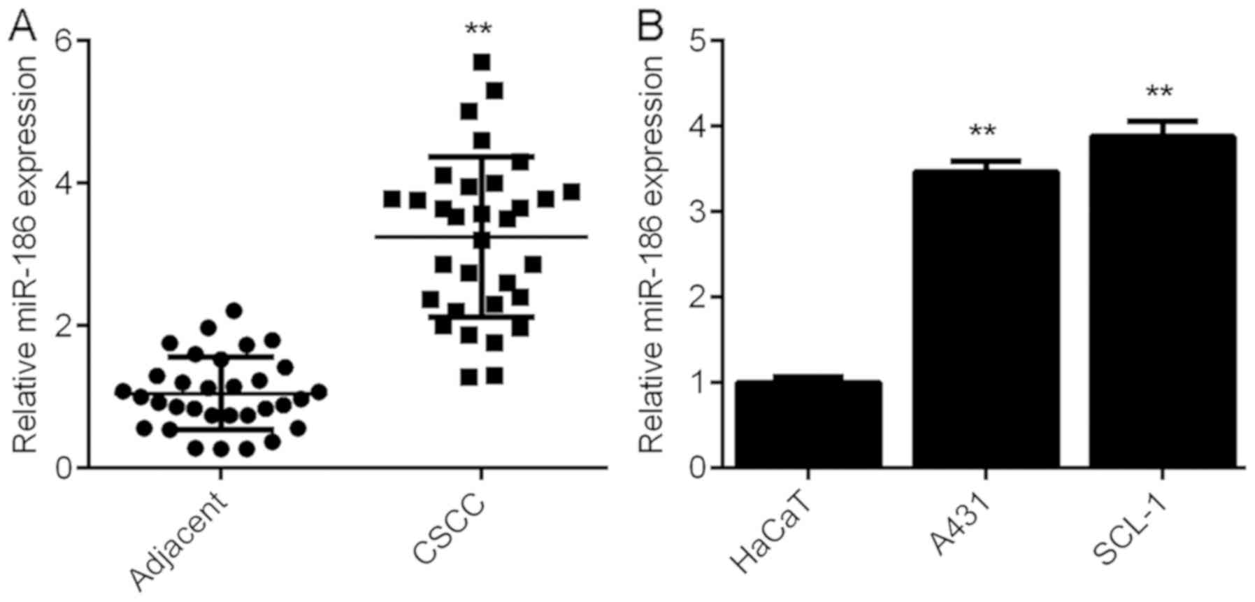
miR-186 is upregulated in CSCC
To investigate the role of miR-186 in CSCC, the
expression levels of miR-186 were determined in 32 CSCC tissues and
their matched adjacent non-tumour tissues. miR-186 was
significantly upregulated in the CSCC tissues compared with the
matched adjacent non-tumour tissues (Fig. 1A). Using the mean expression level of
miR-186 as a cut-off value (3.26), the authors divided the patients
into high and low miR-186 groups. The expression of miR-186 in
tumour tissues exhibited no difference between the male and female
patients with CSCC (P=0.735; Table
I). To further confirm these results, miR-186 expression was
determined in the normal human skin cell line HaCaT and the CSCC
cell lines A431 and SCL-1. miR-186 expression levels increased in
the A431 and SCL-1 cell lines compared with the HaCaT cells
(Fig. 1B). The above results
indicated that miR-186 was upregulated in CSCC.
 | Table I.Association between miR-186 expression
and sex of patients with cutaneous squamous cell carcinoma. |
Table I.
Association between miR-186 expression
and sex of patients with cutaneous squamous cell carcinoma.
| Sex | Low miR-186
(n=15) | High miR-186
(n=17) | P-value |
|---|
| Male | 9 | 9 | 0.735 |
| Female | 6 | 8 |
|
miR-186 affects CSCC cell
proliferation and apoptosis
The function of miR-186 in CSCC was investigated.
SCL-1 and A431 cells were transfected with miR-186 mimics and qPCR
data indicated that the miR-186 levels were significantly increased
in the miR-186 group compared with the miR-NC group in both cell
lines (Fig. 2A). A CCK-8 assay was
subsequently used to study the effects of miR-186 upregulation on
CSCC cell proliferation. The results suggested that overexpression
of miR-186 caused a significant increase in the proliferation of
SCL-1 and A431 cells compared with the miR-NC group (Fig. 2B and C). Flow cytometry was
subsequently conducted to study cell apoptosis and it was not
significantly affected by miR-186 overexpression (data not
shown).
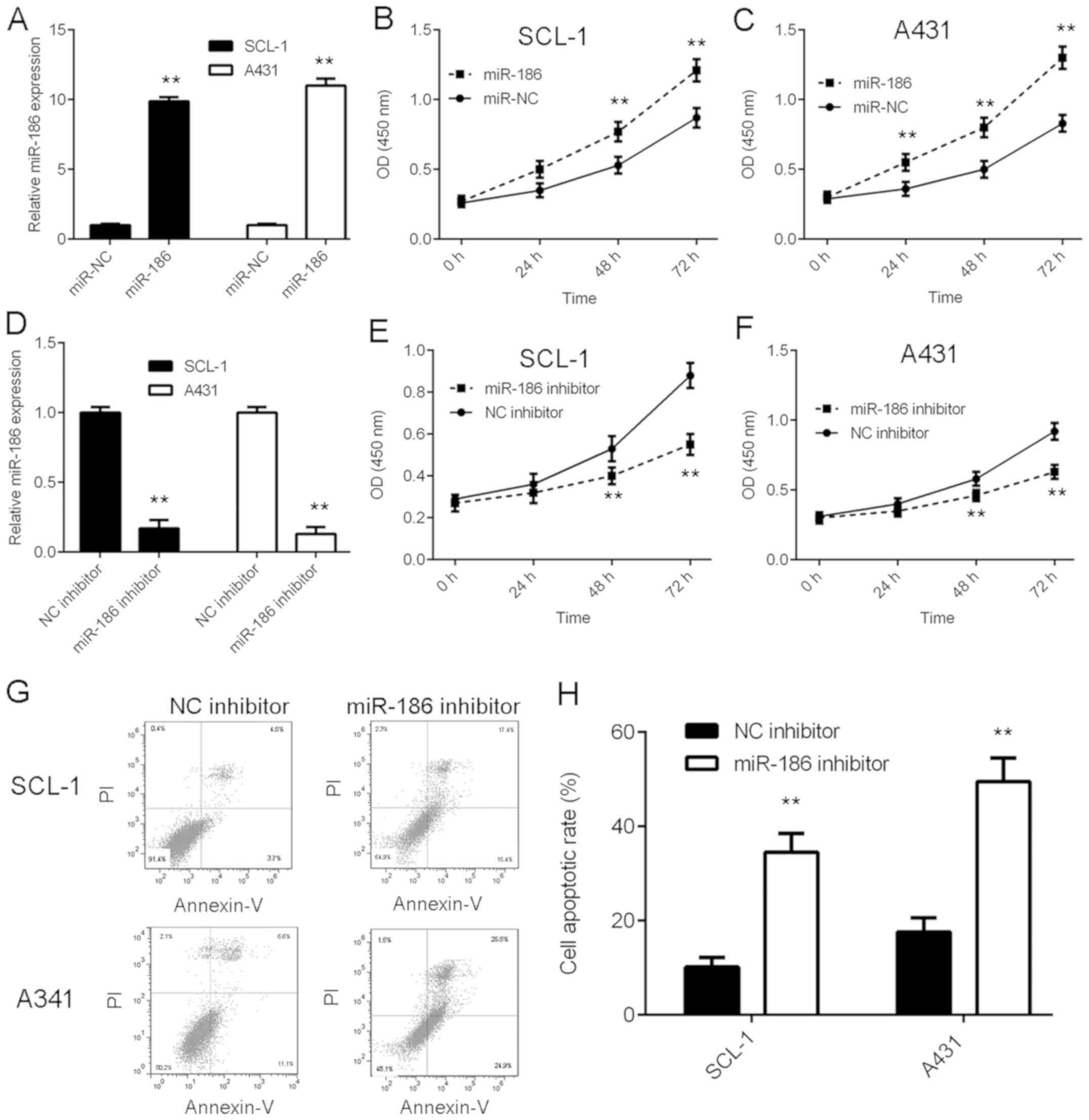 | Figure 2.miR-186 affects CSCC cell
proliferation and apoptosis. SCL-1 and A431 cells were transfected
with miR-186 mimics or miR-NC. Following transfection, (A) RT-qPCR
was used to examine the expression of miR-186, and CCK-8 assay was
used to examine cell proliferation in (B) SCL-1 and (C) A431 cell
lines. **P<0.01 vs. miR-NC. Subsequently, SCL-1 and A431 cells
were transfected with NC inhibitor or miR-186 inhibitor. Following
transfection, (D) RT-qPCR was used to examine the expression of
miR-186, and CCK-8 assay was used to examine cell proliferation in
(E) SCL-1 and (F) A431 cell lines. (G) Flow cytometry was used to
study cell apoptosis. (H) Quantitative analysis of cell apoptotic
rate. **P<0.01 vs. NC inhibitor. Nc, negative control; miR,
micro RNA; CCK-8, cell counting kit-8; CSCC, cutaneous squamous
cell carcinoma; RT-qPCR, reverse transcription-quantitative
polymerase chain reaction; PI, propidium iodide. |
SCL-1 and A431 cells were transfected with miR-186
inhibitor to knockdown miR-186 expression. qPCR data indicated that
the miR-186 levels were significantly reduced in the miR-186
inhibitor group compared with the NC inhibitor group (Fig. 2D). Data from the CCK-8 assay
indicated that inhibition of miR-186 expression caused a
significant reduction in CSCC cell proliferation (Fig. 2E and F). Furthermore, apoptosis of
CSCC cells markedly increased following miR-186 inhibition
(Fig. 2G and H). Therefore, the
results of the current study indicated that miR-186 overexpression
promoted CSCC cell proliferation, and knockdown of miR-186 induced
CSCC cell apoptosis.
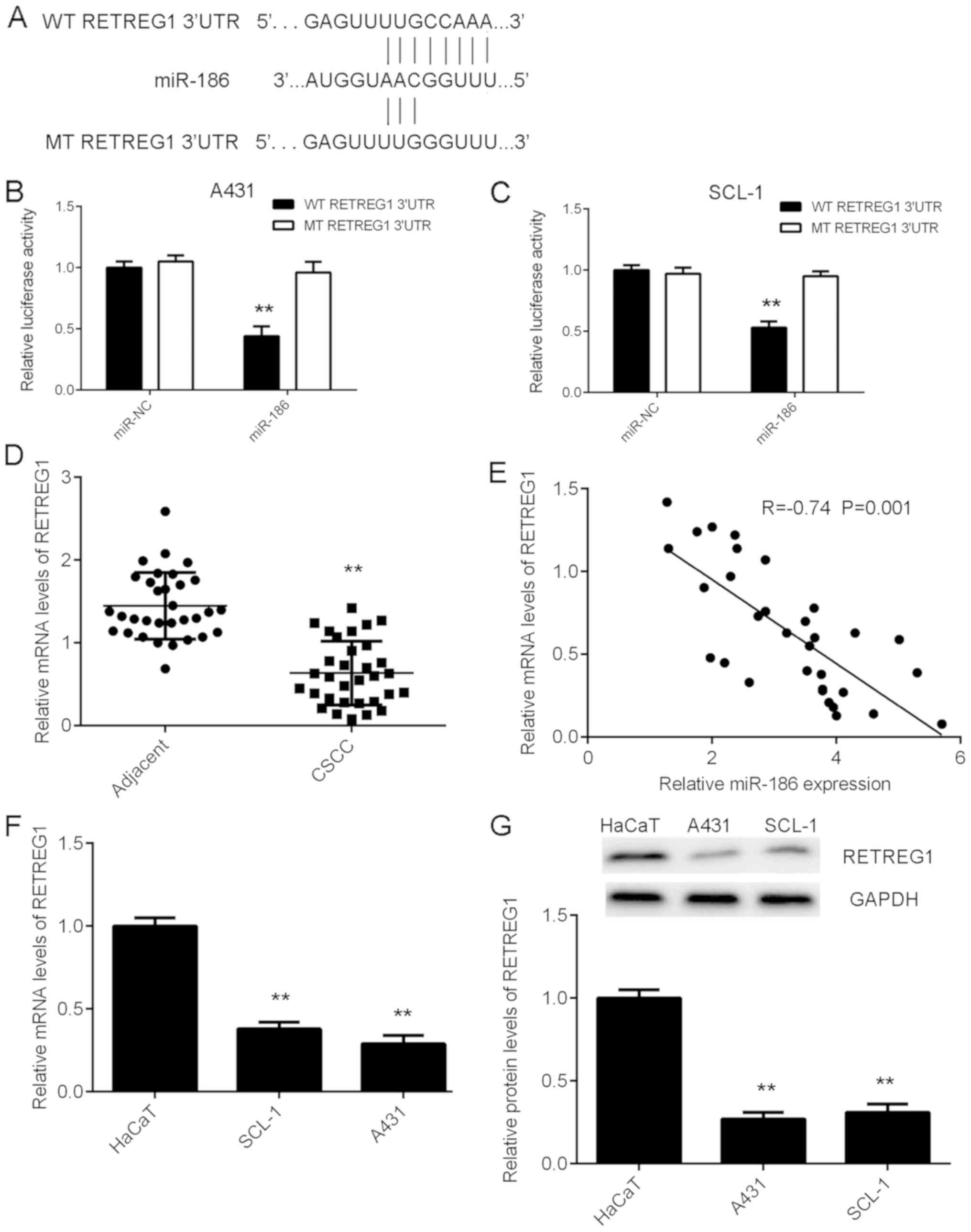
RETREG1 is a target gene of
miR-186
TargetScan software 7.2 was used to predict the
potential target genes of miR-186, and the data indicated that
there were predicted binding sites for miR-186 in the 3′UTR of the
RETREG1 mRNA (Fig. 3A). A
dual-luciferase assay was performed to verify whether RETREG1 was a
target gene of miR-186. The results indicated that overexpression
of miR-186 significantly downregulated the luciferase activity of
the WT 3′-UTR of RETREG1; however, the luciferase activity of the
MT 3′-UTR of RETREG1 was not affected in CSCC cells (Fig. 3B and C). Therefore, the results
indicate that RETREG1 is a direct target gene of miR-186.
Expression level of RETREG1 is reduced
in CSCC tissues and inversely correlated to the miR-186
expression
RT-qPCR data indicated that RETREG1 expression was
significantly lower in the CSCC tissues compared with the matched
adjacent non-tumour tissues (Fig.
3D). Using the mean expression value of RETREG1 as a cut-off
value (0.64), the authors divided the patients into high and low
RETREG1 groups. The expression of RETREG1 in the tumour tissues
exhibited no difference between male and female patients with CSCC
(P=0.473; Table II). Furthermore,
the RETREG1 expression levels were inversely correlated with the
expression levels of miR-186 in CSCC tissues (Fig. 3E). Taken together, these data suggest
that the reduced expression of RETREG1 in CSCC tissues may be due
to the increased expression of miR-186. Furthermore, the mRNA and
protein levels of RETREG1 were lower in CSCC cells compared with
HaCaT cells (Fig. 3F and G).
| Table II.Association between RETREG1
expression and sex of patients with cutaneous squamous cell
carcinoma. |
Table II.
Association between RETREG1
expression and sex of patients with cutaneous squamous cell
carcinoma.
| Sex | Low RETREG1
(n=19) | High RETREG1
(n=13) | P-value |
|---|
| Male | 12 | 6 | 0.473 |
| Female | 7 | 7 |
|
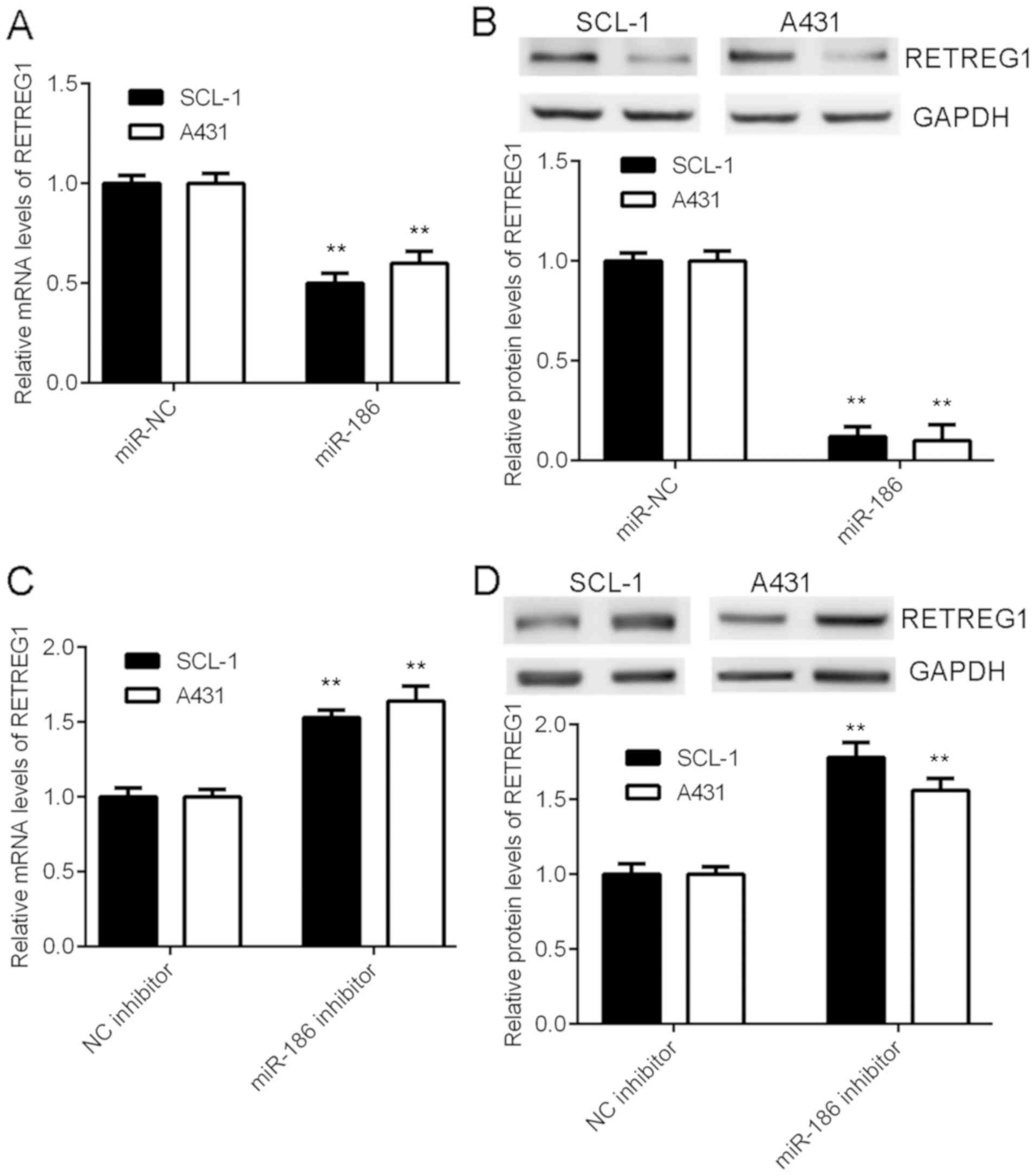
miR-186 negatively regulates the
expression of RETREG1 in CSCC cells
The effects of miR-186 on the expression levels of
RETREG1 in CSCC cells were subsequently studied. Data indicated
that overexpression of miR-186 significantly reduced the mRNA and
protein expression of RETREG1 in CSCC cells (Fig. 4A and B). By contrast, knockdown of
miR-186 significantly increased the expression of RETREG1 in CSCC
cells (Fig. 4C and D). The above
results indicated that RETREG1 expression was negatively regulated
by miR-186 in CSCC.
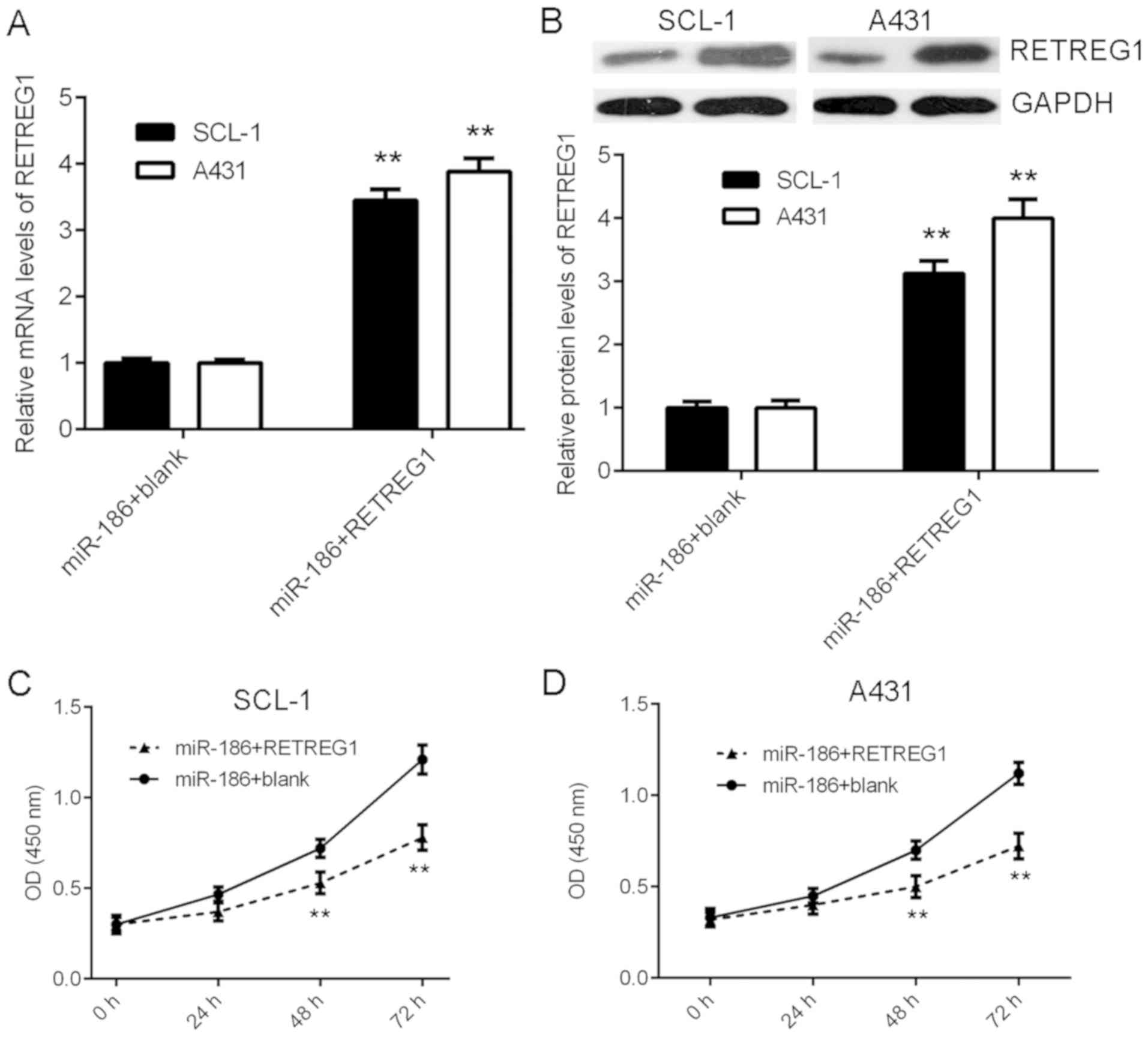
Overexpression of RETREG1 attenuates
the effects of miR-186 on CSCC cell proliferation
Finally, the current study examined whether RETREG1
served a role in the miR-186-mediated proliferation of CSCC cells.
The miR-186-overexpressing CSCC cells were transfected with a
RETREG1 expression plasmid to restore its expression. Following
transfection, the mRNA and protein levels of RETREG1 were
significantly increased in the miR-186 + RETREG1 group compared
with the miR-186 + blank group (Fig. 5A
and B). Further investigation indicated that the proliferation
of CSCC cells was significantly reduced in the miR-186 + RETREG1
group compared with the miR-186 + blank group (Fig. 5C and D). These results suggest that
RETREG1 overexpression attenuated the effects of miR-186 on CSCC
cell proliferation.
Discussion
miR-186 serves a suppressive role in certain common
types of human cancer; however, its exact function in CSCC has not
been previously reported. In the current study, the expression of
miR-186 was significantly increased in CSCC tissues compared with
adjacent non-tumour tissues. Overexpression of miR-186
significantly promoted CSCC cell proliferation and inhibited cell
apoptosis. RETREG1, which is significantly downregulated in CSCC
tissues and cell lines, was identified as a novel target gene of
miR-186. In addition, the expression of RETREG1 was inversely
correlated with the expression level of miR-186 in CSCC tissues.
Furthermore, the expression of RETREG1 was negatively regulated by
miR-186 in CSCC cells, and restoration of RETREG1 attenuated the
effects of miR-186 on CSCC cells.
In recent decades, a large number of miRs,
including, miR-125b (11), miR-365
(20), miR-205 (21), miR-199a-5p (22), and miR-203 (23) have been identified as key regulators
of the development and progression of CSCC and numerous other types
of human cancer. For instance, miR-199a-5p induces CSCC cell
invasion by suppressing E-cadherin expression (22). Among the cancer-associated miRs,
miR-186 has been reported to generally function as a tumour
suppressor (24,25). For instance, Cai et al
(24) demonstrated that miR-186
served as a tumour suppressor in oral squamous cell carcinoma by
negatively regulating the expression of tyrosine-protein
phosphatase non-receptor type 11. Ruan et al (25) revealed that miR-186 suppressed lung
cancer progression by targeting SIRT6. However, whether miR-186
serves a role in CSCC remains unclear. The current study indicated
that the expression of miR-186 was significantly increased in CSCC
tissues compared to adjacent non-tumour tissues. However, the
expression of miR-186 in tumour tissues exhibited no difference
between male and female patients. The present study further
revealed that overexpression of miR-186 significantly promoted CSCC
cell proliferation, and knockdown of miR-186 significantly
inhibited CSCC cell proliferation and induced cell apoptosis. These
results suggest that miR-186 serves an oncogenic role in CSCC.
RETREG1, also known as FAM134B, is a reticulophagy
receptor that regulates the turnover of the endoplasmic reticulum
(ER) by selective phagocytosis (26). Inhibition of RETREG1 contributes to
impaired proteostasis in the ER due to the accumulation of
misfolded or aggregated proteins, leading to compromised neuronal
survival and progressive neuronal degenerative diseases (26). In previous studies, RETREG1 was
downregulated in colorectal cancer, and its low expression was
associated with younger age, larger tumour size, advanced cancer
stages, higher recurrence rate, lymphovascular invasion and poor
prognosis of patients (27,28). Furthermore, overexpression of RETREG1
inhibited the proliferation of colorectal cancer cells in
vitro, and tumour growth in vivo (29). However, the function of RETREG1, and
the regulatory mechanism of RETREG1 expression in CSCC remains
unclear. The current study demonstrated that RETREG1 was a target
gene of miR-186 in CSCC cells and the expression of RETREG1 was
reduced in CSCC tissues and inversely correlated with miR-186
expression. These results suggest that the reduced expression of
RETREG1 in CSCC may be due to the upregulation of miR-186. Similar
observations were also reported in colorectal cancer. Islam et
al (30) demonstrated that
miR-186 overexpression significantly decreased the expression of
RETREG1 in colorectal cancer cells, and the expression levels of
RETREG1 and miR-186 were inversely correlated in colorectal cancer
tissues and cells. The current study expanded the understanding of
the association between miR-186 and RETREG1 in human cancer. In
addition, the expression of miR-186 in tumour tissues exhibited no
difference between male and female patients with CSCC. Furthermore,
overexpression of RETREG1 attenuated the effects of miR-186 on CSCC
cell proliferation, suggesting that RETREG1 may act as a downstream
signalling molecule of miR-186 in CSCC cells.
In conclusion, the present study demonstrated that
miR-186 may promote CSCC cell proliferation and inhibit cell
apoptosis at least partly through direct targeting of RETREG1,
suggesting that targeting the miR-186/RETREG1 axis may be used as a
potential strategy for the treatment of CSCC. Future studies should
further determine the function of the miR-186/RETREG1 axis in CSCC
growth and metastasis in vivo.
Acknowledgements
Not applicable.
Funding
Not applicable.
Availability of data and materials
All data generated or analysed during this study are
included in this published article.
Authors' contributions
SH designed the study. XH collected clinical tissues
and wrote the manuscript. YL, YT, TW, LL and CC performed
experiments. PA conducted statistical analysis.
Ethics approval and consent to
participate
The present study was approved by the Ethics
Committee of First Affiliated Hospital of Hunan College of
Traditional Chinese Medicine. Written informed consent to
participate in the current study were obtained from all
patients.
Patient consent for publication
Written informed consent to publish the current
study were obtained from all patients.
Competing interests
The authors declare that they have no competing
interests.
References
|
1
|
Gordon R: Skin cancer: An overview of
epidemiology and risk factors. Semin Oncol Nurs. 29:160–169. 2013.
View Article : Google Scholar : PubMed/NCBI
|
|
2
|
Linares MA, Zakaria A and Nizran P: Skin
cancer. Prim Care. 42:645–659. 2015. View Article : Google Scholar : PubMed/NCBI
|
|
3
|
Prieto-Granada C and Rodriguez-Waitkus P:
Cutaneous squamous cell carcinoma and related entities:
Epidemiology, clinical and histological features, and basic science
overview. Curr Probl Cancer. 39:206–215. 2015. View Article : Google Scholar : PubMed/NCBI
|
|
4
|
Burton KA, Ashack KA and Khachemoune A:
Cutaneous squamous cell carcinoma: A review of high-risk and
metastatic disease. Am J Clin Dermatol. 17:491–508. 2016.
View Article : Google Scholar : PubMed/NCBI
|
|
5
|
Bartel DP: MicroRNAs: Genomics,
biogenesis, mechanism, and function. Cell. 116:281–297. 2004.
View Article : Google Scholar : PubMed/NCBI
|
|
6
|
Ambros V: The functions of animal
microRNAs. Nature. 431:350–355. 2004. View Article : Google Scholar : PubMed/NCBI
|
|
7
|
Farazi TA, Hoell JI, Morozov P and Tuschl
T: MicroRNAs in human cancer. Adv Exp Med Biol. 774:1–20. 2013.
View Article : Google Scholar : PubMed/NCBI
|
|
8
|
Bai X, Zhou Y, Chen P, Yang M and Xu J:
MicroRNA-142-5p induces cancer stem cell-like properties of
cutaneous squamous cell carcinoma via inhibiting PTEN. J Cell
Biochem. 119:2179–2188. 2018. View Article : Google Scholar : PubMed/NCBI
|
|
9
|
Lin N, Zhou Y, Lian X and Tu Y:
MicroRNA-31 functions as an oncogenic microRNA in cutaneous
squamous cell carcinoma cells by targeting RhoTBT1. Oncol Lett.
13:1078–1082. 2017. View Article : Google Scholar : PubMed/NCBI
|
|
10
|
Zhou M, Zhou L, Zheng L, Guo L, Wang Y,
Liu H, Ou C and Ding Z: miR-365 promotes cutaneous squamous cell
carcinoma (CSCC) through targeting nuclear factor I/B (NFIB). PLoS
One. 9:e1006202014. View Article : Google Scholar : PubMed/NCBI
|
|
11
|
Xu N, Zhang L, Meisgen F, Harada M,
Heilborn J, Homey B, Grandér D, Ståhle M, Sonkoly E and Pivarcsi A:
MicroRNA-125b down-regulates matrix metallopeptidase 13 and
inhibits cutaneous squamous cell carcinoma cell proliferation,
migration, and invasion. J Biol Chem. 287:29899–29908. 2012.
View Article : Google Scholar : PubMed/NCBI
|
|
12
|
Lin J, Ma JC, Yang J, Yin JY, Chen XX, Guo
H, Wen XM, Zhang TJ, Qian W, Qian J and Deng ZQ: Arresting of
miR-186 and releasing of H19 by DDX43 facilitate tumorigenesis and
CML progression. Oncogene. 37:2432–2443. 2018. View Article : Google Scholar : PubMed/NCBI
|
|
13
|
Li J, Xia L, Zhou Z, Zuo Z, Xu C, Song H
and Cai J: MiR-186-5p upregulation inhibits proliferation,
metastasis and epithelial-to-mesenchymal transition of colorectal
cancer cell by targeting ZEB1. Arch Biochem Biophys. 640:53–60.
2018. View Article : Google Scholar : PubMed/NCBI
|
|
14
|
Niinuma T, Kai M, Kitajima H, Yamamoto E,
Harada T, Maruyama R, Nobuoka T, Nishida T, Kanda T, Hasegawa T, et
al: Downregulation of miR-186 is associated with metastatic
recurrence of gastrointestinal stromal tumors. Oncol Lett.
14:5703–5710. 2017.PubMed/NCBI
|
|
15
|
Huang T, Wang G, Yang L, Peng B, Wen Y,
Ding G and Wang Z: MiR-186 inhibits proliferation, migration, and
invasion of non-small cell lung cancer cells by downregulating Yin
Yang 1. Cancer Biomark. 21:221–228. 2017. View Article : Google Scholar : PubMed/NCBI
|
|
16
|
Gou Y, Zhai F, Zhang L and Cui L: RUNX3
regulates hepatocellular carcinoma cell metastasis via targeting
miR-186/E-cadherin/EMT pathway. Oncotarget. 8:61475–61486. 2017.
View Article : Google Scholar : PubMed/NCBI
|
|
17
|
Zhang JJ, Wang DD, Du CX and Wang Y: Long
noncoding RNA ANRIL promotes cervical cancer development by acting
as a sponge of miR-186. Oncol Res. 26:345–352. 2018. View Article : Google Scholar : PubMed/NCBI
|
|
18
|
Liu L, Wang Y, Bai R, Yang K and Tian Z:
MiR-186 inhibited aerobic glycolysis in gastric cancer via HIF-1α
regulation. Oncogenesis. 5:e2242016. View Article : Google Scholar : PubMed/NCBI
|
|
19
|
Livak KJ and Schmittgen TD: Analysis of
relative gene expression data using real-time quantitative PCR and
the 2(-Delta Delta C(T)) method. Methods. 25:402–408. 2001.
View Article : Google Scholar : PubMed/NCBI
|
|
20
|
Zhou L, Gao R, Wang Y, Zhou M and Ding Z:
Loss of BAX by miR-365 promotes cutaneous squamous cell carcinoma
progression by suppressing apoptosis. Int J Mol Sci. 18:E11572017.
View Article : Google Scholar : PubMed/NCBI
|
|
21
|
Stojadinovic O, Ramirez H, Pastar I,
Gordon KA, Stone R, Choudhary S, Badiavas E, Nouri K and
Tomic-Canic M: MiR-21 and miR-205 are induced in invasive cutaneous
squamous cell carcinomas. Arch Dermatol Res. 309:133–139. 2017.
View Article : Google Scholar : PubMed/NCBI
|
|
22
|
Wang S, Cao KE, He Q, Yin Z and Zhou J:
miR-199a-5p induces cell invasion by suppressing E-cadherin
expression in cutaneous squamous cell carcinoma. Oncol Lett.
12:97–101. 2016. View Article : Google Scholar : PubMed/NCBI
|
|
23
|
Canueto J, Cardenoso-Alvarez E,
Garcia-Hernandez JL, Galindo-Villardón P, Vicente-Galindo P,
Vicente-Villardón JL, Alonso-López D, De Las Rivas J, Valero J,
Moyano-Sanz E, et al: MicroRNA (miR)-203 and miR-205 expression
patterns identify subgroups of prognosis in cutaneous squamous cell
carcinoma. Br J Dermatol. 177:168–178. 2017. View Article : Google Scholar : PubMed/NCBI
|
|
24
|
Cai Z, Hao XY and Liu FX: MicroRNA-186
serves as a tumor suppressor in oral squamous cell carcinoma by
negatively regulating the protein tyrosine phosphatase SHP2
expression. Arch Oral Biol. 89:20–25. 2018. View Article : Google Scholar : PubMed/NCBI
|
|
25
|
Ruan L, Chen J, Ruan L, Yang T and Wang P:
MicroRNA-186 suppresses lung cancer progression by targeting SIRT6.
Cancer Biomark. 21:415–423. 2018. View Article : Google Scholar : PubMed/NCBI
|
|
26
|
Islam F, Gopalan V and Lam AK: RETREG1
(FAM134B): A new player in human diseases: 15 years after the
discovery in cancer. J Cell Physiol. 233:4479–4489. 2018.
View Article : Google Scholar : PubMed/NCBI
|
|
27
|
Kasem K, Gopalan V, Salajegheh A, Lu CT,
Smith RA and Lam AK: The roles of JK-1 (FAM134B) expressions in
colorectal cancer. Exp Cell Res. 326:166–173. 2014. View Article : Google Scholar : PubMed/NCBI
|
|
28
|
Islam F, Gopalan V, Pillai S, Lu CT, Kasem
K and Lam AK: Promoter hypermethylation inactivate tumor suppressor
FAM134B and is associated with poor prognosis in colorectal cancer.
Genes Chromosomes Cancer. 57:240–251. 2018. View Article : Google Scholar : PubMed/NCBI
|
|
29
|
Islam F, Gopalan V, Wahab R, Smith RA,
Qiao B and Lam AK: Stage dependent expression and tumor suppressive
function of FAM134B (JK1) in colon cancer. Mol Carcinog.
56:238–249. 2017. View
Article : Google Scholar : PubMed/NCBI
|
|
30
|
Islam F, Gopalan V, Vider J, Wahab R,
Ebrahimi F, Lu CT, Kasem K and Lam AKY: MicroRNA-186-5p
overexpression modulates colon cancer growth by repressing the
expression of the FAM134B tumour inhibitor. Exp Cell Res.
357:260–270. 2017. View Article : Google Scholar : PubMed/NCBI
|