Introduction
Human life expectancy has increased to the age of 90
years and according to statistics obtained from the United States,
it is estimated that by 2050, the number of people aged 60 or above
is expected to more than double from the number in 2017 (1). Owing to the rapid aging of a
considerable proportion of society, there is growing interest in
improving the quality of life, including understanding the
anti-aging effect. Aging, however, has different characteristics,
such as progressive aging of specific organs within individuals.
Although, it is difficult to objectively judge the extent of aging
of an individual, several attempts have been made nonetheless
(2). Piantanelli et al
(3) elaborated on the issue of the
correct choice of uncorrelated markers, Jackson et al
(4) interpreted aging based on its
correspondence with individual life expectancy, and Nakamura et
al (5) used monkeys to study
biomarkers of aging. Thus, objective criteria are required for
effectively evaluating aging.
The mechanism of aging is complex and associated
with various factors. Several theories have been proposed to
explain the process of aging, although none are completely
satisfactory (6). In particular, DNA
damage and cell destruction due to excessive production and
accumulation of superoxide free radicals are known to be important
factors of aging (7). Studies on
antioxidation, such as those on the redox pathway, have been
actively conducted to help prevent aging (8–10).
At present, interest in stem cell research and
experimentation is increasing. Adipose-derived stem cells (ADSCs)
are relatively easy to harvest and can be extracted in large
quantities. Thus, their clinical applications are being studied in
various fields, such as tissue regeneration and wound healing
(11).
ADSCs act by inducing differentiation and releasing
cytokines, such as vascular endothelial growth factor, basic
fibroblast growth factor (bFGF), transforming growth factor-β
(TGF-β), and interleukin-6, which have been reported to exert an
anti-aging effect by acting as antioxidants and inducing
angiogenesis (12–14). Therefore, a model for assessing
anti-aging effects using ADSCs is required.
Chronological age refers to the degree of aging
calculated from the time following birth (10), but it fails to provide an accurate
indication of the aging process (15). One of the typical methods for
evaluating aging is measurement of biological aging (BA), which
represents the overall physiological or functional age of each
individual, and individuals born at the same time may have
different degrees of BA (16).
Therefore, BA is a tool for comprehensive assessment of individual
health status, including genetic and environmental factors, and its
determination is an objective method for assessing disease status
and the degree of functional impairment (17).
Biochemical parameters should be evaluated to
examine BA (18). However, to the
best of our knowledge, biochemical parameters have not been
extensively evaluated in a rat model following ADSC administration
for assessing BA. Furthermore, the efficacy of intravenous systemic
ADSC administration is still debated (19). Therefore, the aim of the present
study was to develop a rat model for measuring BA by evaluating a
number of biochemical parameters post-intravenous ADSC
administration. In addition, the effect of bFGF on ADSC culture was
analyzed by comparing bFGF-supplemented and non-supplemented
groups.
Materials and methods
Animal experiments
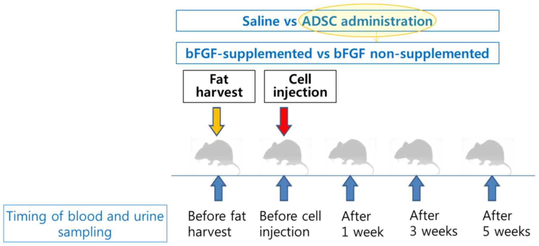
A total of 12 male 6-week-old Sprague Dawley rats
(C57BL/6N; Orient-Bio, Inc., Seongnam, Korea) with mean body weight
of ~350 g were used to evaluate the effects of ADSCs. Animals were
maintained at a controlled temperature (22±2°C) and humidity
(55±5%) with a 12 h light/dark cycle under specific-pathogen-free
conditions with free access to food and water. The rats were housed
in an animal facility and treated in accordance with the Guide for
the Care and Use of Laboratory Animals of Seoul National University
Boramae Hospital. Following a two-week quarantine period, 12 rats
were divided into the ADSC administration (experimental group; n=6)
and saline administration (control group; n=6) groups. The ADSC
administration group was also divided into bFGF supplemented (n=3)
or bFGF non-supplemented (n=3) groups.
Harvesting of autologous inguinal fat
pads and preparation of ADSCs
All surgical procedures were performed under aseptic
conditions. Rats were anesthetized by intraperitoneal
administration of a combination of Zoletil (Zoletil 100 at 20
mg/kg; Virbac Korea Co., Ltd., Seoul, Korea) and xylazine (Rompun
at 10 mg/kg; Bayer, Shanghai, China). Subsequently, the left
inguinal region was shaved and draped, and a 2.0-cm incision was
made along the inguinal fold. Then, the autologous inguinal fat pad
(~3 g) was harvested.
The isolated fat tissues were washed thrice in PBS.
Following rinsing, the fat tissue was digested using 0.2 U/ml
collagenase NB6 GMP grade (SERVA Electrophoresis GmbH, Heidelberg,
Germany) at 37°C for 1 h with shaking. Stromal vascular fraction
was isolated from the digested fat tissue by centrifugation at 252
× g for 5 min at room temperature and filtering through a 100-µm
nylon mesh. The isolated cells were washed twice with PBS and
plated in a T75 flask in α-minimal essential medium (α-MEM;
WelGENE, Inc., Daegu, Korea). Rat ADSCs (rADSCs) were cultured in
α-MEM supplemented with 10% fetal bovine serum (FBS; WelGENE,
Inc.), 2 mM L-glutamine, and 100 U/ml penicillin/streptomycin. The
flasks were maintained in a tissue culture incubator at 37°C and 5%
carbon dioxide. Cultured cells were harvested following
trypsinization when they exceeded 90% confluence and sub-cultured
at a density of 3×10⁵ cells/T75 flask up to passage 3.
The medium was changed every 2–3 days.
bFGF treatment
Recombinant rat bFGF (3339-FB; R&D Systems,
Inc., Minneapolis, MN, USA) was added to the growth medium in the
bFGF supplemented group. The bFGF concentration in the medium was 5
ng/ml. rADSCs at passage 0 and 1 were seeded in a T75 flask at an
initial density of 3×10⁵ cells and cultured for up to 3
passages. The medium was changed every 2–3 days.
Determination of ADSC doubling
time
The doubling time of ADSCs in bFGF supplemented and
α-MEM and 10% FBS was calculated via a cell counting method with
the following formula: PD=t × Log2/(LogC2-LogC1). PD=Population
doubling, t=24 h, Log=10 based Log, C1=1st cell count and C2=2nd
cell count.
Identification of ADSC by flow
cytometry and differentiation assay
A routine protocol was used for identification of
ADSCs. Flow cytometry analysis was performed to analyze the
phenotype of cultured rADSCs. Nonspecific sites were then blocked
in 2% BSA (Thermo Fisher Scientific, Inc., Waltham, MA, USA) with
PBS for 1 h at room temperature. ADSCs at P3/P4 were stained for 2
h at room temperature with antibodies for different cluster of
differentiation (CD) antigens, such as CD90-fluorescein
isothiocyanate (FITC; 1:50; cat. no. REA897; Miltenyi Biotec GmbH,
Bergisch Gladbach, Germany), CD44-FITC (1:100; cat. no. L178; BD
Biosciences, San Jose, CA, USA), and CD45-FITC (1:100; cat. no.
HI30; BD Biosciences) prior to administration. rADSCs were grown
until 80% confluence, trypsinized, and pelleted by centrifugation
at 252 × g for 5 min at 37°C. Subsequently, ~3×10⁵ cells
were resuspended in 100 µl FACS buffer (WelGENE, Inc.) containing
0.5% BSA in PBS. For FACS analysis of surface markers, each sample
was incubated for 30 min at 4°C in the dark. Following incubation,
the labeled cells were diluted with 1 ml FACS buffer, and pelleted
at 252 × g for 5 min at room temperature and resuspended in 500 µl
FACS buffer. Then, ~2×10⁴ cells were analyzed per sample
using the BD FACS Accuri flow cytometer (BD Biosciences).
Alizarin red S staining was performed for detecting
osteogenesis and oil red O staining for detecting adipogenesis
post-ADSC differentiation. Briefly, to explore the potential of
isolated ADSCs for osteogenic differentiation, ADSCs were seeded at
a density of 1×10⁴ cells/cm2 in 10-cm culture
dishes, and were grown with osteogenic induction medium, which was
comprised of Dulbecco's modified Eagle's medium (DMEM; WelGENE,
Inc.) supplemented with 0.1 mM dexamethasone, 50 mM ascorbate, and
10 mM β-glycerophosphate sodium. Mineralization of ADSCs was
determined by alizarin red S (Sigma-Aldrich; Merck KGaA, Darmstadt,
Germany) staining on day 21 post-drug treatment, and the staining
was visualized and photographed using bright-field microscopy
(magnification, ×40).
Additionally, adipogenic differentiation was induced
by incubating ADSCs in adipogenic induction medium, which contained
DMEM supplemented with 10% FBS, 60 µmol/l indomethacin, 0.5 mmol/l
3-isobutyl-1-methylxanthine, 1 µmol/l dexamethasone, 5 µg/ml
insulin, and 5 µg/ml gentamicin sulfate (Sigma-Aldrich; Merck
KGaA). Following 3 weeks of induction at 37°C, cells were washed
once with PBS and fixed in 4% formaldehyde for 15 min at room
temperature. Fixed cells were washed in 60% isopropyl alcohol, and
lipid droplets were stained for 1 h at room temperature using the
oil red O staining kit (Sigma-Aldrich; Merck KGaA). Following three
consecutive washes in deionized water, the stained cells were
photographed using bright-field microscopy (magnification, ×40;
Thermo Fisher Scientific, Inc.).
In addition, chondrogenic differentiation was
assessed. Confluent ADSCs were incubated at 37°C in chondrogenic
differentiation medium containing DMEM supplemented with 10% FBS,
0.1 µM dexamethasone, 10 ng/ml TGF-β1 (R&D Systems, Inc.,
Minneapolis, MN, USA), insulin-transferrin-selenium (Life
Technologies; Thermo Fisher Scientific, Inc.), and 50 µg/ml
ascorbate at 37°C. Medium was changed every 2 days for 3 weeks.
Systemic administration of autologous
ADSCs
The following surgical procedures were performed ~2
weeks following fat harvesting and completion of autologous ADSC
preparation. Following adequate anesthesia, ADSCs (P2,
1×10⁶ cells/300 µl PBS/rat body weight of 300–400 g)
were administered to the experimental group and the control group
was administered with normal saline via the femoral vein. The blood
and urine samples of both groups were analyzed five times (prior to
fat harvest and cell administration, and following 1, 3, and 5
weeks (Fig. 1.)
Assessment of BA
Blood and urine samples were used for biochemical
investigation. Serum was collected by centrifugation at 13,000 rpm
(18,928 × g) for 20 min at 18°C. Whole blood was collected in an
EDTA tube and serum and whole blood was transferred to the Neodin
Veterinary Laboratory (Seoul, Korea) for complete blood count (CBC)
analysis, which included estimation of white blood cells,
neutrophils, lymphocytes, monocytes, eosinophils, basophils, red
blood cells, hemoglobin, hematocrit, mean cell volume, mean cell
hemoglobin, mean cell hemoglobin concentration, red cell
distribution width, platelets and mean platelet volume (MPV).
Chemical investigations included assessment of albumin, total
bilirubin, blood urea nitrogen (BUN), creatinine, uric acid,
aspartate transaminase, alanine transaminase (ALT), cholesterol,
amylase, calcium, phosphorus, lipase, high density lipoprotein
(HDL), and low-density lipoprotein (LDL) levels. Enzyme assays,
such as those for superoxide dismutase (SOD; using the Superoxide
Dismutase Activity Assay kit; cat. no. ab65354; Abcam, Cambridge,
UK) activity, total antioxidant activity (TAC; using the Total
Antioxidant Capacity Assay kit; cat. no. ab65329; Abcam), and
catalase activity (CT; using the Catalase Assay kit; cat. no.
707002; Cayman Chemical Company, Ann Arbor, MI, USA) were performed
according to the manufacturer's instructions.
Urine was collected using metabolic cages. The urine
was stored in plastic test tubes at 4°C prior to analysis following
the scheme presented in the Fig. 1.
The urine samples were weighed at the time of collection from the
cages and transferred to Neodin Veterinary Laboratory for analysis
of urine protein, BUN, creatinine and creatinine clearance.
Statistical analysis
To build the prediction models, all parameters were
clinically classified into biochemical profiles. Repeated measures
of analysis of covariance (RM ANCOVA) were performed to identify
the differences between groups of measured values at each
time-point. In cases when interactions were significant, additional
ANCOVA was performed at each time-point. The non-parametric RM
ANCOVA was performed following correcting the same value at time 1
as the point prior to cell/saline administration. When the
interactions were significant at the significance level of 5%,
non-parametric ANCOVA was performed by correcting the values from
time 1 to time 5. At this time, the adjusted P-value was obtained
using the Sidak method to prevent the increase in Type I errors due
to multiple tests (20). Analyses
were conducted using SAS version 9.2 (SAS Institute, Inc., Cary,
NC, USA). P<0.05 was considered to indicate a statistically
significant result. Results were also expressed using t-test for
parametric data.
Results
Characterization of ADSCs and their
multilineage differentiation ability
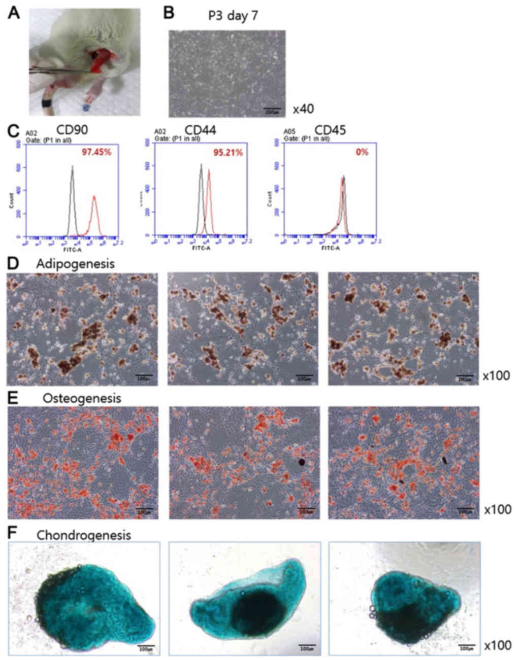
Autologous inguinal fat pad was harvested (Fig. 2A) and ADSCs were cultured, as
presented in Fig. 2B. ADSCs
exhibited stem cell-like features such as strengthened fibers that
could migrate around the cell cluster. Prior to ADSC
administration, flow cytometry was used to evaluate the phenotype
of ADSCs (Fig. 2C). CD90, the marker
for progenitor cells, and CD44, the marker for ADSCs, were present
on 97.45 and 95.21% of the cells, respectively, whereas the
hematopoietic cell marker CD45 was absent. Furthermore, osteogenic,
adipogenic, and chondrogenic differentiation potentials of ADSCs
were monitored. Oil red O staining following three weeks of
induction under adipogenic conditions indicated that ADSCs
underwent adipogenic differentiation (Fig. 2D). Alizarin red S (0.1%) was used to
study osteogenic differentiation potential, and results suggested
that the majority of ADSCs exhibited osteogenic differentiation
potential (Fig. 2E). ADSCs were
incubated in chondrogenic differentiation medium containing TGF-β
for 3 weeks, and alcian blue staining was performed to examine
chondrogenic differentiation of ADSCs (Fig. 2F). Results indicated that ADSCs
successfully underwent chondrogenic differentiation.
ADSCs affected aging-related metabolic
parameters
Aging is a progressive degenerative process tightly
integrated with inflammation. To monitor the inflammatory signals
following fat tissue extraction, CBC was monitored using rat blood
samples. The number of blood monocytes was lower in the ADSC
administration group than in the saline administration group at
each time-point. Additionally, the percentage of monocytes in the
blood increased in the saline administration group until five weeks
following administration (the time of experiment termination), but
it was sustained in the ADSC administration group without any
significant change [Fig. 3A;
P=0.0081 (data not shown)]. MPV level was higher in the ADSC
administration group at the fat pad uptake site, but this was
reversed following the uptake. MPV level decreased following ADSC
administration, but it increased in the saline group (Fig. 3B; P=0.0126), suggesting that
administered ADSCs may be differentiated into adipose cells, which
can reduce inflammation in rats.
 | Figure 3.Alterations in leukocyte variables
and serum or urine kidney signal. Blood and urine were obtained
from the rat model each time-point, and complete blood count and
chemical analyses were performed. Measurements in lean control and
with ADSC administration were evaluated for (A) blood monocytes,
(B) MPV, (C) serum BUN, (D) serum creatinine, and (E) UCCR are
presented. The value of monocytes was sustained in ADSC injected
group compared with that in saline group, which was increased.
Conversely, all others (MPV, BUN, creatinine and UCCR) were
downregulated following ADSC administration. Data are presented as
the mean ± standard error of the mean. ADSC; adipose-derived stem
cells; MPV, mean platelet volume; BUN, blood urea nitrogen; UCCR,
urine cortisol:creatinine ratio. |
A major function of the kidneys is to remove waste
products and excess fluid from the body. Total serum BUN and
creatinine levels were markedly different between saline and ADSC
administered groups in a time-dependent manner. BUN (Fig. 3C; P=0.0029) and creatinine (Fig. 3D; P=0.0095) levels were reduced
following ADSC administration. The urine cortisol:creatinine ratio
(UCCR), which detects renal stress due to illness or pre-diabetes
mellitus, was also markedly decreased following ADSC administration
(Fig. 3E, P=0.0028) (21). Taken together, these findings suggest
that ADSCs can maintain normal renal status.
Cholesterol is an essential component of fats that
can be metabolized in the liver. LDL deposits on blood vessel walls
and adversely affects health. Serum LDL levels were lower in the
ADSC administration group than in the saline administration group
(Fig. 4A; P=0.0078), and serum HDL
level was lower in the ADSC administration group (Fig. 4B; P=0.0069). In the present study,
lower HDL levels in the ADSC administration group could be due to
lower LDL levels than that observed in the saline administration
group.
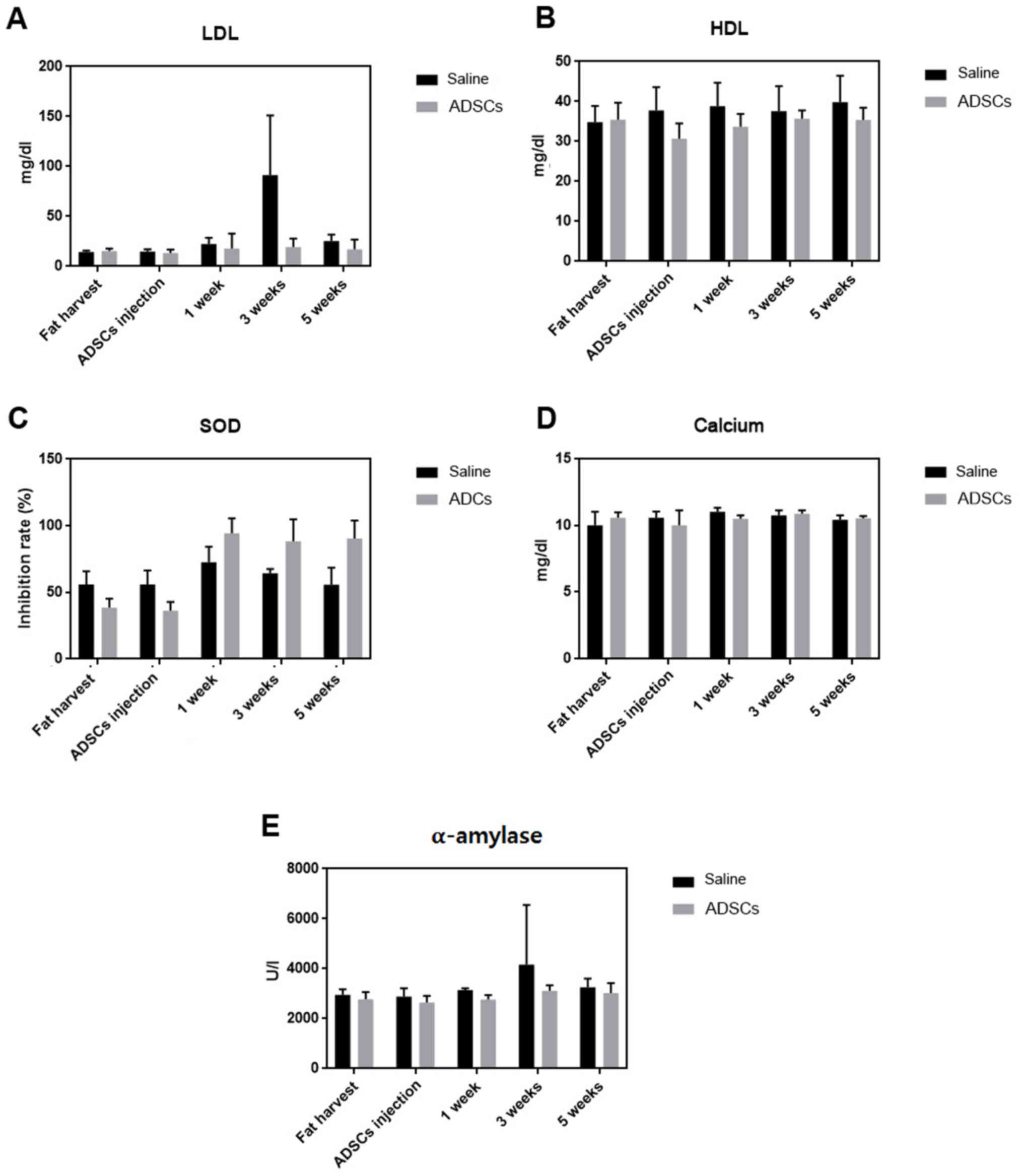 | Figure 4.Serum cholesterol, SOD, calcium, and
α-amylase levels were determined. Bar graphs presenting (A) LDL,
(B) HDL, (C) SOD, (D) calcium and (E) α-amylase levels. Serum LDL
and serum HDL levels were downregulated in the ADSC administration
group, which tends to reduce the risk of heart diseases. SOD level
was increased following ADSC administration. Serum calcium level
was lower in the ADSC administration group in a time-dependent
manner. The level of α-amylase, a calcium metalloenzyme, was also
lower following ADSC administration, thus ADSC can maintain
homeostasis of the blood glucose level. Data are presented as the
mean ± standard error of the mean. SOD, superoxide dismutase; LDL,
low-density lipoprotein; HDL, high-density lipoprotein; ADSC;
adipose-derived stem cells. |
ADSC administration regulates
antioxidant enzyme and calcium and amylase levels
SOD constitutes an important antioxidative line of
defense in nearly all living cells exposed to oxygen (22). SOD activity was lower at the sites of
fat tissue extraction in the ADSC administration group, but it
increased following ADSC administration and was sustained until
five weeks following ADSC administration, which was the time of
experiment termination (Fig. 4C;
P<0.0001).
Total calcium is often measured as a part of routine
health screening. Serum total calcium level was different between
the control and ADSC administration groups [lower in the ADSC
administration group in a time-dependent manner; Fig. 4D; P=0.0002; (data not shown)].
The level of α-amylase, a calcium metalloenzyme that
hydrolyzes starch into sugars and contributes to blood glucose
regulation, was also determined. The level of α-amylase was lower
in the ADSC administration group than in the saline administration
group (Fig. 4E; P=0.0213),
suggesting that ADSCs can maintain homeostasis of blood glucose
level as starch uptake is decreased by α-amylase.
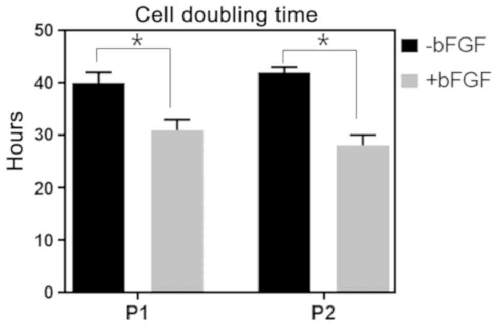
bFGF treatment facilitates stem cell
repopulation
bFGF is the growth factor necessary for maintenance
of undifferentiated state (23). To
compare the effect of bFGF on ADSC culture condition and on the
body following ADSC administration, the effect of ADSCs with or
without bFGF was studied. bFGF markedly increased the cell doubling
rate at passages 1 and 2. The population doubling time was also
markedly shorter in the group with bFGF supplementation than that
in the bFGF non-supplemented group at passages 1 and 2 (Fig. 5; P<0.001).
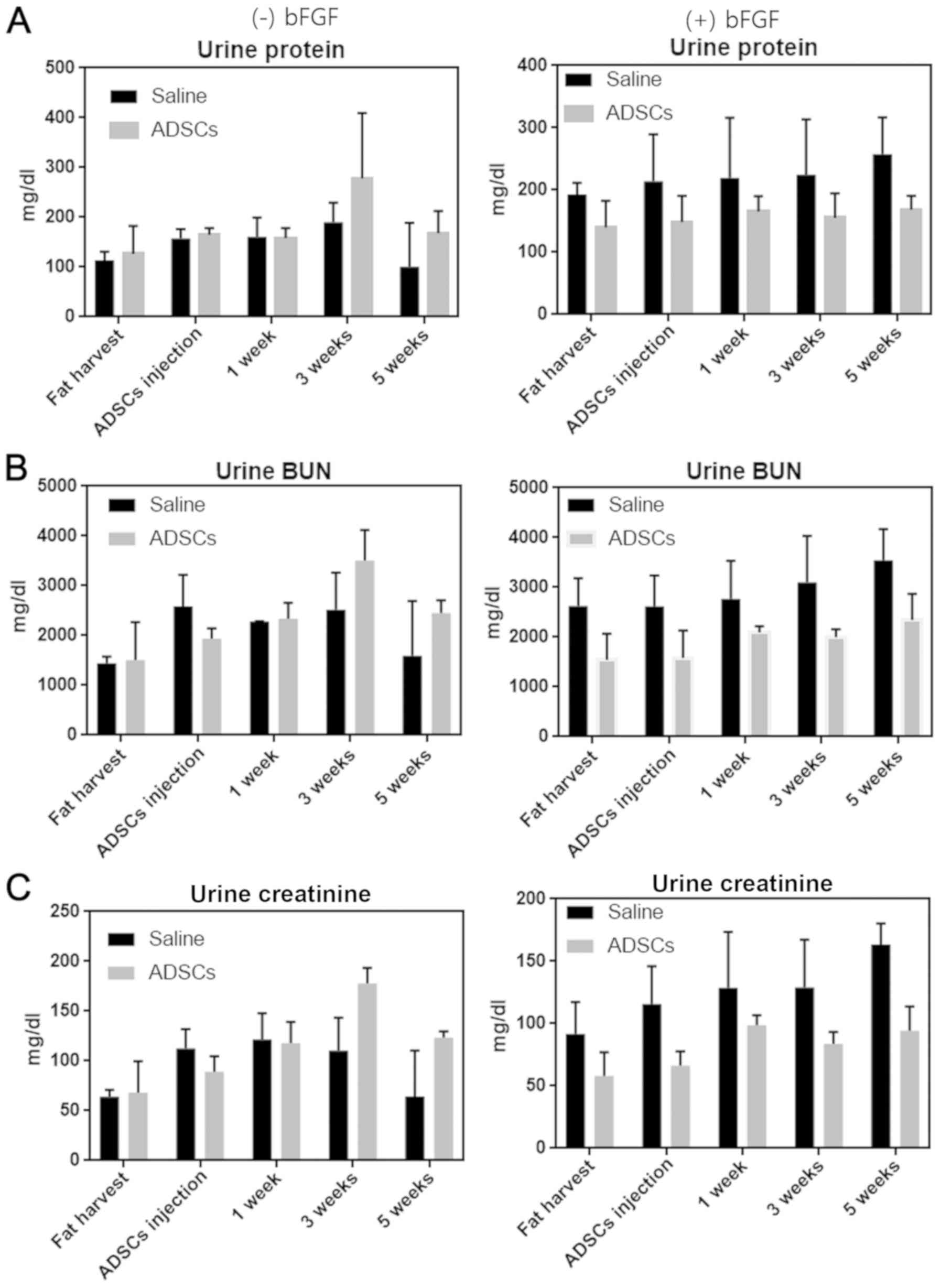
bFGF supplementation affects
biological markers compared with the non-bFGF supplemented
group
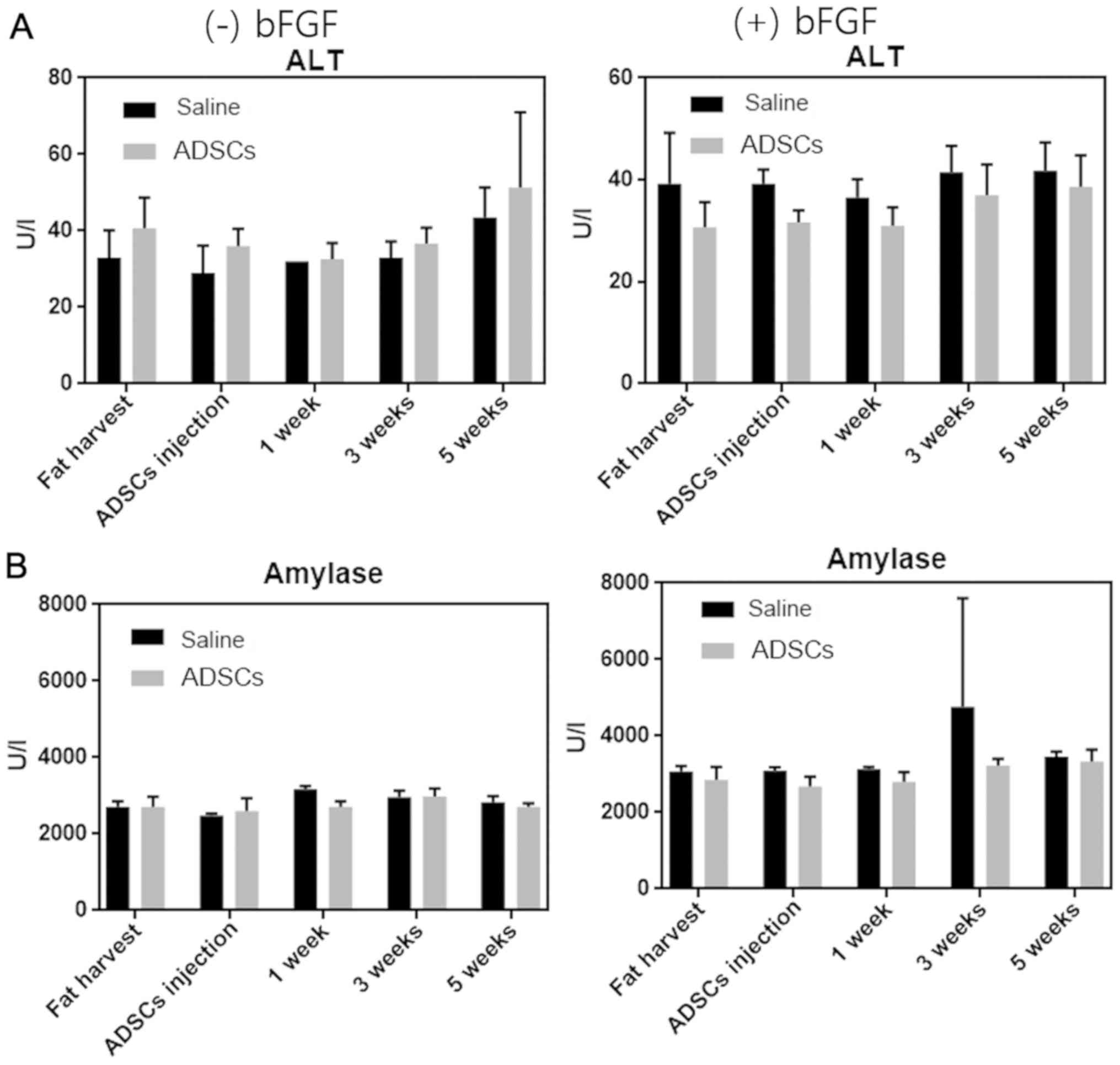
Exposure to D-galactose markedly elevated the levels
of ALT, which may be an aging marker (24). ALT level increased in the bFGF
non-supplemented group, but was lowered in the bFGF-supplemented
ADSC administration group (Fig. 6A;
P=0.0043). Additionally, α-amylase level was unchanged in the bFGF
non-supplemented ADSC administration group, but was lower in the
bFGF-supplemented group (Fig. 6B;
P=0.0032). These results demonstrated that ADSCs serve a role in
the maintenance of liver metabolism in normal and anti-aging
systems by decreasing serum ALT and amylase levels. Kidneys are
associated with the critical regulation of salt, potassium, and
acid content of the body, which are determined by urine protein,
BUN, and creatinine levels (Fig. 7).
Urine protein level increased in the bFGF non-supplemented group,
but decreased in the bFGF-supplemented ADSC administration group
(Fig. 7A; P=0.0005). Urine BUN
(Fig. 7B; P=0.0003) and creatinine
levels (Fig. 7C; P=0.0027)
demonstrated a pattern similar to that of urine protein. These
results demonstrated that bFGF affects the biological markers
associated with hepatic and renal functions.
Discussion
Although immortality continues to be beyond our
understanding, ways of deterring aging and extending life span are
being actively investigated. Owing to these efforts, several
aspects of the anti-aging mechanism, particularly the concept of
BA, have been identified. Nonetheless, the identification of
biomarkers of aging is an ongoing challenge (5). Owing to the various physical, chemical,
and environmental factors associated with aging, research on aging
has encountered several obstacles. Nevertheless, the ability to
quantify aging of an individual numerically and measure the extent
of aging precisely would enable physicians to realistically assess
the efficacy of a therapy. As a prelude to discussing these
approaches and the rationale for their selection, it is appropriate
to first evaluate the biological basis of aging.
In 1998, Nakamura et al (5) used a monkey model for identifying
biomarkers associated with aging, which were divided into three
stages, namely those related to longitudinal, cross-sectional and
stability analyses. In 2008, Dong et al (24) developed equations for evaluating
biological age in Korean men and examined whether pathological
conditions, such as diabetes, affected BA using this equation.
An experimental model is required to assess BA. To
the best of our knowledge, the present study included the first
attempt to develop a rat ADSC model using autologous ADSCs to
realize this objective. In particular, bFGF was further added to
maximize the effect of BA by increasing the differentiation
potential of ADSCs. Furthermore, numerous variables and biomarkers
of aging were categorized and analyzed. As described below, these
physiological variables are considered to represent functions of
vital organs associated with successful aging and maintenance of
life (25). For example, low levels
or reduction in albumin levels have a significant association with
all-cause mortality (26).
Hematology-associated cells undergo coagulation to
resolve the vasculopathy associated with oncology (27). The monocytes and MPV levels in the
CBC of the ADSC administration group were markedly lower than those
of the saline administration group; the low monocyte activity in
the cell group could be due to the immunomodulatory effect of
ADSCs, and the low MPV rate could represent blood viscosity.
Serum BUN, urine creatinine level and UCCR are
indicators of renal function. Routine assessment or screening
programs associated with these indicators were used for incidental
detection of kidney diseases. Comparison of urine parameters
between bFGF-supplemented and non-supplemented groups demonstrated
remarkable results. Uric acid level was markedly elevated, whereas
urine BUN, protein and creatinine levels were markedly reduced in
the bFGF-supplemented group. According to Crasto et al
(28) renal functional was decreased
as concentration of FGF increased. Therefore, development of cell
therapy using bFGF and ADSC targeting the kidneys can be beneficial
for maintaining nephrological homeostasis.
Cholesterol serves an important role in the liver.
LDL assists in transport of metabolites from the liver to regional
cells; however, large quantities of LDL are not metabolized
properly, which may convert into oxidized cholesterol in blood
vessels and cause vasculopathy (29). In the present study, LDL levels in
the ADSC administration group was markedly lower than that in the
saline administration group. This may be caused by ADSCs, which
positively affect the hepatic system and improve BA. Additionally,
a positive association between serum LDL levels and the development
of the first or subsequent attacks of coronary heart disease was
observed in a previous study (30).
Furthermore, cholesterol levels decreased markedly in the bFGF
non-supplemented group over time, but remained unchanged in the
bFGF-supplemented group. Singh et al (31) previously reported that FGF induces
expression of various cytokines in hepatocytes. Conversely, the ALT
level increased in the bFGF non-supplemented group at the late
stage, but was maintained at low levels in the bFGF-supplemented
group. These results suggest that the hepatic organs associated
with ALT are more likely to be affected by bFGF and ADSCs over
time.
Serum calcium levels are essential for the
evaluation of electrolyte-related metabolism. Calcium levels affect
levels of other electrolytes, such as sodium and potassium, through
various pumps and channels (32).
The serum calcium level was markedly affected by ADSC
administration, indicating that ADSCs affect electrolyte
metabolism.
The enzyme parameters were compared between the two
groups in the present study. Three ELISA kit-based assays were
performed for assessing TAC, SOD, and CT activities, which are the
major determinants of the rate of aging associated with endogenous
oxidative stress. In the present study, SOD activity improved with
time following administration of ADSCs, which suggests that ADSCs
can be differentiated into damaged cells that produce and activate
SOD to lower the aging rate. This has also been confirmed in a worm
model and has been identified as an anti-aging target (33). Reactive oxygen species (ROS) and
reactive nitrogen species (RNS) serve important roles in the
regulation of cell survival. In general, moderate levels of ROS/RNS
may function as signals to promote cell proliferation and survival,
whereas severe increases in ROS/RNS levels can induce cell death.
Under physiological conditions, the balance between generation and
elimination of ROS/RNS maintains the proper function of
redox-sensitive signaling proteins. Oxidative stress may lead to
aberrant cell death and contribute to disease development when
redox homeostasis is disturbed (34). The role of ADSCs in these redox
signaling pathways should be further studied from a molecular and
genetic point of view. Assessment of antioxidant factors in the BA
assay is essential considering the anti-aging effects of ADSCs.
It is an important and challenging task to identify
a valid biomarker for each affected individual. Experiments with
monkey model indicated that valid biomarkers differ between
macaques and humans owing to differing environments (5). Furthermore, owing to the requirement of
minimum criteria for a potential biomarker (35), identifying a valid biomarker has
become a highly sophisticated task.
Taken together, these findings suggest that ADSCs
affect the physiological characteristics of an individual. The type
of cell therapy that may be applied to the various BA component
variables can also be determined. The results of the present study
can be used to develop a BA model and customize cell therapy.
However, the present study is not without
limitations. First, biopsy could not be performed following tissue
sampling and human biopsy was not allowed in a clinical setting.
Therefore, blood and urine were sampled. Second, according to the
results of the present study, experiments were planned using adult
rats. Third, formulating a BA equation for ADSCs was challenging. A
number of previous studies have attempted to formulate BA equations
using biomarkers (3,24). In the present study, the aim was to
develop models for assessing human BA and producing optimized stem
cell therapeutics by developing highly efficient stem cell
extraction technology from the human adipose tissue. The rat model
designed in the present study may form an important basis for
further developments in this field.
The present findings suggest that the rat model
developed herein may help researchers elucidate the anti-aging
mechanism associated with ADSCs along with bFGF supplementation.
The present study suggests using a standardized anti-aging rat
model for measuring BA with physical and biochemical parameters
following intravenous administration of ADSCs.
Acknowledgements
Not applicable
Funding
The present study was supported by grant no.
04-2016-0670 from the SNUH Research Fund and a Mi-Rae international
consortium grant (grant no. BR16-0018).
Availability of data and materials
The datasets used and/or analyzed during the current
study are available from the corresponding author on reasonable
request.
Authors' contributions
HSB, HYS, HSH and JUP performed the experiments and
analyzed the data; YS, SK, HSH and JUP designed and supervised the
study; SK, YS and JUP provided crucial input for the project; HSB,
HYS and JUP wrote the manuscript. All authors read and approved the
final manuscript.
Ethics approval and consent to
participate
Animals used in the present study were housed in a
specific pathogen-free animal facility and all experimental
procedures were approved by the Institutional Animal Care and Use
Committee (IACUC) of the Seoul Metropolitan Government-Seoul
National University, Boramae Medical Center (approval no.
2016-0031).
Patient consent for publication
Not applicable.
Competing interests
The authors declare that they have no competing
interests.
References
|
1
|
World Population Prospects: The 2017
Revision. Executive Summary. United Nations Department of Economic
and Social Affairs, Population Division. 2017, https://esa.un.org/unpd/wpp/publications/files/wpp2017_keyfindings.pdfJune
21–2017
|
|
2
|
Klemera P and Doubal B: A new approach to
the concept and computation of biological age. Mech Ageing Dev.
127:204–248. 2006. View Article : Google Scholar
|
|
3
|
Piantanelli L, Rossolini G, Basso A,
Piantanelli A, Malavolta M and Zaia A: Use of mathematical models
of survivorship in the study of biomarkers of aging: The role of
heterogeneity. Mech Ageing Dev. 122:1461–1475. 2001. View Article : Google Scholar : PubMed/NCBI
|
|
4
|
Jackson S, Weale MR and Weale RA:
Biological age-what is it and can it be measured? Arch Gerontol
Geriatr. 36:103–115. 2003. View Article : Google Scholar : PubMed/NCBI
|
|
5
|
Nakamura E, Lane MA, Roth GS and Ingram
DK: A strategy for identifying biomarkers of aging: Further
evaluation of hematology and blood chemistry data from a calorie
restriction study in rhesus monkeys. Exp Gerontol. 33:421–443.
1998. View Article : Google Scholar : PubMed/NCBI
|
|
6
|
Davidovic M, Sevo G, Svorcan P, Milosevic
DP, Despotovic N and Erceg P: Old age as a privilege of the
‘selfish ones’. Aging Dis. 1:139–146. 2010.PubMed/NCBI
|
|
7
|
Liochev SI: Reactive oxygen species and
the free radical theory of aging. Free Radic Biol Med. 60:1–4.
2013. View Article : Google Scholar : PubMed/NCBI
|
|
8
|
Chandrasekaran A, Idelchik MDPS and
Melendez JA: Redox control of senescence and age-related disease.
Redox Biol. 11:91–102. 2017. View Article : Google Scholar : PubMed/NCBI
|
|
9
|
Go YM and Jones DP: Redox theory of aging:
Implications for health and disease. Clin Sci (Lond).
131:1669–1688. 2017. View Article : Google Scholar : PubMed/NCBI
|
|
10
|
Ji LL: Redox signaling in skeletal muscle:
Role of aging and exercise. Adv Physiol Educ. 39:352–359. 2015.
View Article : Google Scholar : PubMed/NCBI
|
|
11
|
Mizuno H: Adipose-derived stem cells for
tissue repair and regeneration: Ten years of research and a
literature review. J Nippon Med Sch. 76:56–66. 2009. View Article : Google Scholar : PubMed/NCBI
|
|
12
|
Park BS, Jang KA, Sung JH, Park JS, Kwon
YH, Kim KJ and Kim WS: Adipose-derived stem cells and their
secretory factors as a promising therapy for skin aging. Dermatol
Surg. 34:1323–1326. 2008. View Article : Google Scholar : PubMed/NCBI
|
|
13
|
Bae CY, Kang YG, Kim S, Cho C, Kang HC, Yu
BY, Lee SW, Cho KH, Lee DC, Lee K, et al: Development of models for
predicting biological age (BA) with physical, biochemical, and
hormonal parameters. Arch Gerontol Geriatr. 47:253–265. 2008.
View Article : Google Scholar : PubMed/NCBI
|
|
14
|
Fan Y, Jeong JH, You GY, Park JU, Choi TH
and Kim S: An experimental model design for photoaging. J Craniofac
Surg. 26:e467–e471. 2015. View Article : Google Scholar : PubMed/NCBI
|
|
15
|
Ries W and Pöthig D: Chronological and
biological age. Exp Gerontol. 19:211–216. 1984. View Article : Google Scholar : PubMed/NCBI
|
|
16
|
Finkel D, Whitifield K and Mcgue M:
Genetic and environmental influences on functional age: A twin
study. J Gerontol B Psychol Sci Soc Sci. 50:P104–P113. 1995.
View Article : Google Scholar : PubMed/NCBI
|
|
17
|
Karasik D, Demissie S, Cupples LA and Kiel
DP: Disentangling the genetic determinants of human aging:
Biological age as an alternative to the use of survival measures. J
Gerontol A Biol Sci Med Sci. 60:574–587. 2005. View Article : Google Scholar : PubMed/NCBI
|
|
18
|
Borkan GA and Norris AH: Biological age in
adulthood: Comparison of active and inactive US males. Human Biol.
52:787–802. 1980.PubMed/NCBI
|
|
19
|
Ra JC, Shin IS, Kim SH, Kang SK, Kang BC,
Lee HY, Kim YJ, Jo JY, Yoon EJ, Choi HJ and Kwon EN: Safety of
intravenous infusion of human adipose tissue-derived mesenchymal
stem cells in animals and humans. Stem Cells Dev. 20:1297–1308.
2011. View Article : Google Scholar : PubMed/NCBI
|
|
20
|
Kahya M, Wood TA, Sosnoff JJ and Devos H:
Increased postural demand is associated with greater cognitive
workload in healthy young adults: A pupillometry study. Front Hum
Neurosci. 12:2882018. View Article : Google Scholar : PubMed/NCBI
|
|
21
|
Kapoor N, Job V, Jayaseelan L and
Rajaratnam S: Spot urine cortisol-creatinine ratio-A useful
screening test in the diagnosis of Cushing's syndrome. Indian J
Endocrinol Metab. 16 (Suppl 2):S376–S377. 2012.PubMed/NCBI
|
|
22
|
ghodaro OM and Akinloye OA: First line
defence antioxidants-superoxide dismutase (SOD), catalase (CAT) and
glutathione peroxidase (GPX): Their fundamental role in the entire
antioxidant defence grid. Alexandria J Med. 13:1–7. 2017.(In
press).
|
|
23
|
Lotz S, Goderie S, Tokas N, Hirsch SE,
Ahmad F, Corneo B, Le S, Banerjee A, Kane RS, Stern JH, et al:
Sustained levels of FGF2 maintain undifferentiated stem cell
cultures with biweekly feeding. PLoS One. 8:e562892013. View Article : Google Scholar : PubMed/NCBI
|
|
24
|
Dong MH, Bettencourt R, Brenner DA,
Barrett-Connor E and Loomba R: Serum levels of alanine
aminotransferase decrease with age in longitudinal analysis. Clin
Gastroenterol Hepatol. 10:285–290.e1. 2012. View Article : Google Scholar : PubMed/NCBI
|
|
25
|
Park J, Cho B, Kwon H and Lee C:
Developing a biological age assessment equation using principal
component analysis and clinical biomarkers of aging in Korean men.
Arch Gerontol Geriatr. 49:7–12. 2009. View Article : Google Scholar : PubMed/NCBI
|
|
26
|
Shaper AG, Wannamethee SG and Whincup PH:
Serum albumin and risk of stroke, coronary heart disease, and
mortatlity: The role of cigarette smoking. J Clin Epidemiol.
57:195–202. 2004. View Article : Google Scholar : PubMed/NCBI
|
|
27
|
Pinto A, De Filippi R, Frigeri F,
Corazzelli G and Normanno N: Aging and the hemopoietic system. Crit
Rev Oncol Hematol. 48 (Suppl):S3–S12. 2003. View Article : Google Scholar : PubMed/NCBI
|
|
28
|
Crasto C, Semba RD, Sun K and Ferrucci L:
Serum fibroblast growth factor 21 is associated with renal function
and chronic kidney disease in community-dwelling adults. J Am
Geriatr Soc. 60:792–793. 2012. View Article : Google Scholar : PubMed/NCBI
|
|
29
|
Kapadia SR, Nissen SE, Ziada KM, Rincon G,
Crowe TD, Boparai N, Young JB and Tuzcu EM: Impact of lipid
abnormalities in development and progression of transplant coronary
disease: A serial intravascular ultrasound study. J Am Coll
Cardiol. 38:206–213. 2001. View Article : Google Scholar : PubMed/NCBI
|
|
30
|
Stone NJ, Bilek S and Rosenbaum S: Recent
national cholesterol education program adult treatment panel III
update: Adjustments and options. Am J Cardiol. 96A:E53–E59. 2005.
View Article : Google Scholar
|
|
31
|
Singh S, Grabner A, Yanucil C, Schramm K,
Czaya B, Krick S, Czaja MJ, Bartz R, Abraham R, Di Marco GS, et al:
Fibroblast growth factor 23 directly targets hepatocytes to promote
inflammation in chronic kidney disease. Kidney Int. 90:985–996.
2016. View Article : Google Scholar : PubMed/NCBI
|
|
32
|
Joborn H, Lundin L, Hvarfner A, Johansson
G, Wide L and Ljunghall S: Serum electrolytes and parathyroid
hormone in patients in a coronary care unit. J Intern Med.
225:9–14. 1989. View Article : Google Scholar : PubMed/NCBI
|
|
33
|
Melov S, Ravenscroft J, Malik S, Gill MS,
Walker DW, Clayton PE, Wallace DC, Malfroy B, Doctrow SR and
Lithgow GJ: Extension of life-span with superoxide
dismutase/catalase mimetics. Science. 289:1567–1569. 2000.
View Article : Google Scholar : PubMed/NCBI
|
|
34
|
Meo SD, Reed TT, Venditti P and Victor PM:
Role of ROS and RNS sources in physiological and pathological
conditions. Oxid Med Cell Longev. 2016:12450492016.PubMed/NCBI
|
|
35
|
Sprott RL: Biomarkers of aging and
disease: Introduction and definitions. Exp Gerontol. 45:2–4. 2010.
View Article : Google Scholar : PubMed/NCBI
|