Introduction
Systemic lupus erythematosus (SLE) is a chronic
autoimmune disease with a variety of clinical outcomes, ranging
from benign illness to progressive disease, which may result in
organ failure and mortality (1). The
heterogeneity of SLE makes the prediction of patient prognosis
challenging. The majority of previous studies investigating SLE
prognosis have focused on assessing its clinical manifestation and
parameters (2–4). At present, high levels of anti-double
stranded DNA antibody titers (dsDNA Ab), hypocomplementemia and
renal dysfunction are the most reliable predictors of SLE prognosis
(5–7). However, there are currently no
biomarkers that accurately predict SLE prognosis.
Toll-like receptor 9 (TLR9) is a transmembrane
receptor widely expressed in the monocytes and T and B cells of
patients with SLE (8,9). TLR9 is a receptor for extracellular DNA
and is therefore capable of nucleic acid recognition (10). Although the role of TLR9 in SLE
remains controversial, the involvement of TLR9 in the pathogenesis
of SLE by identifying DNA-containing immune complexes has been
established (11). It has been
demonstrated that the expression of TLR9 mRNA is associated with
SLE disease activity (12) and high
SLE disease activity (including high SLEDAI score and severe organ
damage) is associated with poor patient prognosis (13–15). It
has been demonstrated that high disease activity at the time of SLE
diagnosis does not prevent remission in patients with SLE (16). Thus, the association between TLR9 and
SLE disease activity and the predictive value of TLR9 in SLE
prognosis requires further investigation.
The current study aimed to determine TLR9 expression
in patients with newly diagnosed and active SLE and investigate the
associations between TLR9 expression and the clinical parameters of
SLE. Furthermore, the potential of using TLR9 as a predictor of SLE
prognosis was determined over a 2-year period.
Materials and methods
Patients and collection of data
A total of 90 patients with newly diagnosed SLE (87
females and 3 males, mean age at enrollment, 38±13.9 years) were
enrolled in the current study at the Department of Rheumatology and
Immunology of the First Hospital of Jilin University (Changchun,
China) between October 2013 and August 2014. All patients with SLE
were followed up until August 2016. A total of 49 age- and
sex-matched healthy controls (47 females, 2 males, mean age at
enrollment, 36.8±12.8 years) were enrolled from the Medical
Examination Center of the First Hospital of Jilin University
between October 2013 and January 2014 and acted as a control
group.
The inclusion criteria for enrollment was newly
diagnosed SLE according to the American College of Rheumatology
(ACR) revised criteria for the classifications of SLE (17). The exclusion criteria were as
follows: i) Use of steroids, antimalarial drugs or
immunosuppressants prior to diagnosis and ii) the presence of
active infection, hypertension, diabetes or neoplastic disease at
the time of SLE diagnosis. Clinical and laboratory parameters were
recorded during each visit, according to a standardized protocol
(18) and the clinical
manifestations of SLE were defined using definitions outlined by
the ACR (17).
The current study was approved by the Institutional
Medical Ethics Review Board of the First Hospital of Jilin
University. Written informed consent was obtained from all patients
in compliance with the Declaration of Helsinki.
Disease activity and definitions
Disease activity was monitored using the SLE Disease
Activity Index (SLEDAI)-2K (19) and
calculated at each visit. Patients were divided into SLEDAI ≤9 and
SLEDAI >9 groups, according to the SLEDAI-2K index. Due to the
limited number of kidney biopsies available from patients, ‘renal
involvement’ in the disease was defined as persistent proteinuria
of >0.5 g/day with no infection (19). The Crithidia luciliae indirect
immunofluorescence test is the typical method used to measure
levels of anti-dsDNA antibodies (Abs) in the clinic (20) and was used in the current study as it
is regarded as a reference method due to its high specificity
(21). An anti-dsDNA Ab of ≥1:100
was considered to indicate high-dsDNA Ab whereas ≤1:32 was defined
as a low-dsDNA Ab. Patients were categorized into two groups during
the 2-year follow-up period: A favorable prognosis group and a poor
prognosis group, according to disease activity. Patients in the
favorable prognosis group had an SLEDAI-2K <4; an active
serological profile consisting of a low complement C3 levels
(defined as C3 level <0.9 g/l; normal range is 0.9–1.8 g/l)
and/or high anti-dsDNA Ab levels; were defined as clinically
quiescent, serologically active or quiescent (22,23)
during each visit over the consecutive 2-year period and were
treated with a daily dose of prednisone (<5 mg),
immunosuppressants (cyclophosphamide, mycophenolate, cyclosporine,
methotrexate, azathioprine or leflunomide) and antimalarials. Poor
prognosis was defined as situations other than favorable prognosis,
including mortality, persistent active disease (PAD; defined as
SLEDAI-2K ≥4 excluding serology alone, on ≥2 consecutive visits)
(24) and flare. Flare was defined
as the presence of ≥1 of the following features: i) >3-point
change in the SLEDAI score, ii) new or worsening clinical lupus
symptoms and iii) an increase in the steroid dose (25) during follow-up visits.
Treatment and follow-up
All 90 patients with SLE were treated with 0.5
mg/kg/day methylprednisolone (Pfizer, Inc., New York, NY, USA) and
400 mg/day hydroxychloroquine (Sanofi-Synthelabo Ltd., Guildford,
UK). A proportion of patients also underwent treatment with
immunosuppressants, including cyclophosphamide (100 mg on alternate
days; Maoxiang Pharmaceutical Co., Ltd., Tonghua, China),
mycophenolate (25 mg/kg/day; Roche Diagnostics GmbH, Mannheim,
Germany), cyclosporine (3 mg/kg/day; North China Pharmaceutical
Group Corp., Hebei, China), methotrexate (10–15 mg/week; Xinyi
Pharmaceutical Co., Ltd., Shanghai, China), azathioprine (50 mg,
twice/day; Excella GmbH, Feucht, Germany) or leflunomide (20
mg/day; Xinkai Pharmaceutical Co., Ltd., Suzhou, China) (Table I).
 | Table I.The demographic and clinical
characteristics and therapies administered to the high-TLR9 and
low-TLR9 groups at baseline. |
Table I.
The demographic and clinical
characteristics and therapies administered to the high-TLR9 and
low-TLR9 groups at baseline.
| Characteristics | Low-TLR9 group | High-TLR9 group | P-values |
|---|
| TLR9 mRNA levels,
median (LQ, UQ) | 18,402 (10,687,
23,734) | 40,450 (32,510,
49,442) | <0.0001 |
| Patients, n | 67 | 23 |
|
| Age, years | 40.6±13.5 | 30.4±12.4 |
|
| Female, N (%) | 66 (98.5) | 23 (100) | n.s. |
| Lag-time
onset-diagnosis, months | 16.8±16.3 | 17.1±15.2 | n.s. |
| SLEDAI | 13.7±9.1 | 17.4±10.6 | n.s. |
| Clinical
manifestations |
| Fever,
n (%) | 25 (37.3) | 10 (43.5) | n.s. |
| Skin
involvement, n (%) | 32 (47.8) | 11 (47.8) | n.s. |
|
Arthritis, n (%) | 43 (64.2) | 16 (69.6) | n.s. |
|
Serositis, n (%) | 19 (29.7) | 7 (30.4) | n.s. |
|
Persistent proteinuria of
>0.5 g/day, n (%) | 23 (34.3) | 12 (52.2) | n.s. |
|
Neuropsychiatric
manifestations, n (%) | 5 (7.5) | 6 (26.1) | 0.029 |
|
Vasculitis, n (%) | 13 (19.4) | 4 (17.4) | n.s. |
|
Haematological involvement, n
(%) | 30 (44.8) | 15 (65.2) | n.s. |
| Laboratory
parameters |
| White
blood cell counts | 5.8±3.6 | 5.7±3.3 | n.s. |
| Red
blood cell counts | 3.8±0.7 | 3.3±0.7 | n.s. |
|
Platelet counts | 163.3±90.5 | 168.1±95.9 | n.s. |
|
IgM | 1.3±0.7 | 1.1±0.6 | n.s. |
|
IgA | 3.2±1.5 | 2.8±1.5 | n.s. |
|
IgG | 17.6±9.1 | 18.8±9.3 | n.s. |
| CRP,
median (LQ, UQ) | 3 (1, 18.3) | 4 (1, 12) | n.s. |
|
ESR | 42.6±35.2 | 35.4±35.1 | n.s. |
| Serum
C3 level | 0.71±0.31 | 0.55±0.28 | n.s. |
| Serum
C4 level | 0.13±0.13 | 0.09±0.06 | n.s. |
| Low C3
serum level, n (%) | 49 (73.1) | 20 (87) | n.s. |
| Low C4
serum level, n (%) | 41 (61.2) | 14 (60.9) | n.s. |
| ANA
positivity, n (%) | 67 (100) | 23 (100) | n.s. |
|
Anti-dsDNA Ab positivity, n
(%) | 49 (73.1) | 20 (87.0) | n.s. |
| Drugs |
|
Glucocorticoids, n (%) | 67 (100) | 23 (100) | n.s. |
|
Hydroxychloroquine, n (%) | 67 (100) | 23 (100) | n.s. |
|
Immunosuppressants |
|
Cyclophosphamide, n (%) | 13 (19.4) | 5 (21.7) | n.s. |
|
Mycophenolate, n (%) | 29 (43.3) | 14 (60.9) | n.s. |
|
Cyclosporine, n (%) | 0 (0) | 8 (34.8) | <0.0001 |
|
Methotrexate, n (%) | 0 (0) | 2 (8.7) | n.s. |
|
Azathioprine, n (%) | 1 (0) | 1 (2.8) | n.s. |
|
Leflunomide, n (%) | 6 (11.1) | 2 (5.6) | n.s. |
| >1
immunosuppressant, n (%) | 11 (16.42) | 4 (17.39) | n.s. |
| Prognosis |
|
Mortality rate, n (%) | 2 (3.0) | 4 (17.4) | 0.035 |
| Poor
prognosis over 2-years, n (%) | 22 (32.8) | 16 (69.6) | 0.003 |
All patients with SLE were followed-up until August
2016 and evaluated for clinical manifestations and laboratory
parameters during ≥4 annual visits over the 2-year follow-up
period. Situations in which patients were classified as having
‘poor prognosis’ were as follows: i) Patients who succumbed, ii)
patients that experienced a flare, of which the date was assumed to
be the midpoint between the visit at which an SLE flare was
initially diagnosed and the previous follow-up visit and iii)
patients with PAD, in which the date was defined as the midpoint
between the first visit at which SLE patients exhibited PAD and the
previous follow-up visit.
All 90 patients with SLE were divided into two
groups according to the level of TLR9 mRNA expression at SLE onset.
The high-TLR9 group included patients with a TLR9 mRNA level higher
than the mean TLR9 mRNA level in healthy controls + two standard
deviations. The remaining patients were classified as the low-TLR9
group. Additionally, 30 patients with SLE (20 from the low-TLR9
group and 10 from the high-TLR9 group) were randomly selected using
a random number table to measure TLR9 mRNA expression 2 years
following the induction of immunosuppressive treatment.
Blood sample collection and reverse
transcription-quantitative polymerase chain reaction (RT-qPCR)
Peripheral venous blood samples (2 ml) were
collected from all patients prior to and 2 years following the
induction of immunosuppressive treatment. Following centrifugation
(4°C, 1,000 × g, 10 min), whole blood samples were suspended in 1
ml TRIzol reagent (Invitrogen; Thermo Fisher Scientific, Inc.,
Waltham, MA, USA) and stored at −80°C. Total RNA was isolated from
whole blood using TRIzol. The purity of RNA was determined using a
Synergy™ H1 Hybrid Reader (BioTek China, Beijing, China). RNA
samples were reverse transcribed into cDNA using a PrimeScript RT
Reagent kit (Takara Biotechnology Co., Ltd., Dalian, China). The
levels of TLR9 mRNA relative to GAPDH were determined by qPCR using
a SYBR Green PCR Master Mix kit and specific primers on an ABI 7500
Real-Time PCR system (Applied Biosystems; Thermo Fisher Scientific,
Inc.). The primer sequences used were as follows: TLR9 forward,
5′-CTGCCTTCCTACCCTGTGAG-3′ and reverse, 5′-GGATGCGGTTGGAGGACAA-3′;
GAPDH forward, 5′-CGGATTTGGTCGTATTGGG-3′ and reverse,
5′-TCTCGCTCCTGGAAGATGG-3′. Samples were incubated at 95°C for 10
min, followed by 40 cycles of denaturation at 95°C for 10 sec and
annealing and extension at 60°C for 30 sec. The reactions were
performed in duplicate. Relative TLR9 mRNA expression was
calculated using the 2−ΔΔCq method (26).
Statistical analysis
Statistical analysis was performed using GraphPad
Prism 5.0 (GraphPad Software, Inc., San Diego, CA, USA) and SPSS
20.0 (IBM Corp., Armonk, NY, USA). Categorical variables were
analyzed using a Fisher's exact test. Continuous variables were
analyzed using a Student's t-test (if the data were normally
distributed), a Wilcoxon signed rank test for paired samples or a
Mann-Whitney U test for unpaired samples (if the data were not
normally distributed). One-way analysis of variance with Tukey's
test (for non-parametric data) was used for comparisons among
multiple groups. The Kaplan-Meier method was used to analyze the
association between TLR9 mRNA expression and poor prognosis of SLE
and the log-rank test was used to compare survival curves. A Cox
regression model was used to perform multivariable survival
analysis. The difference between TLR9 mRNA expression at baseline
(time of SLE diagnosis) and after 2 years of immunosuppressive
treatment, was compared by paired analysis using the Wilcoxon
signed rank test. P<0.05 was considered to indicate a
statistically significant difference.
Results
TLR9 mRNA expression is increased in
the whole blood cells of patients with SLE and is significantly
different in the SLEDAI, anti-dsDNA Ab and C3 patient subgroups but
not in the renal involvement subgroup
A total of 87 female and 3 male patients with SLE
with a mean age of 38±13.9 years were enrolled in the current
study. The healthy control group consisted of 47 females and 2
males. The average age of subjects in the control group at study
enrollment was 36.8±12.8 years. There were no significant
differences in the age or sex of patients with SLE compared with
healthy controls.
The expression of TLR9 mRNA in the whole blood
samples of the 90 patients with SLE and 49 healthy controls was
compared to identify the role of TLR9 in the pathogenesis of SLE.
Levels of TLR9 mRNA in patients newly diagnosed with SLE were
significantly higher than in healthy controls (P=0.004; Fig. 1A). The baseline demographic and
clinical characteristics of patients in the high-TLR9 and low-TLR9
groups as well as the treatments administered are presented in
Table I.
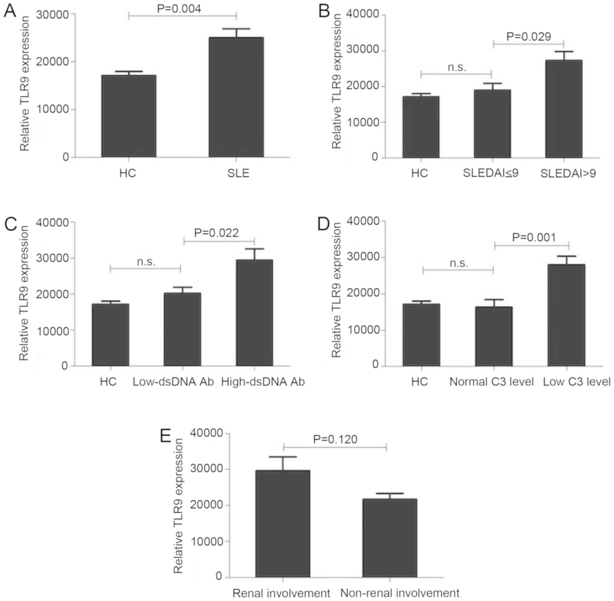 | Figure 1.The expression of TLR9 mRNA is
increased in whole blood samples from patients with SLE and is
associated with SLEDAI scores, anti-dsDNA Ab levels and C3 levels,
but not with renal involvement. (A) Expression of TLR9 mRNA in the
whole blood of newly diagnosed patients with SLE (n=90) compared
with HCs (n=49). (B) Expression of TLR9 mRNA in the whole blood of
patients with SLE in the SLEDAI ≤9 and SLEDAI >9 groups, as well
as HCs. (C) Expression of TLR9 mRNA in the whole blood of patients
with SLE in the high- and low-dsDNA Ab groups, as well as HCs. (D)
Expression of TLR9 mRNA in whole blood from patients with SLE in
the normal- and low-C3 level groups, as well as HCs. (E) Expression
of TLR9 mRNA in the whole blood of patients with SLE with renal
involvement and those with no renal involvement. Data are expressed
as the mean ± standard deviation. TLR9, toll-like receptor 9; HCs,
healthy controls; SLE, systemic lupus erythematosus; SLEDAI, SLE
Disease Activity Index; C3, complement C3; dsDNA Ab, double
stranded DNA antibody titer. |
Differences in the expression of TLR9 mRNA in the
different subgroups of patients with SLE were subsequently
determined. Levels of TLR9 mRNA expression in the whole blood
samples of patients with SLE were significantly higher in the
SLEDAI >9 group compared with the SLEDAI ≤9 group (P=0.029,
Fig. 1B). Similarly, TLR9 mRNA
expression was significantly increased in the high-dsDNA Ab group
compared with the low-dsDNA Ab group (P=0.022; Fig. 1C) and the low-C3 level group compared
with the normal-C3 level group (P=0.001; Fig. 1D). The difference in the levels of
TLR9 mRNA in patients with renal involvement compared with patients
with no renal involvement was not significant (Fig. 1E). The difference in the percentage
of patients with renal involvement (persistent proteinuria of
>0.5 g/day) between the low-TLR9 and high-TLR9 group was also
not significant (Table I).
High TLR9 mRNA expression is
independently associated with the poor prognosis of patients with
SLE
During the 2-year follow-up period, 52 patients
(57.8%) experienced clinical remission and the remaining 38
patients (42.2%) were categorized into the poor prognosis group.
The difference in age, sex, onset-diagnosis lag-time, laboratory
analysis findings, other clinical manifestations and the therapies
administered to the high and low-TLR9 groups was not significant
(Table I). However, there was a
significant increase in neuropsychiatric manifestations (P=0.029),
application of cyclosporine (P<0.001), mortality rates (P=0.035)
and poor prognosis (P=0.003) in the high-TLR9 group compared with
the low-TLR9 group over the 2-year follow-up period (Table I).
Univariate analyses demonstrated that persistent
proteinuria >0.5 g per day [hazard ratio (HR), 3.81; 95%
confidence interval (CI), 1.96–7.40], serum C3 (HR, 0.31; 95% CI,
0.10–1.0), SLEDAI (HR, 1.05; 95% CI, 1.02–1.09), C-reactive protein
(HR, 1.006; 95% CI, 1.001–1.012) and high-TLR9 mRNA levels (HR,
3.60; 95% CI, 1.88–6.90) were predictive of poor patient prognosis
over the 2-year follow-up period (Table
II). Following adjustment for several factors, including age,
persistent proteinuria (>0.5 g/day), serum C3 serum level,
SLEDAI, C-reactive protein, treatment with >1 immunosuppressant
therapy and high-TLR9 mRNA level, the multivariate analysis
demonstrated that persistent proteinuria (>0.5 g/day; HR, 6.314;
95% CI, 2.858–13.947), C-reactive protein (HR, 1.013; 95% CI,
1.007–1.019) and high-TLR9 mRNA levels (HR 3.852; 95% CI,
1.931–7.684) were independent risk factors for poor prognosis,
whereas treatment with >1 immunosuppressant (HR, 0.374; 95% CI,
0.173–0.808) was indicative of favorable prognosis (Table II).
| Table II.Univariate and multivariate analyses
of risk factors for SLE prognosis. |
Table II.
Univariate and multivariate analyses
of risk factors for SLE prognosis.
|
| Univariate
analysis | Multivariate
analysis |
|---|
|
|
|
|
|---|
| Variable | HR | 95% CI | P-values | HR | 95% CI | P-values |
|---|
| Persistent
proteinuria of >0.5 g per day | 3.600 | 1.879–6.898 |
<0.0001a | 6.314 | 2.858–13.947 |
<0.0001a |
| High-TLR9
level | 3.812 | 1.963–7.400 |
<0.0001a | 3.852 | 1.931–7.684 |
<0.0001a |
| >1
immunosuppressant for therapy | 0.775 | 0.401–1.499 | 0.450 | 0.374 | 0.173–0.808 | 0.012a |
| CRP | 1.006 | 1.001–1.012 | 0.027a | 1.013 | 1.007–1.019 |
<0.0001a |
| Serum C3 level | 0.311 | 0.098–0.989 | 0.048a |
|
|
|
| SLEDAI | 1.051 | 1.016–1.087 | 0.004a |
|
|
|
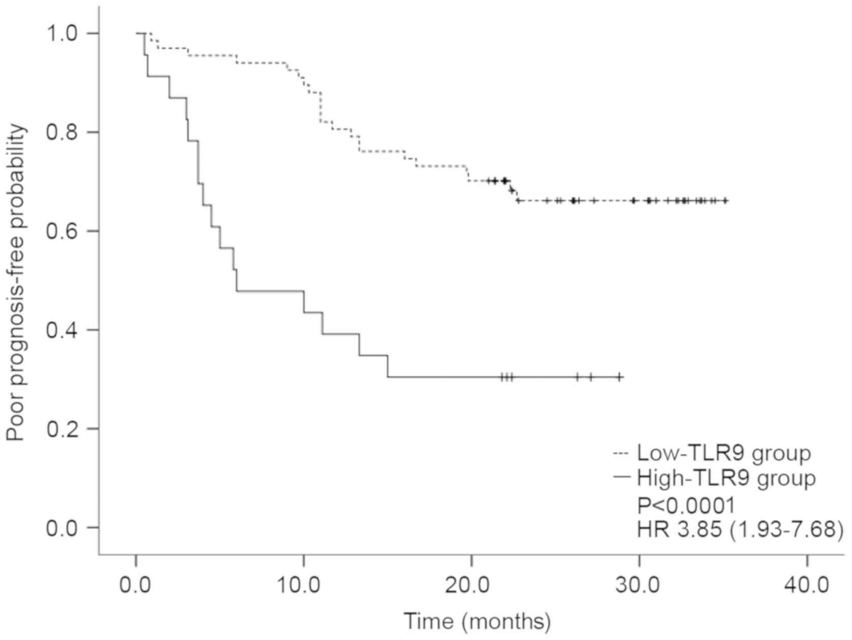
The median time of patients with SLE developing poor
prognosis was 6 months following diagnosis (range, 0–13.83 months).
Kaplan-Meier survival curves indicated that the 2-year poor
prognosis-free probability was 66% (95% CI, 54.2–77.8) in the
low-TLR9 group and 30% (95% CI, 10.4–49.6) in the high-TLR9 group
(HR, 3.85; 95% CI, 1.93–7.68; Fig.
2). This difference between prognosis-free probability was
significant (P<0.0001; all Fig.
2).
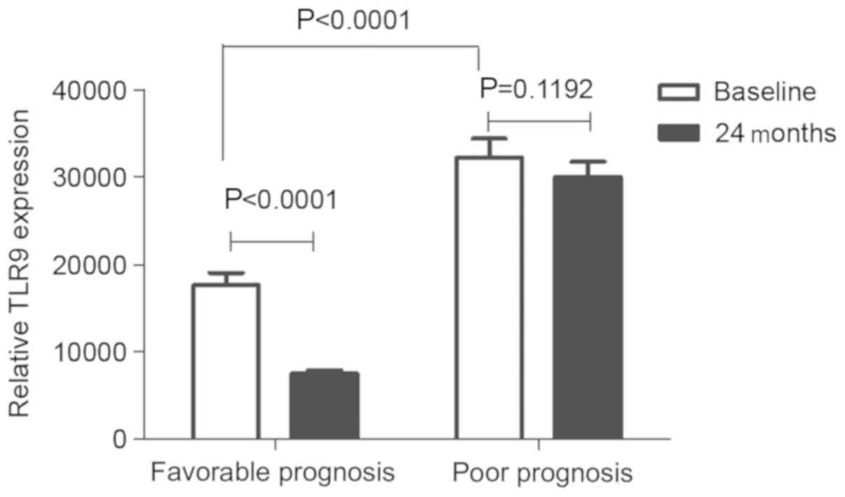
TLR9 mRNA levels remain high in the
poor prognosis group following treatment for 2 years with immune
suppressants
At the end of the 2-year follow-up period, the
expression of TLR9 mRNA in the favorable prognosis group (n=20) was
compared with the poor prognosis group (n=10) at baseline and 2
years following diagnosis of SLE (Fig.
3). There was a significant increase in the baseline expression
of TLR9 mRNA in the poor prognosis group compared with the
favorable prognosis group (P<0.0001). Compared with baseline
levels, the levels of TLR9 mRNA expression 2 years following
induction of immunosuppressant treatment decreased significantly in
the favorable prognosis group (P<0.0001) but remained high in
the poor prognosis group (P=0.1192; both Fig. 3).
Discussion
The current study demonstrated that levels of TLR9
mRNA were significantly increased in the whole blood samples from
patients with SLE and that these levels were associated with
disease activity. Furthermore, to the best of our knowledge this is
the first study to demonstrate that increased expression of TLR9
mRNA indicates the poor prognosis of patients with SLE over a
2-year period. Thus, over the short term at least, TLR9 may predict
the successful recovery of patients from SLE without remission and
determine the most effective therapeutic strategy for treating
patients.
In humans, TLR9 is expressed in the spleen, lymph
nodes, tonsils and peripheral blood mononuclear cells (27,28).
Previous studies have demonstrated that TLR9 is highly expressed in
SLE monocytes, T and B cells (8,12,29,30)
and that TLR9 expression in patients with active SLE is increased
compared with healthy controls (31). The results of the current study are
consistent with this, as they indicate that the expression of TLR9
mRNA in patients with active SLE was significantly increased
compared with healthy controls. The current study also measured the
expression of TLR9 mRNA in peripheral whole blood cells. TLR9 is
expressed by monocytes, T and B cells in patients with SLE;
therefore, measuring the expression of TLR9 mRNA in the whole blood
may provide a better indication of the in vivo environment
and avoids the potential adverse effects of separating cells in
collected blood samples.
It has been suggested that a deficiency in the
clearance of cellular debris causes the accumulation of
extracellular nucleic acids, which may be recognized by TLR9 in
patients with SLE (32). This is
consistent with the results of the current study, as it was
demonstrated that patients with SLE and an increased titer of
anti-dsDNA Ab exhibited increased expression of TLR9 mRNA.
The association between TLR9 and different clinical
parameters remains controversial. Wong et al (8) demonstrated that there was no
correlation between TLR9 expression and SLEDAI in patients with SLE
(mean SLEDAI, 3.13±2.29). By contrast, Nakano et al
(33) identified a positive
correlation between the expression of TLR9 mRNA and SLEDAI in
patients with active SLE. Furthermore, Papadimitraki et al
(12) reported a positive
correlation between levels of TLR9 expression and anti-dsDNA Ab in
the B cells of patients with active SLE. The results of the current
study demonstrated that the expression of TLR9 mRNA in whole blood
cell samples from patients with newly diagnosed active SLE differed
significantly different among the SLEDAI, anti-dsDNA Ab and
complement C3 subgroups. This suggests that TLR9 is involved in the
pathogenesis of active SLE.
The optimal criteria for predicting SLE remission
remain unknown, as the results of previous studies provide
differing views (16,24,34).
Varying definitions of remission are likely to be the main reason
for the divergent results. Unlike other studies that define SLE
disease activity as complete remission, serologically active
clinically quiescent disease or persistently active disease and
flare, the current study categorized SLE outcomes as favorable or
poor. At the end of the study, the proportion of patients with
favorable prognosis and poor prognosis were 57.8 and 42.2%,
respectively. Additionally, the expression of TLR9 mRNA in the
whole blood cells of patients at the onset of SLE was closely
associated with prognosis at 2 years. The increased expression of
TLR9 mRNA at SLE onset was associated with a poorer outcome of SLE
during the 2-year follow-up period. The current study demonstrated
that the proportion of patients with poor prognosis was 69.6% in
the high-TLR9 group and 32.8% in the low-TLR9 group. There were 4
(17.4%) patient mortalities in the high-TLR9 group and 2 (3.0%) in
the low-TLR9 group. Thus, the increased expression of TLR9 mRNA may
indicate an increased likelihood of mortality in patients with
SLE.
Currently, there are no reliable predictors of SLE
prognosis and the majority of studies concerning SLE prognosis have
been clinical reports. It has been demonstrated that
glomerulonephritis, vasculitis and hematological abnormalities are
independent risk factors for the absence of clinical remission in
patients with SLE (4) and a recent
study, demonstrated that renal and neurological involvement
decreased the likelihood of remission and favorable prognosis
(35). The results of the current
study are partially consistent with those from these previous
studies, as it was demonstrated that renal involvement is an
independent risk factor and increased levels of TLR9 mRNA and
C-reactive protein in the whole blood are risk factors for poor
prognosis in patients with SLE. The effects of renal involvement
(persistent proteinuria of >0.5 g/day; HR, 6.31) and high TLR9
mRNA levels (HR, 3.85) on patient prognosis were greater than those
of C-reactive protein (HR, 1.01). It has been demonstrated that
high levels of TLR9 mRNA predict poor outcomes in breast cancer
(36); however, to the best of our
knowledge, the current study is the first to identify an
association between TLR9 expression and SLE prognosis. The
expression of TLR9 mRNA was significantly decreased 2 years
following the induction of immunosuppressive treatment in the
favorable prognosis group but remained unchanged in the poor
prognosis group.
Administration of >1 type of immunosuppressant
for SLE therapy promoted the favorable prognosis of patients with
SLE in the current study. Although the administration of multiple
immunosuppressants is an acceptable practice, it is necessary to
evaluate the side effects of immunosuppressants and their
cost-effectiveness in the long term.
The role of TLR9 in SLE pathogenesis remains
unclear. A previous study suggested that activation of TLR9 induces
the progression of nephritis in MRL-Fas lupus prone mice (37), which may lead to the progression of
autoimmune disease. Other studies have demonstrated that TLR9
induces cytokine secretion in B cells and plasmacytoid dendritic
cells (29,38). Additionally, DNA-containing immune
complexes activate TLR9 in SLE (12)
and excessive dsDNA fragments may induce high TLR9 expression in
patients with SLE. This is consistent with the results of the
current study, which identified that TLR9 expression was positively
associated with the SLEDAI score in patients with SLE. Previous
studies have indicated that SLE disease activity is the primary
factor affecting SLE prognosis in the 5 years following diagnosis
(13,14). High expression of TLR9 mRNA is
suggestive of a severe autoimmune response in patients with SLE and
high SLEDAI (37). A more severe
autoimmune response makes recovery more difficult, leading to
poorer patient prognosis. This has been confirmed in previous
studies, which demonstrated that high SLE disease activity is a
factor that predicts poor disease prognosis (13,14).
Organ damage may also be a risk factor for poor SLE prognosis,
however this was not assessed during the follow-up period in the
present study, which is a limitation. Furthermore, the potential
role of TLR9 expression in SLE prognosis remains unclear.
In conclusion, TLR9 is involved in the pathogenesis
of SLE, associated with disease activity and may predict SLE
outcome within a 2-year period. This suggests that the detection of
TLR9 mRNA at SLE onset should be used to identify patients with
poor prognosis who may require more frequent monitoring in the
short-term. Thus, TLR9 may be a useful biomarker for predicting SLE
prognosis.
Acknowledgements
The authors wish to thank the members of the
Department of Rheumatology and Immunology in the First Hospital of
Jilin University for their contributions to the immunological
characterization of the patients. The authors also wish to thank
patients and their families for their support and cooperation.
References
|
1
|
O'Neill S and Cervera R: Systemic lupus
erythematosus. Best Pract Res Clin Rheumatol. 24:841–855. 2010.
View Article : Google Scholar : PubMed/NCBI
|
|
2
|
Kasitanon N, Magder LS and Petri M:
Predictors of survival in systemic lupus erythematosus. Medicine
(Baltimore). 85:147–156. 2006. View Article : Google Scholar : PubMed/NCBI
|
|
3
|
Ward MM, Pyun E and Studenski S: Causes of
death in systemic lupus erythematosus. Long-term followup of an
inception cohort. Arthritis Rheum. 38:1492–1499. 1995. View Article : Google Scholar : PubMed/NCBI
|
|
4
|
Zen M, Iaccarino L, Gatto M, Bettio S,
Nalotto L, Ghirardello A, Punzi L and Doria A: Prolonged remission
in Caucasian patients with SLE: Prevalence and outcomes. Ann Rheum
Dis. 74:2117–2122. 2015. View Article : Google Scholar : PubMed/NCBI
|
|
5
|
Lloyd W and Schur PH: Immune complexes,
complement, and anti-DNA in exacerbations of systemic lupus
erythematosus (SLE). Medicine (Baltimore). 60:208–217. 1981.
View Article : Google Scholar : PubMed/NCBI
|
|
6
|
Kao AH, Navratil JS, Ruffing MJ, Liu CC,
Hawkins D, McKinnon KM, Danchenko N, Ahearn JM and Manzi S:
Erythrocyte C3d and C4d for monitoring disease activity in systemic
lupus erythematosus. Arthritis Rheum. 62:837–844. 2010. View Article : Google Scholar : PubMed/NCBI
|
|
7
|
Bootsma H, Spronk PE, Ter Borg EJ, Hummel
EJ, de Boer G, Limburg PC and Kallenberg CG: The predictive value
of fluctuations in IgM and IgG class anti-dsDNA antibodies for
relapses in systemic lupus erythematosus. A prospective long-term
observation. Ann Rheum Dis. 56:661–666. 1997. View Article : Google Scholar : PubMed/NCBI
|
|
8
|
Wong CK, Wong PT, Tam LS, Li EK, Chen DP
and Lam CW: Activation profile of Toll-like receptors of peripheral
blood lymphocytes in patients with systemic lupus erythematosus.
Clin Exp Immunol. 159:11–22. 2010. View Article : Google Scholar : PubMed/NCBI
|
|
9
|
Wu O, Chen GP, Chen H, Li XP, Xu JH, Zhao
SS, Sheng J, Feng JB, Cai J, Fang XH, et al: The expressions of
Toll-like receptor 9 and T-bet in circulating B and T cells in
newly diagnosed, untreated systemic lupus erythematosus and
correlations with disease activity and laboratory data in a Chinese
population. Immunobiology. 214:392–402. 2009. View Article : Google Scholar : PubMed/NCBI
|
|
10
|
Clancy RM, Markham AJ and Buyon JP:
Endosomal Toll-like receptors in clinically overt and silent
autoimmunity. Immunol Rev. 269:76–84. 2016. View Article : Google Scholar : PubMed/NCBI
|
|
11
|
Means TK, Latz E, Hayashi F, Murali MR,
Golenbock DT and Luster AD: Human lupus autoantibody-DNA complexes
activate DCs through cooperation of CD32 and TLR9. J Clin Invest.
115:407–417. 2005. View
Article : Google Scholar : PubMed/NCBI
|
|
12
|
Papadimitraki ED, Choulaki C, Koutala E,
Bertsias G, Tsatsanis C, Gergianaki I, Raptopoulou A, Kritikos HD,
Mamalaki C, Sidiropoulos P and Boumpas DT: Expansion of toll-like
receptor 9-expressing B cells in active systemic lupus
erythematosus: Implications for the induction and maintenance of
the autoimmune process. Arthritis Rheum. 54:3601–3611. 2006.
View Article : Google Scholar : PubMed/NCBI
|
|
13
|
Cervera R, Khamashta MA, Font J,
Sebastiani GD, Gil A, Lavilla P, Mejía JC, Aydintug AO,
Chwalinska-Sadowska H, de Ramón E, et al: Morbidity and mortality
in systemic lupus erythematosus during a 10-year period: A
comparison of early and late manifestations in a cohort of 1,000
patients. Medicine (Baltimore). 82:299–308. 2003. View Article : Google Scholar : PubMed/NCBI
|
|
14
|
Abu-Shakra M, Urowitz MB, Gladman DD and
Gough J: Mortality studies in systemic lupus erythematosus. Results
from a single center. II. Predictor variables for mortality. J
Rheumatol. 22:1265–1270. 1995.PubMed/NCBI
|
|
15
|
Abu-Shakra M, Urowitz MB, Gladman DD and
Gough J: Mortality studies in systemic lupus erythematosus. Results
from a single center. I. Causes of death. J Rheumatol.
22:1259–1264. 1995.PubMed/NCBI
|
|
16
|
Formiga F, Moga I, Pac M, Mitjavila F,
Rivera A and Pujol R: High disease activity at baseline does not
prevent a remission in patients with systemic lupus erythematosus.
Rheumatology (Oxford). 38:724–727. 1999. View Article : Google Scholar : PubMed/NCBI
|
|
17
|
Hochberg MC: Updating the American College
of Rheumatology revised criteria for the classification of systemic
lupus erythematosus. Arthritis Rheum. 40:17251997. View Article : Google Scholar : PubMed/NCBI
|
|
18
|
Zen M, Bassi N, Nalotto L, Canova M,
Bettio S, Gatto M, Ghirardello A, Iaccarino L, Punzi L and Doria A:
Disease activity patterns in a monocentric cohort of SLE patients:
A seven-year follow-up study. Clin Exp Rheumatol. 30:856–863.
2012.PubMed/NCBI
|
|
19
|
Bombardier C, Gladman DD, Urowitz MB,
Caron D and Chang CH: Derivation of the SLEDAI. A disease activity
index for lupus patients. The Committee on Prognosis Studies in
SLE. Arthritis Rheum. 35:630–640. 1992. View Article : Google Scholar : PubMed/NCBI
|
|
20
|
Somerfield SD, Roberts MW and Booth RJ:
Double-stranded DNA antibodies: A comparison of four methods of
detection. J Clin Pathol. 34:1032–1035. 1981. View Article : Google Scholar : PubMed/NCBI
|
|
21
|
Haugbro K, Nossent JC, Winkler T,
Figenschau Y and Rekvig OP: Anti-dsDNA antibodies and disease
classification in antinuclear antibody positive patients: The role
of analytical diversity. Ann Rheum Dis. 63:386–394. 2004.
View Article : Google Scholar : PubMed/NCBI
|
|
22
|
Ng KP, Manson JJ, Rahman A and Isenberg
DA: Association of antinucleosome antibodies with disease flare in
serologically active clinically quiescent patients with systemic
lupus erythematosus. Arthritis Rheum. 55:900–904. 2006. View Article : Google Scholar : PubMed/NCBI
|
|
23
|
Gladman DD, Urowitz MB and Keystone EC:
Serologically active clinically quiescent systemic lupus
erythematosus: A discordance between clinical and serologic
features. Am J Med. 66:210–215. 1979. View Article : Google Scholar : PubMed/NCBI
|
|
24
|
Nikpour M, Urowitz MB, Ibañez D and
Gladman DD: Frequency and determinants of flare and persistently
active disease in systemic lupus erythematosus. Arthritis Rheum.
61:1152–1158. 2009. View Article : Google Scholar : PubMed/NCBI
|
|
25
|
Tseng CE, Buyon JP, Kim M, Belmont HM,
Mackay M, Diamond B, Marder G, Rosenthal P, Haines K, Ilie V and
Abramson SB: The effect of moderate-dose corticosteroids in
preventing severe flares in patients with serologically active, but
clinically stable, systemic lupus erythematosus: Findings of a
prospective, randomized, double-blind, placebo-controlled trial.
Arthritis Rheum. 54:3623–3632. 2006. View Article : Google Scholar : PubMed/NCBI
|
|
26
|
Livak KJ and Schmittgen TD: Analysis of
relative gene expression data using real-time quantitative PCR and
the 2(-Delta Delta C(T)) method. Methods. 25:402–408. 2001.
View Article : Google Scholar : PubMed/NCBI
|
|
27
|
Nishimura M and Naito S: Tissue-specific
mRNA expression profiles of human toll-like receptors and related
genes. Biol Pharm Bull. 28:886–892. 2005. View Article : Google Scholar : PubMed/NCBI
|
|
28
|
Chuang TH and Ulevitch RJ: Cloning and
characterization of a sub-family of human toll-like receptors:
HTLR7, hTLR8 and hTLR9. Eur Cytokine Netw. 11:372–378.
2000.PubMed/NCBI
|
|
29
|
Rao H, Zeng Q, Liang Y, Xiao C, Xie S and
Xu X: Correlation between TLR9 expression and cytokine secretion in
the clinical diagnosis of systemic lupus erythematosus. Mediators
Inflamm. 2015:7107202015. View Article : Google Scholar : PubMed/NCBI
|
|
30
|
Komatsuda A, Wakui H, Iwamoto K, Ozawa M,
Togashi M, Masai R, Maki N, Hatakeyama T and Sawada K: Up-regulated
expression of Toll-like receptors mRNAs in peripheral blood
mononuclear cells from patients with systemic lupus erythematosus.
Clin Exp Immunol. 152:482–487. 2008. View Article : Google Scholar : PubMed/NCBI
|
|
31
|
Mortezagholi S, Babaloo Z, Rahimzadeh P,
Namdari H, Ghaedi M, Gharibdoost F, Mirzaei R, Bidad K and Salehi
E: Evaluation of TLR9 expression on PBMCs and CpG ODN-TLR9 ligation
on IFN-α production in SLE patients. Immunopharmacol Immunotoxicol.
39:11–18. 2017. View Article : Google Scholar : PubMed/NCBI
|
|
32
|
Kruse K, Janko C, Urbonaviciute V, Mierke
CT, Winkler TH, Voll RE, Schett G, Muñoz LE and Herrmann M:
Inefficient clearance of dying cells in patients with SLE:
Anti-dsDNA autoantibodies, MFG-E8, HMGB-1 and other players.
Apoptosis. 15:1098–1113. 2010. View Article : Google Scholar : PubMed/NCBI
|
|
33
|
Nakano S, Morimoto S, Suzuki J, Nozawa K,
Amano H, Tokano Y and Takasaki Y: Role of pathogenic auto-antibody
production by Toll-like receptor 9 of B cells in active systemic
lupus erythematosus. Rheumatology (Oxford). 47:145–149. 2008.
View Article : Google Scholar : PubMed/NCBI
|
|
34
|
Laustrup H, Voss A, Green A and Junker P:
SLE disease patterns in a Danish population-based lupus cohort: An
8-year prospective study. Lupus. 19:239–246. 2010. View Article : Google Scholar : PubMed/NCBI
|
|
35
|
Medina-Quiñones CV, Ramos-Merino L,
Ruiz-Sada P and Isenberg D: Analysis of complete remission in
systemic lupus erythematosus patients over a 32-year period.
Arthritis Care Res (Hoboken). 68:981–987. 2016. View Article : Google Scholar : PubMed/NCBI
|
|
36
|
Qiu J, Shao S, Yang G, Shen Z and Zhang Y:
Association of Toll like receptor 9 expression with lymph node
metastasis in human breast cancer. Neoplasma. 58:251–255. 2011.
View Article : Google Scholar : PubMed/NCBI
|
|
37
|
Anders HJ, Vielhauer V, Eis V, Linde Y,
Kretzler M, Perez de Lema G, Strutz F, Bauer S, Rutz M, Wagner H,
et al: Activation of toll-like receptor-9 induces progression of
renal disease in MRL-Fas (lpr) mice. FASEB J. 18:534–536. 2004.
View Article : Google Scholar : PubMed/NCBI
|
|
38
|
He B, Qiao X and Cerutti A: CpG DNA
induces IgG class switch DNA recombination by activating human B
cells through an innate pathway that requires TLR9 and cooperates
with IL-10. J Immunol. 173:4479–4491. 2004. View Article : Google Scholar : PubMed/NCBI
|