Introduction
Acute coronary syndrome (ACS) is a severe type of
coronary heart disease, which is a pathological symptom caused by
obstructed coronary arteries. It occurs primarily in older people
and is a leading cause of poor prognosis and sudden death of
patients with coronary heart disease. ACS patients often die
suddenly without any sign, so it causes great harms (1). As a common type of cardiovascular
system disease, atherosclerosis (AS) is a chronic inflammatory
disease of the arterial wall induced by lipid metabolism disorder
in the body (2). AS is the major
pathological basis of ACS which is a clinical pathological syndrome
resulting from total and non-total occlusive thrombus secondary to
coronary AS plaque rupture or invasion. Vascular inflammation
reactions triggered by inflammatory factors are the main reason of
ACS occurrence, which destroys the stability of the coronary AS
plaque and causes rupture and hemorrhage of the plaque, thus
leading to thrombus (3,4).
The pathogenesis of coronary AS has not been
clarified thus far, and studies have demonstrated that both immune
cells and inflammatory factors are involved in the process of
coronary AS formation (5).
Programmed cell death protein 4 (PDCD4) is a novel
suppressor gene discovered in recent years, which cannot only
control the proliferation of tumor cells but also suppress the
generation of tumor cells, having a close correlation with the
occurrence and development of tumors (6). According to the study by Liang et
al (7), PDCD4 is expressed in
myocardial cells and vascular smooth muscle cells, and it can
inhibit the expression of the inflammatory factor interleukin-10
(IL-10) by activating nuclear factor-kappa B (NF-κB) in vascular
smooth muscle cells. In addition to its inhibitory effects in the
occurrence and development of multiple tumors, PDCD4 also
participates in immune response, inflammatory reaction and other
pathophysiological processes.
Research on PDCD4 in recent years was mainly focused
on the mechanism of tumors, but there are rare studies on its role
in coronary AS. This study aimed to analyze the function and
mechanism of PDCD4 in the process of coronary AS formation by means
of observing the PDCD4 expression in coronary AS plaque of
rats.
Materials and methods
Laboratory animals
A total of 80 healthy, clean and specific
pathogen-free (SPF) Wistar rats, aged 6–8 weeks, with a body mass
of 170–190 g, were purchased from Shanghai Jia Ke Biotechnology
Co., Ltd. (Shanghai, China) [animal certification no.
SCXK(Shanghai)2016-18]. The rats were maintained in a clean
environment, with indoor temperature of 21–25°C and humidity of
52–57%. All the rats were fed adaptively for 2 weeks prior to the
experiment. This animal experiment was approved by the Ethics
Committee of Yidu Central Hospital of Weifang (Weifang, China).
Main instruments and reagents
Rabbit anti-rat PDCD4 and glyceraldehyde-3-phosphate
dehydrogenase (GAPDH) monoclonal antibodies were purchased from
Cell Signaling Technology, Inc. (cat nos. 9535 and 2118; Danvers,
MA, USA), bicinchoninic acid (BCA) protein assay kit was obtained
from Beijing Solarbio Science & Technology Co., Ltd. (Beijing,
China), and terminal deoxynucleotidyl transferase-mediated dUTP
nick end-labeling (TUNEL) apoptosis assay kit was purchased from
Beijing Jiamay Biotech Co., Ltd. (Beijing, China). Real-time
quantitative polymerase chain reaction (PCR) instrument as well as
real-time quantitative PCR kits for interleukin-6 (IL-6) and IL-8
were purchased from Shanghai HuaGen Biotech Co., Ltd. (Shanghai,
China). Total ribonucleic acid (RNA) extraction kit (TRIzol reagent
method) was obtained from Thermo Fisher Scientific Inc. (Waltham,
MA, USA). Promega reverse transcription kit was from ABclonal
Biotech Co., Ltd. (Woburn, MA, USA) and the internal reference
primers for IL-6, IL-8 and β-actin in reverse-transcription
quantitative PCR were purchased from Shanghai Gefan Biotechnology
Co., Ltd. (Shanghai, China). Primer sequences are listed in
Table I.
 | Table I.Primer sequences for IL-6, IL-8 and
β-actin genes. |
Table I.
Primer sequences for IL-6, IL-8 and
β-actin genes.
| Genes | Forward primer
sequence | Reverse primer
sequence |
|---|
| IL-6 |
5′-CTCTCCGCAAGAGACTTCCA-3′ |
5′-TGGTCTTCTGGAGTTCCGTT-3′ |
| IL-8 |
5′-CTTTGTCCATTCCCACTTCTGA-3′ |
5′-TCCCTAACGGTTGCCTTTGTAT-3′ |
| β-actin | 5′-
TGAAGTGTGACGTGGACATC-3′ |
5′-TAGAAGCATTTGCGGTGGAC-3′ |
Construction and grouping of animal
models
The healthy SPF Wistar rats with similar body weight
were selected and divided into control group (n=40) and research
group (n=40), and rats in the control group were fed with normal
diet. By reference to the modeling methods in the literature by
Ganzetti et al (8), the rats
in the research group were injected with vitamin D3 from the right
lower extremity and raised with high-fat diet (recipe: 0.2%
propylthiouracil, 10% lard, 1.5% sodium cholate, 4% cholesterol and
84.3% basic diet) provided by Guangzhou SeBiona Bio-Tech Co., Ltd.
at 30 days before the modeling. At 3 and 6 weeks of feeding, 10%
bovine serum albumin (250 mg/kg) was injected into the rats from
the tail veins for immune damage. At 15 weeks after feeding, the
rats were sacrificed by decapitation. In the research group, the
tissues of coronary plaque were extracted to examine the cardiac
pathology, and the existence of AS plaque suggested successful
model establishment. The harvested coronary artery tissues of all
the rats were placed into liquid nitrogen immediately and then
stored in a refrigerator at −80°C.
Index detection
PDCD4 detection
Western blotting was utilized to measure the PDCD4
in the coronary arteries. The coronary artery tissues were taken
and fully ground into tissue homogenates, followed by
centrifugation at 3,000 × g at 4°C for 8 min and preparation of 50
µg protein extracts. Next, proteins with 8% loading sample were
extracted for polyacrylamide gel electrophoresis experiment, and
were transferred to a polyvinylidene fluoride (PVDF) membrane at
the end of electrophoresis. Then the proteins were blocked in 5%
skim milk powder for 1 h, followed by incubation with PDCD4 primary
antibody (1:1,000) and GAPDH polyclonal antibody (1:1,000) at 4°C
overnight. Subsequently, goat anti-rabbit horseradish peroxidase
(HRP)-labeled secondary polyclonal antibody (1:1,000; cat. no.
7074; Cell Signaling Technology, Inc.) was added and reacted at
37°C for 1.5 h, and diaminobenzidine was used for staining and
development. Images were captured and stored for analysis and
processing. The gel analysis software Quantity One 4.6.2 (Bio-Rad
Laboratories, Inc., Hercules, CA, USA) was applied to measure the
relative expression of PDCD4.
Detection of IL-6 and IL-8
Reverse transcription PCR (RT-PCR) was performed to
detect IL-6 and IL-8 in the coronary arteries, of which the primer
and probe sequences are shown in Table
I. Experimental procedures involved use of phenol method
(TRIzol reagent method) to extract the total RNA in the coronary
artery tissues in strict accordance with the TRIzol kit
instructions. The concentration and purity of the total RNA were
measured using a micro-spectrophotometer (Bio-Rad Laboratories,
Inc.), and the integrity was detected by virtue of denaturing
agarose gel electrophoresis. The RNA was reverse transcribed into
complementary deoxyribonucleic acid (cDNA) via the Promega reverse
transcription kit strictly according to the steps in the
instructions. The PCR system was prepared as described in the
instructions. Reaction conditions were: 35 cycles of
pre-denaturation at 94°C for 10 min, denaturation at 94°C for 30
sec, annealing at 60°C for 45 sec, extension at 72°C for 45 sec and
final extension at 65°C for 10 min. The software offered by
manufacturers was applied to analyze the amplification data, and
the relative expression of internal references of IL-6, IL-8 and
β-actin were calculated by using the formula of 2−ΔΔCq
(9).
Apoptosis rate of coronary artery
smooth muscle cells
A portion of coronary artery tissues was fixed in 4%
formaldehyde solution and then embedded in paraffin. After dewaxing
and rehydration, TUNEL assay was conducted to detect the apoptosis
rate of smooth muscle cells in the coronary artery in strict
accordance with the instructions of TUNEL assay kit. Five
consecutive high-power fields were examined using a light
microscope (Olympus, Tokyo, Japan), and the apoptotic cells were
counted in every 100 cells, to calculate the apoptosis rate as per
the formula: (Number of apoptotic cells/total number of cells) ×
100% = apoptosis rate.
Statistical analysis
Statistical Product and Service Solutions (SPSS)
17.0 software (Tianjin Ksoft Tech. Co., Ltd., Kerala, India) was
used for statistical analysis. Measurement data were presented as
mean ± standard deviation (SD). The Student's t-test was performed
for comparison of measurement data, and the Chi-square test was
used for comparison of enumeration data. P<0.05 was considered
to indicate a statistically significant difference.
Results
General conditions of rats in the
control group and the research group
In this experiment, coronary AS models were
established in the Wistar rats, and all 40 rats in the research
group were sacrificed by decapitation at 15 weeks after feeding.
There were 36 successful models, with a success rate of 90.00%
(36/40). The sex, age and body mass of the rats, indoor temperature
and indoor humidity had no impacts on the experiment (P>0.05)
(Table II).
| Table II.General information of rats in the two
groups. |
Table II.
General information of rats in the two
groups.
| Category | Control group
(n=40) | Research group
(n=36) | t/χ2 | P-value |
|---|
| Sex (%) |
|
| 0.038 | 0.846 |
| Male | 22 (55.00) | 19 (52.78) |
|
|
|
Female | 18 (45.00) | 17 (47.22) |
|
|
| Age (weeks) |
|
| 0.261 | 0.650 |
| ≤8 | 19 (47.50) | 15 (41.67) |
|
|
|
>8 | 21 (52.50) | 21 (58.33) |
|
|
| Body mass (g) |
|
| 2.880 | 0.121 |
| ≤180 | 21 (52.50) | 22 (61.11) |
|
|
|
>180 | 19 (47.50) | 14 (38.89) |
|
|
| Indoor temperature
(°C) | 23.12±1.63 | 22.89±0.98 | 0.735 | 0.464 |
| Indoor | 54.45±1.03 | 54.16±1.37 | 1.049 | 0.297 |
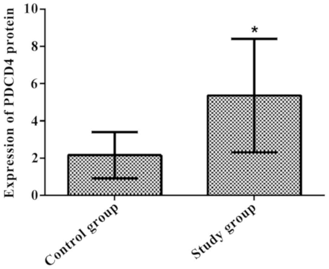
PDCD4 expression in coronary artery
tissues of rats in the control group and research group
The relative expression of PDCD4 in coronary artery
tissues was 2.16±1.24 in the control group and 5.37±3.05 in the
research group. The relative expression of PDCD4 in coronary artery
tissues in the research group was obviously higher than that in the
control group, and the difference was statistically significant
(t=6.121, P<0.01) (Fig. 1).
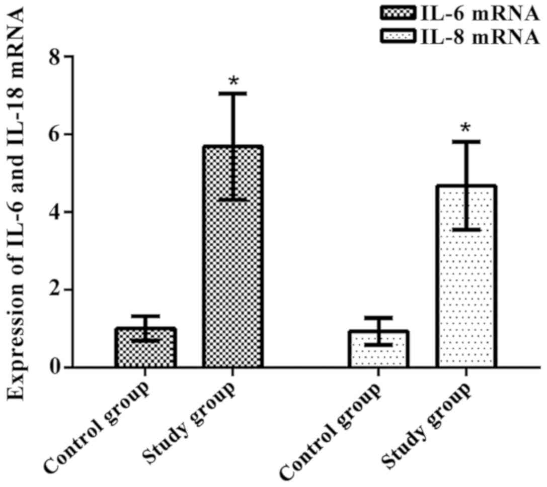
Messenger RNA (mRNA) expression of
IL-6 and IL-8 in coronary artery tissues of rats in the control and
research groups
The relative expression of IL-6 mRNA in coronary
artery tissues was 1.01±0.31 in the control group, and it was
5.69±1.37 in the research group. The research group had a
significantly higher level of IL-6 mRNA relative expression than
that of the control group, with a statistically significant
difference (t=21.03, P<0.01). The relative expression of IL-8
mRNA in coronary artery tissues were 0.93±0.35 and 4.68±1.13 in the
control and research groups, respectively. The difference in the
relative expression of IL-8 mRNA between the two groups was
statistically significant, and a much higher level was evident in
the research group (t=19.96, P<0.01) (Fig. 2).
Apoptotic rates of smooth muscle cells
in the control and research groups
The apoptosis rate of smooth muscle cells was
12.56±9.63% in the control group and 28.36±13.26% in the research
group. The apoptosis rate of smooth muscle cells in the research
group was increased notably compared with that in the control
group, and the difference was statistically significant (t=5.985,
P<0.001) (Table III).
| Table III.Comparison of apoptosis rates of
smooth muscle cells between the two groups (mean ± SD). |
Table III.
Comparison of apoptosis rates of
smooth muscle cells between the two groups (mean ± SD).
| Group | n | Apoptosis rate
(%) |
|---|
| Control group | 40 | 12.56±9.63 |
| Research group | 36 | 28.36±13.26 |
| t |
| 5.985 |
| P-value |
| <0.001 |
Discussion
Approximately 18 million individuals succumb to
atherosclerotic cardiovascular diseases annually worldwide, and ACS
is the primary cause of sudden cardiac death of patients. ACS is
mainly a consequence of coronary occlusion caused by AS plaque
rupture and thrombosis (10).
Therefore, AS is a non-linear process alternating between stable
phase and unstable phase, and the vulnerability of AS plaque is an
initiating agent of ACS (11).
Current treatments of AS tend to eliminate the plaques and control
vulnerable ones (12). The rupture
of unstable AS plaques is one of the causes for ACS, and
inflammatory responses are vital factors for the rupture of AS
plaques and thrombosis (13).
Several theories of the AS pathogenesis have been put forward
successively, of which the inflammation theory was the most
acceptable, suggesting that inflammatory response plays an
important role in various stages of an AS event (14).
As a kind of apoptosis-related gene, PDCD4
can bind to eukaryotic initiation factor-4A (eIF4A) via the
functional domain of MA3 protein and repress the combination of
eIF4C with eIF4A, thus suppressing the synthesis of ribosome and
protein (15). A study indicated
that PDCD4 is involved in the reaction of pro-inflammatory
Toll-like receptor 4 (TLR4) signaling pathway induced by
lipopolysaccharide (LPS); thus, PDCD4 is also considered as a
pro-inflammatory protein that may have functions in inflammatory
diseases (16). It has been proven
in recent years that IL-6 and IL-8 are cytokines closely associated
with the pathological progression of ACS. IL-6 is a category of
inflammatory factors capable of promoting the proliferation and
differentiation of B lymphocytes, which participates in the
reaction processes of multiple inflammatory diseases. IL-6 can act
as an inflammatory marker for local and peripheral blood
circulations of AS. In addition, its expression level may reflect
the degree of instability of AS plaque (17). IL-8, one of the cytokines with the
highest chemotactic activity, can accelerate local thrombosis
around the AS plaque and enhance a series of mechanisms of the AS
plaque, such as local oxidative stress response (18). The early pathological changes of AS
involve in a series of reactions, including changes in endothelial
function and aggregation of monocytes and T cells. Moreover, with
the progression of the disease, the vascular smooth muscle cells
are getting involved. The proliferation and apoptosis always play a
crucial role in the formation of plaques (19). Apoptosis of vascular smooth muscle
cells participates in the pathological processes of plaque and
thrombus formation. Moreover, it can lead to release of various
inflammatory factors, including IL-1, IL-6 and IL-8, thus
aggravating inflammation symptoms of the AS plaque (20). Research by Green et al
(21) manifested that PDCD4 is
downregulated remarkably in the arterial smooth muscle cells of
rats with acute balloon injury, thus facilitating the proliferation
of these cells. Yu and Li (22)
argued that high PDCD4 expression can inhibit the proliferation of
vascular smooth muscle cells. Apoptosis of vascular smooth muscle
cells is significantly decreased in PDCD4 knockout rats, while the
highly expressed PDCD4 can increase such apoptosis notably.
Therefore, PDCD4 can regulate the proliferation and apoptosis of
smooth muscle cells. However, the results of this research
indicated that the relative expressions of PDCD4, IL-6 and IL-8 in
coronary artery tissues in the research group were elevated
remarkably compared with those in the control group, suggesting
that PDCD4 may participate in the formation of AS plaque. The
research group had a significantly higher apoptosis rate of smooth
muscle cells than that of the control group, and it was considered
through further analysis that PDCD4 may increase the expression of
inflammatory factors and then upregulate the IL-6 and IL-8
expression by means of suppressing the proliferation of vascular
smooth muscle cells during the formation of AS plaque.
Considering the repeatability and reliability of
animal experiment, the rats in this study were screened strictly to
control the differences in age, body mass, health and other aspects
of the Wistar rats. It was shown that the sex, age and body mass of
the rats, indoor temperature and humidity had no impacts on the
experiment. Since PDCD4 knockout was not performed for the rats and
the expression of inflammatory factors in PDCD4 knockout rats were
not analyzed, there were certain limitations in this experiment.
Therefore, it is expected that the experiment should be conducted
in the rats next time, so as to provide more evidence for these
findings.
In conclusion, PDCD4 may participate in the
formation of coronary AS plaque, and its possible function in the
process is to inhibit the proliferation of vascular smooth muscle
cells and promote the upregulation of IL-6 and IL-8.
Acknowledgements
Not applicable.
Funding
No funding was received.
Availability of data and materials
The datasets used and/or analyzed during the present
study are available from the corresponding author on reasonable
request.
Authors' contributions
YG and HLi wrote the manuscript and assisted in the
construction and grouping of animal models. YZ and HLv performed
western blotting and RT-PCR. YC was responsible for TUNEL assay.
All authors read and approved the final manuscript.
Ethics approval and consent to
participate
The study was approved by the Ethics Committee of
Yidu Central Hospital of Weifang (Weifang, China).
Patient consent for publication
Not applicable.
Competing interests
The authors declare that they have no competing
interests.
References
|
1
|
Mega JL, Braunwald E, Wiviott SD, Bassand
JP, Bhatt DL, Bode C, Burton P, Cohen M, Cook-Bruns N, Fox KA, et
al ATLAS ACS 2–TIMI 51 Investigators, : Rivaroxaban in patients
with a recent acute coronary syndrome. N Engl J Med. 366:9–19.
2012. View Article : Google Scholar : PubMed/NCBI
|
|
2
|
Aronow HD and Beckman JA: Parsing
atherosclerosis: The unnatural history of peripheral artery
disease. Circulation. 134:438–440. 2016. View Article : Google Scholar : PubMed/NCBI
|
|
3
|
Lee T, Murai T, Isobe M and Kakuta T:
Impact of coronary plaque morphology assessed by optical coherence
tomography on cardiac troponin elevation in patients with non-ST
segment elevation acute coronary syndrome. Catheter Cardiovasc
Interv. 90:905–914. 2017. View Article : Google Scholar : PubMed/NCBI
|
|
4
|
De Ronde M, Kok MGM, Beijk MAM, De Winter
RJ, Van Der Wal AC, Sondermeijer BM, Meijers JCM, Creemers EE and
Pinto-Sietsma SJ: Tissue-derived circulating miRNAs can identify
atherosclerosis and plaque instability. Atherosclerosis.
263:e372017. View Article : Google Scholar
|
|
5
|
Barbato E, Toth GG, Johnson NP, Pijls NH,
Fearon WF, Tonino PA, Curzen N, Piroth Z, Rioufol G, Jüni P, et al:
A prospective natural history study of coronary atherosclerosis
using fractional flow reserve. J Am Coll Cardiol. 68:2247–2255.
2016. View Article : Google Scholar : PubMed/NCBI
|
|
6
|
Jo SH, Kim DE, Clocchiatti A and Dotto GP:
PDCD4 is a CSL associated protein with a transcription repressive
function in cancer associated fibroblast activation. Oncotarget.
7:58717–58727. 2016. View Article : Google Scholar : PubMed/NCBI
|
|
7
|
Liang X, Xu Z, Yuan M, Zhang Y, Zhao B,
Wang J, Zhang A and Li G: MicroRNA-16 suppresses the activation of
inflammatory macrophages in atherosclerosis by targeting PDCD4. Int
J Mol Med. 37:967–975. 2016. View Article : Google Scholar : PubMed/NCBI
|
|
8
|
Ganzetti GS, Busnelli M, Parolini C,
Manzini S, Dellera F, Sirtori CR and Chiesa G: ApoA-I depletion in
chow-fed ApoEKO mice severely worsens coronary atherosclerosis
development. Atherosclerosis. 252:e1042016. View Article : Google Scholar
|
|
9
|
Livak KJ and Schmittgen TD: Analysis of
relative gene expression data using real-time quantitative PCR and
the 2(-Delta Delta C(T)) method. Methods. 25:402–408. 2001.
View Article : Google Scholar : PubMed/NCBI
|
|
10
|
Rodriguez F, Maron DJ, Knowles JW, Virani
SS, Lin S and Heidenreich PA: Association between intensity of
statin therapy and mortality in patients with atherosclerotic
cardiovascular disease. JAMA Cardiol. 2:47–54. 2017. View Article : Google Scholar : PubMed/NCBI
|
|
11
|
Wierer M, Prestel M, Schiller HB, Yan G,
Schaab C, Azghandi S, Werner J, Kessler T, Malik R, Murgia M, et
al: Compartment-resolved proteomic analysis of mouse aorta during
atherosclerotic plaque formation reveals osteoclast-specific
protein expression. Mol Cell Proteomics. 17:321–334. 2018.
View Article : Google Scholar : PubMed/NCBI
|
|
12
|
Vogel ME, Idelman G, Konaniah ES and
Zucker SD: Bilirubin prevents atherosclerotic lesion formation in
low-density lipoprotein receptor-deficient mice by inhibiting
endothelial VCAM-1 and ICAM-1 signaling. J Am Heart Assoc. 6:62017.
View Article : Google Scholar
|
|
13
|
Tsivgoulis G, Katsanos AH, Giannopoulos G,
Panagopoulou V, Jatuzis D, Lemnens R, Deftereos S and Kelly PJ: The
role of colchicine in the prevention of cerebrovascular ischemia.
Curr Pharm Des. 24:668–674. 2018. View Article : Google Scholar : PubMed/NCBI
|
|
14
|
Kurdi A, De MG and Martinet W: Everolimus
attenuates atherosclerotic plaque progression, intraplaque
neovascularization, myocardial infarction and sudden death in a
mouse model of advanced atherosclerosis. Atherosclerosis.
263:e592017. View Article : Google Scholar
|
|
15
|
Maeda N, Abdullahi A, Beatty B, Dhanani Z
and Adegoke OAJ: Depletion of the mRNA translation initiation
inhibitor, programmed cell death protein 4 (PDCD4), impairs L6
myotube formation. Physiol Rep. 5:e133952017. View Article : Google Scholar : PubMed/NCBI
|
|
16
|
Wang L, Jiang Y, Song X, Guo C, Zhu F,
Wang X, Wang Q, Shi Y, Wang J, Gao F, et al: Pdcd4 deficiency
enhances macrophage lipoautophagy and attenuates foam cell
formation and atherosclerosis in mice. Cell Death Dis. 7:e20552016.
View Article : Google Scholar : PubMed/NCBI
|
|
17
|
Dongze W, Jiang Y and Tam LS: FRI0523
Short-term efficacy and safety of new biological agents targeting
the IL-6, IL-12/23 and IL-17 pathways for active psoriatic
arthritis: A network meta-analysis of randomised controlled trials.
Annals of the Rheumatic Diseases. 76:6892017.
|
|
18
|
Gualtero DF, Viafara-Garcia SM, Morantes
SJ, Buitrago DM, Gonzalez OA and Lafaurie GI: Rosuvastatin inhibits
interleukin (IL)-8 and IL-6 production in human coronary artery
endothelial cells stimulated with Aggregatibacter
actinomycetemcomitans serotype b. J Periodontol. 88:225–235.
2017. View Article : Google Scholar : PubMed/NCBI
|
|
19
|
Shankman LS, Gomez D, Cherepanova OA,
Salmon M, Alencar GF, Haskins RM, Swiatlowska P, Newman AA, Greene
ES, Straub AC, et al: Corrigendum: KLF4-dependent phenotypic
modulation of smooth muscle cells has a key role in atherosclerotic
plaque pathogenesis. Nat Med. 22:2172016. View Article : Google Scholar : PubMed/NCBI
|
|
20
|
Fakhry M, Roszkowska M, Briolay A,
Bougault C, Guignandon A, Diaz-Hernandez JI, Diaz-Hernandez M,
Pikula S, Buchet R, Hamade E, et al: TNAP stimulates vascular
smooth muscle cell trans-differentiation into chondrocytes through
calcium deposition and BMP-2 activation: Possible implication in
atherosclerotic plaque stability. Biochim Biophys Acta.
1863:643–653. 2017. View Article : Google Scholar
|
|
21
|
Green DE, Murphy T C and Hart CM:
MicroRNA-21 and PDCD4 mediate antiproliferative effects of PPAR in
hypoxia-exposed human pulmonary artery smooth muscle cells. Am J
Respir Crit Care Med. 193:72792016.
|
|
22
|
Yu X and Li Z: MicroRNAs regulate vascular
smooth muscle cell functions in atherosclerosis (Review). Int J Mol
Med. 34:923–933. 2014. View Article : Google Scholar : PubMed/NCBI
|