Introduction
Spinal tuberculosis is the most common type of
osteoarticular tuberculosis accounting for ~75% of cases and
typically induces the destruction of the vertebral body and
intervertebral disc, and further leads to loss of spinal stability
and compression of the spinal cord, resulting in paraplegia and
kyphosis (1). Destruction and
disability of the human body induced by spinal tuberculosis are
more severe compared with other types of bone and extrapulmonary
tuberculosis (1). A previous study
has assessed the mechanism of spinal tuberculosis-induced damages
in the vertebral body and intervertebral disc (2). It has been demonstrated that matrix
metalloproteinases (MMPs) are closely associated with the
degeneration of intervertebral discs (3). A previous study has revealed that MMPs
serve important roles in the disruption of intervertebral discs
caused by spinal tuberculosis (4).
MMPs are a group of endopeptidases that share high
structural homology and can degrade extracellular matrix (ECM)
proteins, usually containing metal ions, including zinc and calcium
(5). Under physiological conditions,
the normal intervertebral disc is located between upper and lower
endplates, which consists of the outer annulus fibrosus (AF) and
the inner jelly-like nucleus pulposus (NP), which is rich in
proteoglycan (6). Normal
intervertebral discs are composed of a large number of ECMs and a
small number of cells, with cells accounting for 1% of the disc
volume. NP cells synthesize type II collagen and fibroblasts
synthesize type I and II collagen (7). MMP13 is derived from chondrocytes, the
former of which mainly degrades type II collagen and aggrecan,
therefore serving a dual role in matrix ECM degradation (8).
Along with in-depth analyses of microRNAs (miRNAs or
miRs), certain studies have suggested that upregulation of miRNAs
can inhibit MMP13 expression (9,10).
miRNAs are associated with regulating differentiation and
maturation of immune cells, including T cells and B cells (11); osteogenic differentiation of stromal
cells (12); expression of
inflammatory cytokines and immune responses (13); host-virus interactions (14); and the sugar metabolism in the body
(15). miR-155 has been predicted as
an upstream regulator for MMP13, which serves an important role in
the regulation of the immune system, inflammation and apoptosis in
the body (16). However, there are
few studies concerning the expression of miR-155 in the
intervertebral disc in patients with spinal tuberculosis and its
association with MMP13 in the disease pathogenesis.
The aim of the present study was to investigate the
role of MMP13 in the pathogenesis of spinal tuberculosis, and MMP13
mRNA and protein expression were measured in patients with spinal
tuberculosis-induced intervertebral disc destruction. The
association between miR-155 and MMP13 was further analyzed. This
present study may provide the theoretical basis for the diagnosis
and treatment of disc damages caused by spinal tuberculosis.
Materials and methods
Study subjects
A total of 26 patients, 10 males and 16 females,
aged 22–62 years (43.6+18.6 years) with spinal tuberculosis-induced
intervertebral disc destruction were included in the current study.
Patients were admitted to the First Affiliated Hospital of Guangxi
Medical University (Nanning, China) between August 2015 and June
2017. A total of 31 healthy subjects, 13 males and 18 females, aged
20–60 years (41.8+15.8 years) were included as control. Subjects
had no medical history of treatments with hormones, Traditional
Chinese Medicine, radio- or chemotherapy. Patients from the control
group were admitted to Guangxi Liuzhou Workers Hospital (Liuzhou,
China), between August 2015 and June 2017, and were diagnosed by
two independent pathologists. Written and informed consent was
obtained from every patient and the study was approved by the local
Ethics Review Boardof the First Affiliated Hospital of Guangxi
Medical University (Nanning, China).
Sample preparation
A total of 10–15 ml peripheral venous blood was
drawn from the subjects and stored at 4°C for 1–2 h. Peripheral
blood serum was obtained by density gradient centrifugation.
Samples were then centrifuged at a speed of 400 × g for 10 min at
4°C, following which the upper serum was collected and stored at
−70°C.
Intervertebral disc tissues and blood samples were
harvested from the 26 patients with spinal tuberculosis-induced
intervertebral disc destruction via surgery. Corresponding tissue
and blood were removed from the 31 healthy controls with fresh disc
trauma as control. Tissue was transferred to a mortar filled with
liquid nitrogen, ground into a powder, collected into 1.5-ml tubes
and stored in liquid nitrogen.
Reverse transcription-quantitative
polymerase chain reaction (RT-qPCR)
MMP13 mRNA expression levels were detected by
RT-qPCR. Total RNA was extracted from patient tissue or serum with
TRIzol (cat. no. 10606ES60; Equitech-Bio, Inc., Kerrville, TX,
USA). RT was performed to obtain a cDNA template using the
TIANScriptII cDNA first strand synthesis kit (cat. no. KR107;
Tiangen Biotech Co., Ltd., Beijing, China) according to the
manufacturer's protocol. qPCR was performed with the Super Real Pre
mix (SYBR Green; FP204; Tiangen Biotech Co., Ltd.) on the PCR-iQ5
system (Bio-Rad Laboratories, Inc., Hercules, CA, USA). The primer
sequences used for qPCR were as follows: MMP13, forward
5′-TCAGGAAACCAGGTCTGGAG-3′ and reverse 5′-TCACCAATTCCTGGGAAGTCT-3′;
and β-actin, forward 5′-CTAAGTCATAGTCCGCCTAGAAGCA-3′ and reverse
5′-TGGCACCCAGCACAATGAA-3′. The 25-µl PCR system consisted of 2 µl
cDNA, 10 µl qRT-PCR-mix, 0.5 µl each primer and 7 µl
ddH2O. Reaction conditions were as follows: 95°C for 2
min; 95°C for 30 sec, 55°C for 30 sec, 72°C for 1 min, 40 cycles;
72°C for 10 min. β-actin was used as control.
For the detection of miR-155 expression, miRNA was
isolated from patient tissue and serum using the miRcute miRNA
isolation kit (Tiangen Biotech Co., Ltd.) and qPCR was performed
using the miRcute miRNA kit (FP401; Tiangen Biotech Co., Ltd.) in
accordance with the manufacturers protocol. The primer sequences
were as follows: miR-155, forward 5′-GTGCTGCAAACCAGGAAGG-3′ and
reverse 5′-CTGGTTGAATCATTGAAGATGG-3′; and U6, forward
5′-GCTTCGGCAGCACATATACTAAAAT-3′ and reverse
5′-CGCTTCACGAATTTGCGTGTCAT-3′. The reaction conditions were as
follows: 95°C for 5 min; 95°C for 15 sec, 60°C for 30 sec, 72°C for
10 sec, 40 cycles. U6 was used as control. Expression levels of
target genes in mRNA and miRNA were calculated using the
2−ΔΔCq method (17).
Bioinformatics analysis
The upstream regulatory miRNA of MMP13 was predicted
using a bioinformatics analysis, performed with mirwalk3.0
(http://129.206.7.150/) and miRecords (http://c1.accurascience.com/miRecords/)
software.
Isolation, culturing, and transfection
of annulus fibrosus cells of the intervertebral disc
Harvested intervertebral disc tissue was carefully
isolated and cut into pieces (tissues were pulverized upon
collection and stored liquid nitrogen). Following digestion with
0.25% trypsin (Gibco; Thermo Fisher Scientific, Inc.) at 37°C for
40 min, tissues were washed and digested with 0.025% type II
collagenase (Invitrogen; Thermo Fisher Scientific, Inc.) at 37°C
for 4 h. Following filtering (aperture, 100 µm; 160 mesh) and
centrifugation (at a speed of 500 × g, at 4°C for 5 min), the
supernatant was discarded. Cells were seeded in culture dishes
(1×10⁵ cells/dish) and grown at 37°C and 5%
CO2 for 3 weeks in Dulbecco's modified Eagle's medium
(DMEM)/F12 medium (Thermo Fisher Scientific, Inc.), containing 15%
fetal bovine serum (FBS; Gibco; Thermo Fisher Scientific, Inc.) and
1% penicillin/streptomycin (Invitrogen; Thermo Fisher Scientific,
Inc.). Culture medium was changed twice weekly. For transfection,
cells were plated in the 24-well plates (3×10⁵
cells/well) and cultured at 37°C in antibiotic-free DMEM/F12
containing 10% FBS. At 70% confluence, transfection was performed.
Plasmid/siRNA/agomiR (1 µg/µl)a nd 1 µl lipo2000 (cat. no.
11668019; Invitrogen; Thermo Fisher Scientific, Inc.) was added to
50 µl OptiMemi medium (cat. no. 31985062; Thermo Fisher Scientific,
Inc.). Following 20 min at room temperature, the mixture was added
to the cells in the wells and incubated at 37°C for 6 h. DMEM/F12
was then replaced with fresh medium containing 10% FBS. Following
48 h (at 37°C), mRNA and protein were isolated from the cells. The
cell culture supernatant was kept for further analysis.
Western blot analysis
Cells (annulus fibrosus cells) were lysed
with lysis (cat. no. P0013B; Beyotime Institute of Biotechnology,
Haimen, China). Total protein concentration was determined using a
bicinchoninic acid assay (RTP7102; Real-TimesBiotechnology Co.,
Ltd., Beijing, China). A total of 20 µg protein was separated on
10% SDS-PAGE gels and transferred onto a polyvinylidene dilfuoride
membrane. Membranes were blocked with 5% non-fat milk at room
temperature for 1 h and then incubated with rabbit anti-human
anti-MMP13 (dilution, 1:1,000; ab39012; Abcam, Cambridge, MA, USA),
rabbit anti-human anti-type II collagen (dilution, 1:1,000;
ab34712; Abcam) and rabbit anti-human anti-β-actin (dilution,
1:5,000; ab129348; Abcam) primary antibodies at 4°C overnight.
Membranes were then incubated with goat anti-rabbit horseradish
peroxidase-conjugated polyclonal secondary antibody (dilution,
1:3,000; ab6721; Abcam) at room temperature for 1 h. Protein was
visualized using an enhanced chemiluminescence kit (ab65623; Abcam)
and protein bands were imaged and analyzed using Image Lab software
(Version 3.0; Bio-Rad Laboratories, Inc.). β-actin was used as
control. Molecular weights were as follows: MMP-13, 60 kDa; type II
collagen, 142 kDa; and β-actin, 43 kDa.
Dual-luciferase reporter assay
Wild-type and mutant MMP13 seed regions for miR-155
were synthesized by Sangon Biotech Co., Ltd. (Shanghai, China) with
SpeI and HindIII restriction sites. Wild-type and
mutant DNA fragments were cloned into the pMIR-REPORT luciferase
reporter plasmids (Ambion; Thermo Fisher Scientific, Inc., Waltham,
MA, USA). Mutant 3′-untranslated region (UTR) served as control
(NC, normal control group, transfected with the blank plasmid;
wild-type, the group containing the wild-type fragment; and mutant,
the group containing the mutant fragment; All groups were
stimulated with agomir-155). A total of 0.8 µg plasmid containing
wild-type and mutant 3′-UTR were transfected into the 293 cells
(Type Culture Collection of the Chinese Academy of Sciences,
Shanghai, China) with the Lipofectamine® 2000 (cat. no.
11668019; Invitrogen; Thermo Fisher Scientific, Inc.) for 24 h,
followed by a transfection with 100 nM agomiR-155
(5′-UUAAUGCUAAUCGUGAUAGGGGUU-3′; Sangon Biotech Co., Ltd.) for 24
h. Cells were lysed and luciferase activity was detected using the
Dual-Luciferase Reporter® kit (cat. no. E1980;
PromegaCorporation, Madison, WI, USA) on the GloMax 20/20
luminometer. Renilla was used as internal reference.
ELISA
MMP13 levels in cell culture supernatants were
detected using an MMP13 ELISA kit (ab100605; Abcam) according to
the manufacturer's instructions. Cell culture supernatants were
collected by centrifugation (500 × g at 4°C for 10 min) and 10 µl
sample was added to the wells of the 96-well plate, followed by the
addition of 40 µl dilution solution (as provided by the kit).
Standards at indicated concentrations (50 µl; provided by the kit)
were added to the standard wells. With an exception of the blank
well, 100 µl horseradish peroxidase-conjugated detection antibody
(provided by the kit) was added into the standard and sample wells.
The plate was sealed and incubated at 37°C for 1 h. Following
washing, substrates A and B (50 µl each) were added to each well
and the plate was incubated at 37°C for 15 min. A total of 50 µl
stop solution was added into each well and the OD at 450 nm was
detected within 15 min at 37°C, using the Thermo Fisher Multiskan
FC microplate reader (Thermo Fisher Scientific, Inc).
Statistical analysis
Data are presented as mean ± standard deviation.
SPSS 18.0 software (SPSS, Inc., Chicago, IL, USA) was used for
statistical analysis. Following a normality test, one-way analysis
of variance was performed for multiple comparisons, followed by a
least significant difference or a Student-Newman-Keuls post-hoc
test for the homogenous variance, or a Tamhane's T2 or Dunnett's T3
post-hoc test for heterogeneous variance. P<0.05 was considered
to indicate a statistically significant difference.
Results
MMP13 expression in patients with
spinal tuberculosis
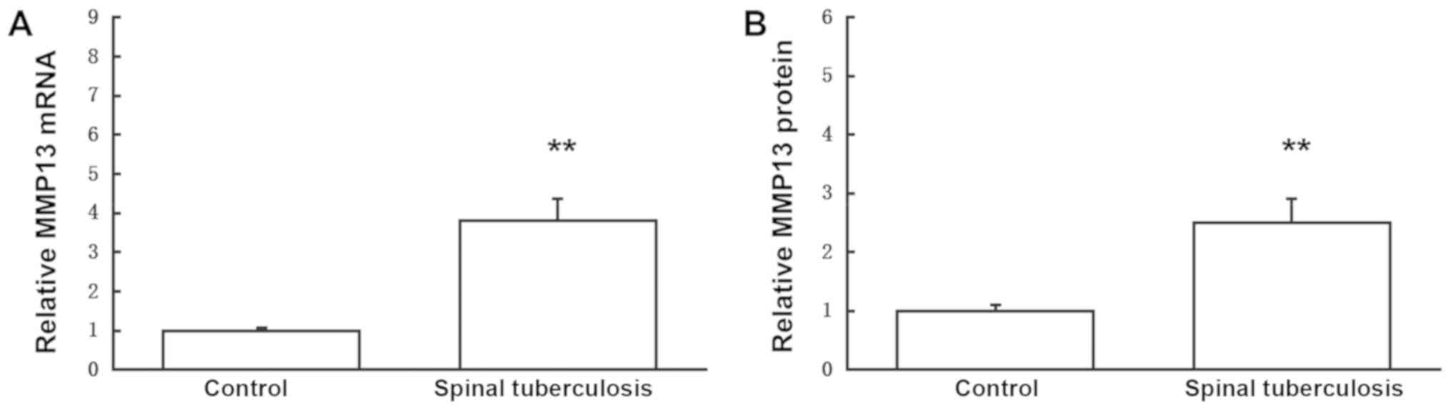
MMP13 mRNA and protein expression in the
intervertebral disc of patients with spinal tuberculosis were first
investigated by RT-qPCR and western blot analysis, respectively.
Results suggested that, compared with the control group, MMP13 mRNA
and protein expression in the intervertebral disc were
significantly increased in patients with spinal tuberculosis
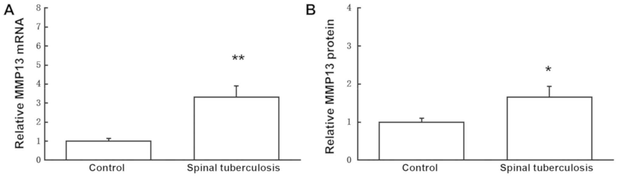
(P<0.01; Fig. 1). To investigate
MMP13 mRNA and protein levels in the serum of patients with spinal
tuberculosis, RT-qPCR and ELISA were performed, respectively. The
results suggested that, compared with the control group, serum
MMP13 mRNA and protein levels were significantly increased in
patients with spinal tuberculosis (P<0.05; Fig. 2). These results demonstrated that
MMP13 may serve a regulatory role in the pathogenesis of spinal
tuberculosis-induced intervertebral disc destruction.
miR-155 expression in patients with
spinal tuberculosis
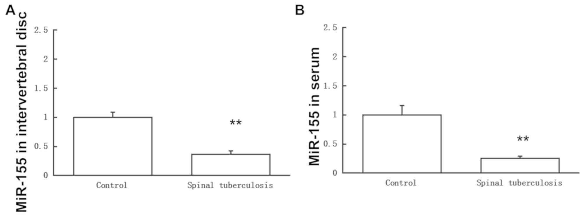
Based on the bioinformatics analysis, miR-155 was
predicted as an upstream regulator for MMP13 (Fig. 3). To investigate miR-155 expression
in the intervertebral disc and levels in the serum in patients with
spinal tuberculosis, RT-qPCR was performed. The results suggested
that compared with the control group, miR-155 expression in all
samples was significantly decreased in patients with spinal
tuberculosis (P<0.05; Fig. 4).
Combined with the data on MMP13 expression in patients with spinal
tuberculosis, the results suggested that miR-155 may serve a
regulatory role in the spinal tuberculosis-induced intervertebral
disc destruction and potentially negatively regulates the
transcription levels of the target gene MMP13.
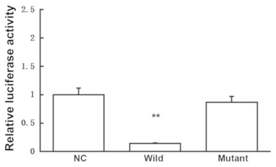
miR-155 and MMP13 interaction
To confirm interactions between miR-155 and MMP13, a
dual-luciferase reporter assay was performed. The results suggested
that compared with the negative control (NC) group (transfected
with the blank control), luciferase activity was significantly
reduced for cells cotransfected with agomiR-155 and pMIR-REPORT
plasmids (P<0.01), whereas no significant difference in
luciferase activity was observed in the mutant group (Fig. 5). The results suggested that miR-155
may directly interact with MMP13 to regulate gene expression.
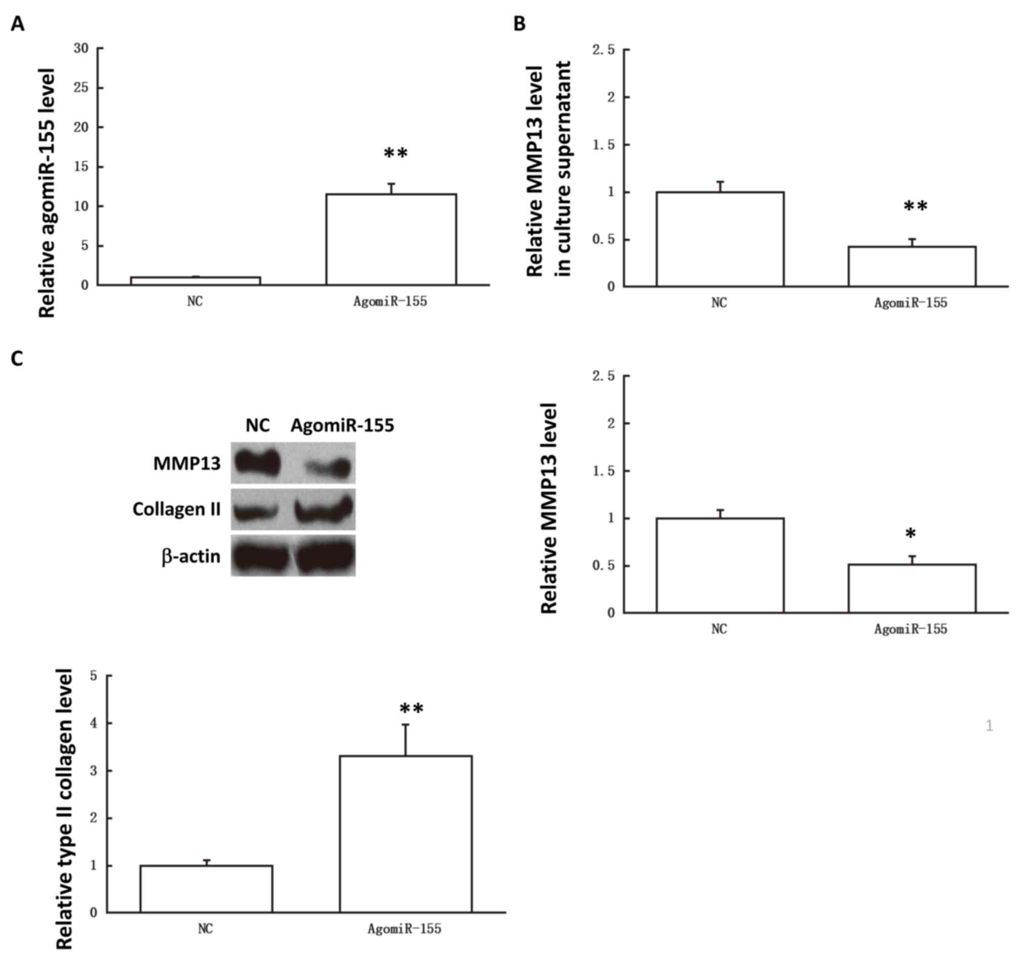
Effects of agomiR-155 on primary
annulus fibrosus cells
It has been demonstrated that agomir-155
significantly upregualtes miR-155 levels in cells (18). To further investigate interactions
between miR-155 and MMP13, the effects of agomiR-155 transfection
on primarily cultured annulus fibrosus cells were analyzed. The
results suggested that following transfection with agomiR-155,
MMP13 protein expression in the fibroblasts and MMP13 levels in the
cell culture supernatant were significantly reduced compared with
the NC (P<0.01; Fig. 6).
Corresponding expression levels of type II collagen were
significantly upregulated compared with the NC (P<0.05; Fig. 6C). The results suggested that
upregulated miR-155 expression may regulate MMP13 and affect type
II collagen expression.
Discussion
In the current study, MMP13 expression in patients
with spinal tuberculosis-induced intervertebral disc was
investigated and expression levels of miR-155, an upstream
regulator, were analyzed. The biological functions of miR-155 and
MMP13 were studied in vitro. The molecular mechanism through
which miR-155 may regulate protein expression affecting patients
with spinal tuberculosis-induced intervertebral disc destruction
was also analyzed. Herein, a negative regulatory association
between miR-155 and MMP-13 expression was proposed for the first
time. Following the reduced miR-155 expression in the spinal
tuberculosis tissue, the regulation of MMP13 expression was
reduced, resulting in changes in type II collagen expression.
The majority of the ECM of the NP is composed of
water, type II collagen (20%) and proteoglycan (50%, mainly
aggrecan) (11). Other minor
components include types III, IV, IX and XI collagen and small
proteoglycans (11). The majority of
the intervertebral disc is formed of ECM, including a variety of
collagen. At ≥10 years, majority of cells disappear and adult NP
cells represent a type of cartilage-like cells (19). Chondrocytes are characterized by the
expression of type II collagen (19). Type II collagen is one of the most
important collagen components in the intervertebral disc, also
known as cartilage collagen, present mainly in the cartilage and
disc nucleus (20). Sive et
al (21) described in
situ hybridization experiments that confirmed an increasing
degree of degeneration associated with decreasing type II collagen
expression in NP cells.
Under physiological and pathological conditions,
MMPs participate in various biological processes, including
embryonic and neovascular formation, wound healing, and
pathogenesis of cardiovascular diseases and cancers (21). ECMs account for ≥95% of the
intervertebral disc tissue, whereas other tissues rarely contain
ECM levels this high (7). Due to the
structural characteristics of the intervertebral disc, MMPs may
serve an important role in the pathogenesis and development of
spinal tuberculosis-induced intervertebral disc destruction
(7). It has been suggested that
MMP13 is expressed in the intervertebral disc in animals and humans
(22,23). Anderson et al (24) established a rabbit model for
intervertebral disc degeneration by damaging the annulus fibrosus
and reported that MMP13 levels in the intervertebral disc are
elevated (3–4 times). Roberts et al (25) compared patients with degenerated and
normal intervertebral disc using immunohistochemical methods and
demonstrated that compared with patients with normal intervertebral
disc, MMP13 is significantly increased in patients with degenerated
disc. In the current study, intervertebral disc lesions in patients
with spinal tuberculosis were investigated and, in accordance with
the previous findings, the results suggested that MMP13 expression
was significantly upregulated in tissues of patients with spinal
tuberculosis-induced intervertebral disc destruction, suggesting
that MMP13 may serve an important role in disease pathogenesis.
Regulation of mRNA transcription and expression is a
complex process involving multiple factors. To investigate MMP13
upstream regulators, the present study focused on recently
discovered endogenous, small, non-coding miRNAs, which exerted
negative regulatory roles on target mRNAs to inhibit gene
translation (26–28). These miRNAs are important regulators
in normal development, physiological and pathological processes
(29,30). Additionally, several miRNAs have been
recognized as the biomarkers for various diseases (29,30).
Based on a bioinformatics analysis, an upstream regulator of MMP13
was predicted and the results suggested miR-155. Wang et al
(31) have reported that
downregulated miR-155 expression may be the underlying mechanism
through which Fas mediates cell apoptosis in degenerated NP cells.
In addition, it has been demonstrated that in mouse models of liver
cancer, miR-155 expression and nuclear factor-kB activity are
significantly elevated (32). In
estrogen-receptor positive breast cancer cells, miR-155 has been
revealed to activate the mitogen-activated protein kinase signaling
pathway to regulate transcription levels of cytokines (33). Additionally, miR-155 has been
demonstrated to inhibit mothers against decapentaplegic homolog 2
expression to influence cellular responses to transforming growth
factor-β (34). In the current
study, the results suggested that miR-155 expression was
significantly downregulated in patients with spinal
tuberculosis-induced intervertebral disc destruction compared with
the normal control group. Due to various roles of miR-155 in
regulating the body immune system, inflammation and cellular
apoptosis (15), it is speculated
that in intervertebral disc destruction influenced by spinal
tuberculosis, miR-155 may affect the release and biological
function of MMP13 in the intervertebral disc. Results from the
dual-luciferase reporter assay suggested that MMP13 was a direct
target for miR-155. Additionally, in the primary culture of annulus
fibrosus cells, which were transfected with agomiR-155, MMP13
levels in the cells and cultured supernatant were significantly
reduced, whereas type II collagen expression was significantly
elevated. These results suggested that miR-155 negatively regulates
MMP13 expression to influence the intracellular expression of type
II collagen and further inducing lesions in intervertebral
discs.
A limitation of the present study is the limited
sample size and it should only be regarded as a preliminary study.
Further in-depth analyses are required to address the roles of
further MMPs, including MMP-8 and MMP-9. Furthermore, a majority of
the current study focused on the regulatory association between
miR-155 and MMP-13, whereas tissue inhibitors of metalloproteinases
were not investigated, which should be considered as a focus of
further studies. In addition, MMP-13 and/or miR-155 expression may
be studied in patients with spondylitis to complete the presented
conclusions.
In conclusion, the results suggested that there was
negative association between miR-155 and MMP13. In patients with
spinal tuberculosis-induced intervertebral disc destruction,
miR-155 expression was decreased, affecting its target MMP13 by
downregulating expression and further influencing type II collagen
expression. These findings may contribute to the understanding of
the roles of miR-155 and MMP13 in spinal tuberculosis-induced
intervertebral disc destruction.
Acknowledgements
Not applicable.
Funding
The current study was supported by the Scientific
and Technological Breakthroughs and New Product Trial Project from
the Liuzhou Science and Technology Bureau (grant no. 2017BH20308)
and the Key Research and Development Programs of the Department of
Science and Technology of Guangxi (grant no. Guike AB17129001).
Availability of data and materials
All data generated or analyzed during this study are
included in this published article.
Authors' contributions
CY designed the current study, performed the
experiments, and prepared the manuscript. ZS, JH, RW, GY, and DZ
contributed to the data collection and analysis, and manuscript
preparation. All authors read and approved the final
manuscript.
Ethics approval and consent to
participate
Written and informed consent was obtained from every
patient and the study was approved by the local Ethics Review Board
of the First Affiliated Hospital of Guangxi Medical University
(Nanning, China).
Patient consent for publication
Written and informed consent was obtained from every
patient.
Competing interests
The authors declare that they have no competing
interests.
References
|
1
|
Wang Y, Wang Q, Zhu R, Yang C, Chen Z, Bai
Y, Li M and Zhai X: Trends of spinal tuberculosis research
(1994-2015): A bibliometric study. Medicine (Baltimore).
95:e49232016. View Article : Google Scholar : PubMed/NCBI
|
|
2
|
Wang XW, Liu JJ, Wu QN, Wu SF and Hao DJ:
The in vitro and in vivo effects of microRNA-133a on intervertebral
disc destruction by targeting MMP9 in spinal tuberculosis. Life
Sci. 188:198–205. 2017. View Article : Google Scholar : PubMed/NCBI
|
|
3
|
Tsarouhas A, Soufla G, Katonis P, Pasku D,
Vakis A and Spandidos DA: Transcript levels of major MMPs and
ADAMTS-4 in relation to the clinicopathological profile of patients
with lumbar disc herniation. Eur Spine J. 20:781–790. 2011.
View Article : Google Scholar : PubMed/NCBI
|
|
4
|
Xiong C, Zhan X and Xiao Z: Transcript
levels of major MMPs and ADAMTS-4 in relation to the
clinicopathological profile of patients with tuberculous
intervertebral discs and healthy controls. Clin Biochem.
46:603–611. 2013. View Article : Google Scholar : PubMed/NCBI
|
|
5
|
Bonnans C, Chou J and Werb Z: Remodelling
the extracellular matrix in development and disease. Nat Rev Mol
Cell Biol. 15:786–801. 2014. View
Article : Google Scholar : PubMed/NCBI
|
|
6
|
Roughley PJ, Geng Y and Mort JS: The
non-aggregated aggrecan in the human intervertebral disc can arise
by a non-proteolytic mechanism. Eur Cell Mater. 28:129–136. 2014.
View Article : Google Scholar : PubMed/NCBI
|
|
7
|
Wang SZ, Rui YF, Lu J and Wang C: Cell and
molecular biology of intervertebral disc degeneration: Current
understanding and implications for potential therapeutic
strategies. Cell Prolif. 47:381–390. 2014. View Article : Google Scholar : PubMed/NCBI
|
|
8
|
Wang J, Ma J, Gu JH, Wang FY, Shang XS,
Tao HR and Wang X: Regulation of type II collagen, matrix
metalloproteinase-13 and cell proliferation by interleukin-1β is
mediated by curcumin via inhibition of NF-κB signaling in rat
chondrocytes. Mol Med Rep. 16:1837–1845. 2017. View Article : Google Scholar : PubMed/NCBI
|
|
9
|
Guo Y, Li W, Qin J, Lu C and Fan W:
Kaposi's sarcoma-associated herpesvirus (KSHV)-encoded microRNAs
promote matrix metalloproteinases (MMPs) expression and
pro-angiogenic cytokine secretion in endothelial cells. J Med
Virol. 89:1274–1280. 2017. View Article : Google Scholar : PubMed/NCBI
|
|
10
|
Schneider A, Victoria B, Lopez YN,
Suchorska W, Barczak W, Sobecka A, Golusinski W, Masternak MM and
Golusinski P: Tissue and serum microRNA profile of oral squamous
cell carcinoma patients. Sci Rep. 8:6752018. View Article : Google Scholar : PubMed/NCBI
|
|
11
|
Eleme K, Taner SB, Onfelt B, Collinson LM,
McCann FE, Chalupny NJ, Cosman D, Hopkins C, Magee AI and Davis DM:
Cell surface organization of stress-inducible proteins ULBP and
MICA that stimulate human NK cells and T cells via NKG2D. J Exp
Med. 199:1005–1010. 2004. View Article : Google Scholar : PubMed/NCBI
|
|
12
|
Iliopoulos D, Kavousanaki M, Ioannou M,
Boumpas D and Verginis P: The negative costimulatory molecule PD-1
modulates the balance between immunity and tolerance via miR-21.
Eur J Immunol. 41:1754–1763. 2011. View Article : Google Scholar : PubMed/NCBI
|
|
13
|
Bird L: Immune regulation: MicroRNAs keep
microglia quiet. Nat Rev Immunol. 11:762011. View Article : Google Scholar : PubMed/NCBI
|
|
14
|
Amin I, Patil BL, Briddon RW, Mansoor S
and Fauquet CM: A common set of developmental miRNAs are
upregulated in Nicotiana benthamiana by diverse begomoviruses.
Virol J. 8:1432011. View Article : Google Scholar : PubMed/NCBI
|
|
15
|
Jordan SD, Krüger M, Willmes DM, Redemann
N, Wunderlich FT, Brönneke HS, Merkwirth C, Kashkar H, Olkkonen VM,
Böttger T, et al: Obesity-induced overexpression of miRNA-143
inhibits insulin-stimulated AKT activation and impairs glucose
metabolism. Nat Cell Biol. 13:434–446. 2011. View Article : Google Scholar : PubMed/NCBI
|
|
16
|
Tili E, Michaille JJ, Wernicke D, Alder H,
Costinean S, Volinia S and Croce CM: Mutator activity induced by
microRNA-155 (miR-155) links inflammation and cancer. Proc Natl
Acad Sci USA. 108:4908–4913. 2011. View Article : Google Scholar : PubMed/NCBI
|
|
17
|
Livak KJ and Schmittgen TD: Analysis of
relative gene expression data using real-time quantitative PCR and
the 2(-Delta Delta C(T)) method. Methods. 25:402–408. 2001.
View Article : Google Scholar : PubMed/NCBI
|
|
18
|
Chen X, Gu S, Chen BF, Shen WL, Yin Z, Xu
GW, Hu JJ, Zhu T, Li G, Wan C, et al: Nanoparticle delivery of
stable miR-199a-5p agomir improves the osteogenesis of human
mesenchymal stem cells via the HIF1a pathway. Biomaterials.
53:239–250. 2015. View Article : Google Scholar : PubMed/NCBI
|
|
19
|
Specchia N, Pagnotta A, Toesca A and Greco
F: Cytokines and growth factors in the protruded intervertebral
disc of the lumbar spine. Eur Spine J. 11:145–151. 2002. View Article : Google Scholar : PubMed/NCBI
|
|
20
|
Cauci S, Viganò M, de Girolamo L, De Luca
P, Perucca Orfei C, Banfi G, Lombardi G, Brayda-Bruno M and
Colombini A: High levels of circulating type II collagen
degradation marker (CTx-II) are associated with specific VDR
polymorphisms in patients with adult vertebral osteochondrosis. Int
J Mol Sci. 18:E20732017. View Article : Google Scholar : PubMed/NCBI
|
|
21
|
Sive JI, Baird P, Jeziorsk M, Watkins A,
Hoyland JA and Freemont AJ: Expression of chondrocyte markers by
cells of normal and degenerate intervertebral discs. Mol Pathol.
55:91–97. 2002. View Article : Google Scholar : PubMed/NCBI
|
|
22
|
Loreto C, Leonardi R, Musumeci G, Pannone
G and Castorina S: An ex vivo study on immunohistochemical
localization of MMP-7 and MMP-9 in temporomandibular joint discs
with internal derangement. Eur J Histochem. 57:e122013. View Article : Google Scholar : PubMed/NCBI
|
|
23
|
Loreto C, Musumeci G, Castorina A, Loreto
C and Martinez G: Degenerative disc disease of herniated
intervertebral discs is associated with extracellular matrix
remodeling, vimentin-positive cells and cell death. Ann Anat.
193:156–62. 2011. View Article : Google Scholar : PubMed/NCBI
|
|
24
|
Anderson DG, Izzo MW, Hall DJ, Vaccaro AR,
Hilibrand A, Arnold W, Tuan RS and Albert TJ: Comparative gene
expression profiling of normal and degenerative discs: Analysis of
a rabbit annular laceration model. Spine (Phila Pa 1976).
27:1291–1296. 2002. View Article : Google Scholar : PubMed/NCBI
|
|
25
|
Roberts S, Caterson B, Menage J, Evans EH,
Jaffray DC and Eisenstein SM: Matrix metalloproteinases and
aggrecanase: Their role in disorders of the human intervertebral
disc. Spine (Phila Pa 1976). 25:3005–3013. 2000. View Article : Google Scholar : PubMed/NCBI
|
|
26
|
Inukai S, Pincus Z, de Lencastre A and
Slack FJ: A microRNA feedback loop regulates global microRNA
abundance during aging. RNA. 24:159–172. 2018. View Article : Google Scholar : PubMed/NCBI
|
|
27
|
Williams AE, Moschos SA, Perry MM, Barnes
PJ and Lindsay MA: Maternally imprinted microRNAs are
differentially expressed during mouse and human lung development.
Dev Dyn. 236:572–580. 2007. View Article : Google Scholar : PubMed/NCBI
|
|
28
|
Leonardi R, Loreto C, Barbato E,
Caltabiano R, Lombardo C, Musumeci G and Lo Muzio L: MMP-13
(collagenase 3) localization in human temporomandibular joint discs
with internal derangement. Acta Histochem. 110:314–318. 2008.
View Article : Google Scholar : PubMed/NCBI
|
|
29
|
Paliouras AR, Monteverde T and Garofalo M:
Oncogene-induced regulation of microRNA expression: Implications
for cancer initiation, progression and therapy. Cancer Lett.
421:152–160. 2018. View Article : Google Scholar : PubMed/NCBI
|
|
30
|
Lv ZC, Fan YS, Chen HB and Zhao DW:
Investigation of microRNA-155 as a serum diagnostic and prognostic
biomarker for colorectal cancer. Tumour Biol. 36:1619–1625. 2015.
View Article : Google Scholar : PubMed/NCBI
|
|
31
|
Wang HQ, Yu XD, Liu ZH, Cheng X, Samartzis
D, Jia LT, Wu SX, Huang J, Chen J and Luo ZJ: Deregulated miR-155
promotes Fas-mediated apoptosis in human intervertebral disc
degeneration by targeting FADD and caspase-3. J Pathol.
225:232–242. 2011. View Article : Google Scholar : PubMed/NCBI
|
|
32
|
Wang B, Majumder S, Nuovo G, Kutay H,
Volinia S, Patel T, Schmittgen TD, Croce C, Ghoshal K and Jacob ST:
Role of microRNA-155 at early stages of hepatocarcinogenesis
induced by choline-deficient and amino acid-defined diet in C57BL/6
mice. Hepatology. 50:1152–1161. 2009. View Article : Google Scholar : PubMed/NCBI
|
|
33
|
Martin EC, Krebs AE, Burks HE, Elliott S,
Baddoo M, Collins-Burow BM, Flemington EK and Burow ME: miR-155
induced transcriptome changes in the MCF-7 breast cancer cell line
leads to enhanced mitogen activated protein kinase signaling. Genes
Cancer. 5:353–364. 2014.PubMed/NCBI
|
|
34
|
Louafi F, Martinez-Nunez RT and
Sanchez-Elsner T: MicroRNA-155 targets SMAD2 and modulates the
response of macrophages to transforming growth factor-{beta}. J
Biol Chem. 285:41328–41336. 2010. View Article : Google Scholar : PubMed/NCBI
|