Introduction
Stem cells are recognized as potential tools for use
in the development of novel therapeutic strategies (1). All stem cells share two common
characteristics, long-term self-renewal and plasticity and the
ability to differentiate into specialized cells, which have
therapeutic potential for the repair of different tissues and
organs (2). Bone marrow-derived stem
cells (BMSCs) are widely used for cell therapy in regenerative
medicine (3). In a previous study,
the age-associated osteogenic potential of BMSCs was investigated,
and a decrease in osteogenic differentiation potential was observed
during aging in humans (4). However,
another study demonstrated that there were no age-associated
changes in the osteoblastic differentiation potential or the steady
state levels of messenger RNA (mRNA) of osteogenic gene markers
(5). A previous study demonstrated
that the capacity of BMSCs to form bone in vivo was
maintained with age, which suggests that the observed
senescence-associated decrease in bone formation may be due to a
defect in bone microenvironment (6).
A previous study demonstrated that the expression levels of
bone-associated genes under osteogenic culture conditions were
similar in BMSCs isolated from females compared with BMSCs isolated
from males (7). However, the number
of the colony-forming units which express alkaline phosphatase from
bone marrow decreased significantly with age for women, but not for
men (8). In addition, there was no
significant difference observed in in vitro osteogenic
activity in cultures of patient-derived mesenchymal stem cells
compared with that in normal donor cultures (9). BMSCs have the ability to undergo
adipogenic and chondrogenic differentiation (10). Few studies have investigated whether
age- and gender-associated differences in the adipogenic and
chondrogenic potential of BMSCs exist. The aim of the current study
was to investigate the effect of demographic factors on adipogenic
and chondrogenic differentiation in BMSC spheroids.
Materials and methods
Human bone marrow-derived stem
cells
Human bone marrow-derived mesenchymal stem cells
(Catholic MASTER Cells) were obtained from the Catholic Institute
of Cell Therapy (Seoul, South Korea). BMSC isolation and
characterization was performed, as previously reported (11). Tests performed by the Catholic
Institute of Cell Therapy confirmed high expression levels of CD73
and CD90 (>90% positive; data not shown). The current study was
approved by the Institutional Review Board of Seoul St. Mary's
Hospital (approval no. KC17SNSI0606). Written informed consent was
obtained from the participants as specified in the Declaration of
Helsinki. The methods used in this study were performed in
accordance with the relevant guidelines and regulations.
Cell culture
Human BMSCs were seeded in 24-well plates at a
density of 2×10⁴ cells/well and cultured in a-minimal
essential medium (Invitrogen; Thermo Fisher Scientific, Inc.,
Waltham, MA, USA) supplemented with 15% fetal bovine serum
(Invitrogen; Thermo Fisher Scientific, Inc.), 100 U/ml penicillin,
100 µg/ml streptomycin (Sigma-Aldrich; Merck KGaA, Darmstadt,
Germany), 200 mM L-glutamine (Sigma-Aldrich; Merck KGaA) and 10 mM
ascorbic acid 2-phosphate (Sigma-Aldrich; Merck KGaA) at 37°C.
Adipogenic differentiation
To determine the adipocyte differentiation potential
of BMSCs, isolated cells were cultured using a StemPro®
Adipogenesis Differentiation kit (Invitrogen; Thermo Fisher
Scientific, Inc.), according to the manufacturer's protocol.
Adipogenic induction medium and adipogenic maintenance medium were
supplied. On day 8 and 16 respectively, cells were rinsed twice
with phosphate buffered saline (PBS) and fixed with 4%
paraformaldehyde (Biosesang Inc., Seongnam, Korea) at 20°C for 5
min. Cells were subsequently washed with distilled water, rinsed
with 60% isopropanol and covered with oil red O solution
(Sigma-Aldrich; Merck KGaA) for 10 min (12). Following incubation, cells were
rinsed in 60% isopropanol and washed with distilled water. Cell
morphology was observed using an inverted microscope (Leica DM IRM;
Leica Microsystems GmbH, Wetzlar, Germany) and relative values of
adipogenesis were determined by measuring the intensity of oil red
O staining using ImageJ (version 1.8.0, National Institutes of
Health, Bethesda, MD, USA) analysis software (magnification, ×100
for day 8; ×200 for day 16).
Following induction of adipocyte differentiation,
1.5×10⁵ cells were collected on day 8 and 16,
respectively, and incubated with specific fluorescein
isothiocyanate-conjugated mouse monoclonal human CD44 antibody
(dilution 1:200, cat. no. 11-0441-81; Invitrogen; Thermo Fisher
Scientific, Inc.) at 20°C for 20 min. Cells were analyzed using a
flow cytometer (FACSCanto II; BD Biosciences, San Jose, CA, USA),
and FACSDiva software (Version 8.0.1, BD Biosciences). Human BD Fc
Block™ (564219, BD Biosciences) was used as a blocking reagent and
1% bovine serum albumin/PBS served as the washing reagent.
Chondrogenic differentiation
To determine the chondrogenic potential of BMSCs,
isolated cells were cultured using a StemPro®
Chondrogenesis Differentiation kit (Invitrogen; Thermo Fisher
Scientific, Inc.), according to the manufacturer's protocol. On day
8 and 16 respectively, cells were rinsed twice with PBS and fixed
with 4% paraformaldehyde at 20°C for 5 min. Cells were subsequently
washed with distilled water, rinsed with 60% isopropanol and
covered with Alcian blue solution (Sigma-Aldrich; Merck KGaA) for
10 min (13). Following incubation,
cells were rinsed in 3% acetic acid and washed with distilled
water. Cell morphology was observed using an inverted microscope
and relative values of chondrogenesis were determined by measuring
the intensity of Alcian blue staining using ImageJ analysis
software (magnification, ×100 for day 8; ×200 for day 16).
Statistical analysis
Data are presented as the mean ± standard deviation.
All analyses were performed using SPSS software (version 12.0; SPSS
Inc., Chicago, IL, USA). A test for normality was performed, and
all statistical comparisons between groups were determined using
Student's t-test or one-way analysis of variance with Tukey's
post-hoc test. P<0.05 was considered to indicate a statistically
significant difference.
Results
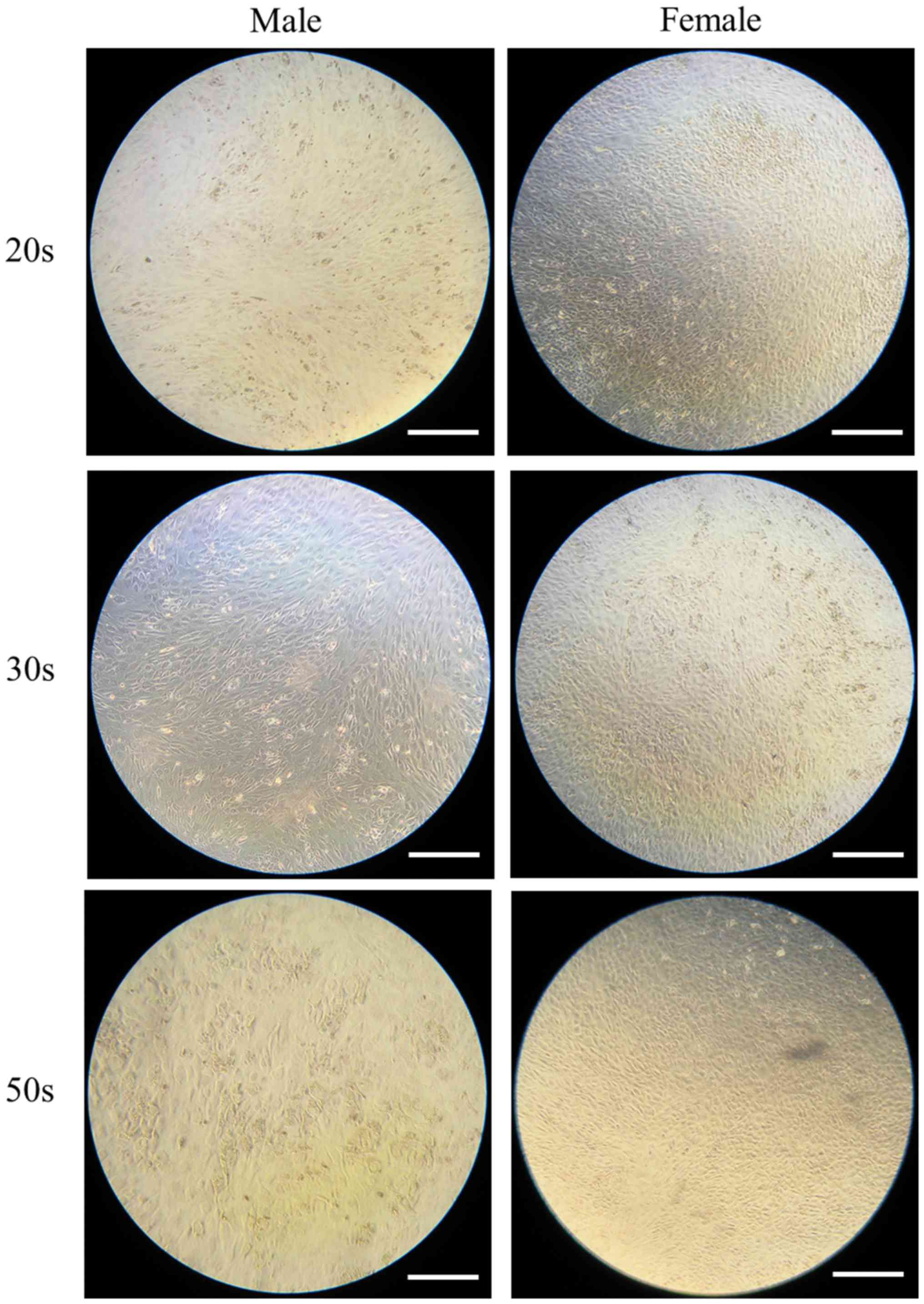
Morphologic evaluation of adipogenic
differentiation
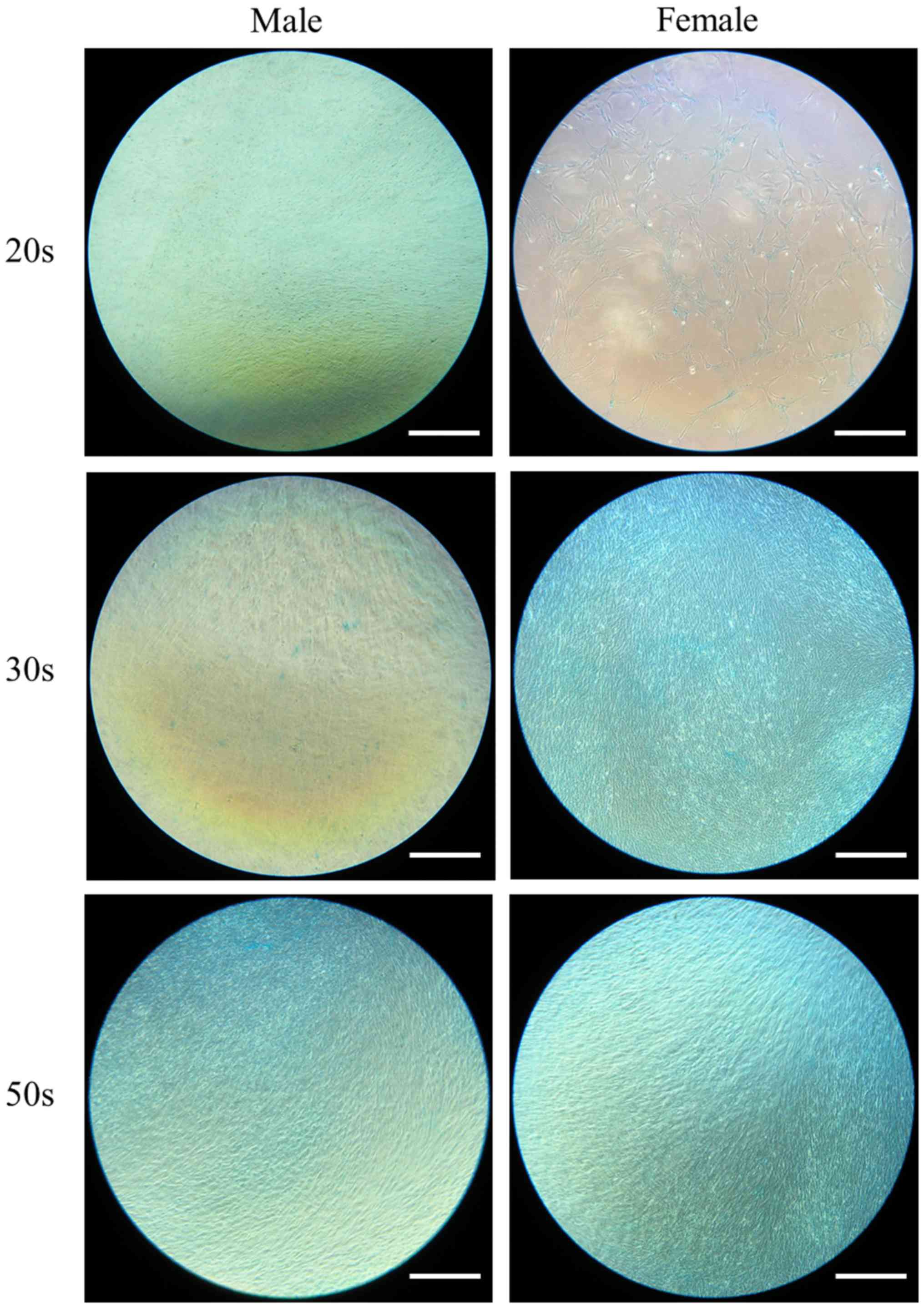
The ability of the isolated BMSCs to differentiate
into adipocytes was examined in BMSCs isolated from male and female
participants in their 20s, 30s and 50s and grown in adipogenic
media. Following eight days of growth in adipogenic media, observed
changes in cell morphology were very similar among the different
age groups. In addition, similar changes in oil red O staining
intensity were also observed (Fig.
1). Similarly, following 16 days of growth in adipogenic media,
observed changes in cell morphology were very similar among the
different age groups. In addition, similar changes in oil red O
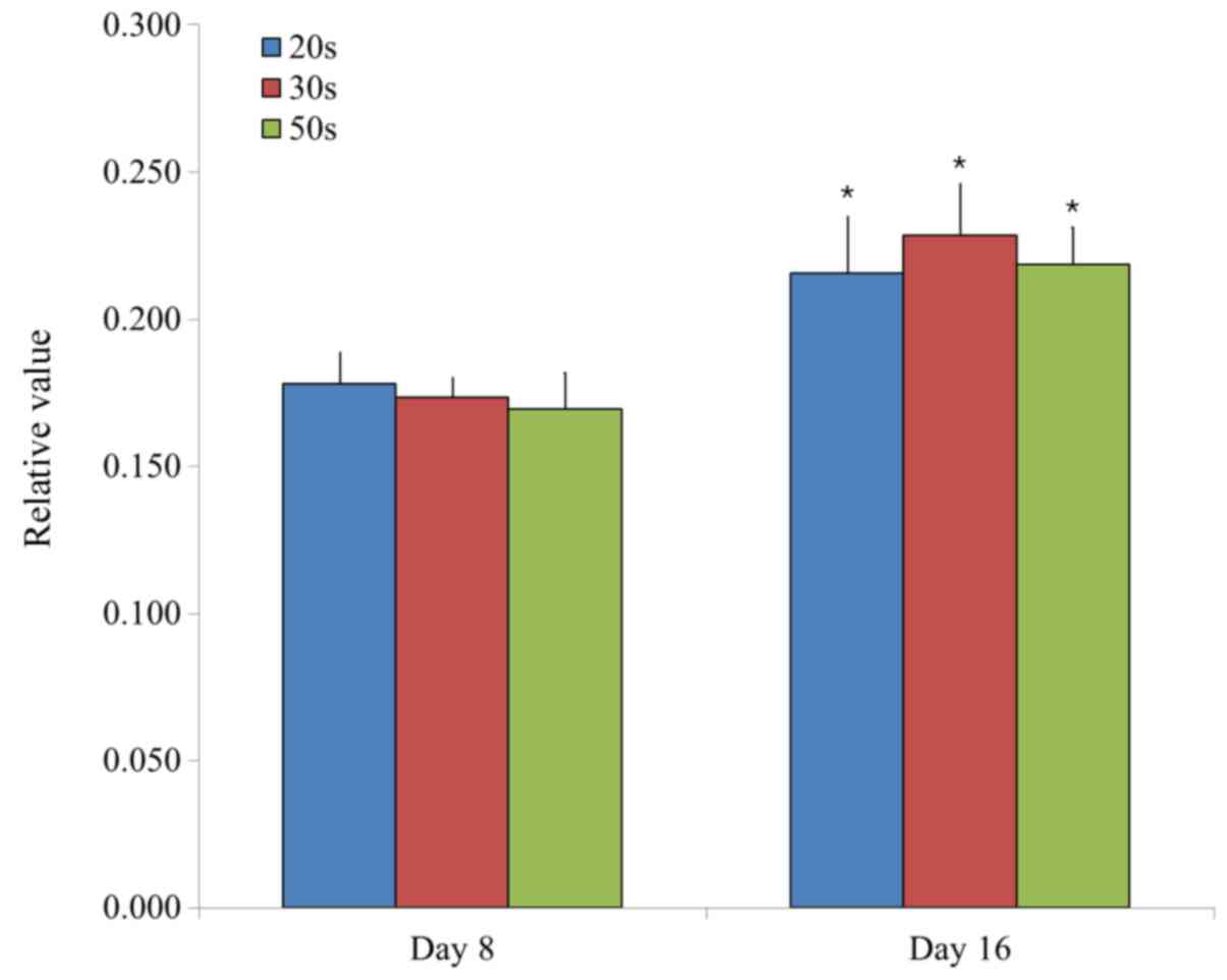
staining intensity were also observed (Fig. 2). In general, oil red O staining
intensity was significantly increased at day 16 compared with day 8
in each group (P<0.05; Fig.
3).
Adipogenesis was evaluated by measuring the relative
intensity of oil red O staining in BMSCs. Relative values of
adipogenesis were 0.178±0.010, 0.173±0.007 and 0.170±0.012 for the
20s, 30s and 50s age groups on day 8, respectively, whilst the
relative values of adipogenesis were 0.216±0.019, 0.228±0.017 and
0.219±0.013 for the 20s, 30s, and 50s age groups on day 16,
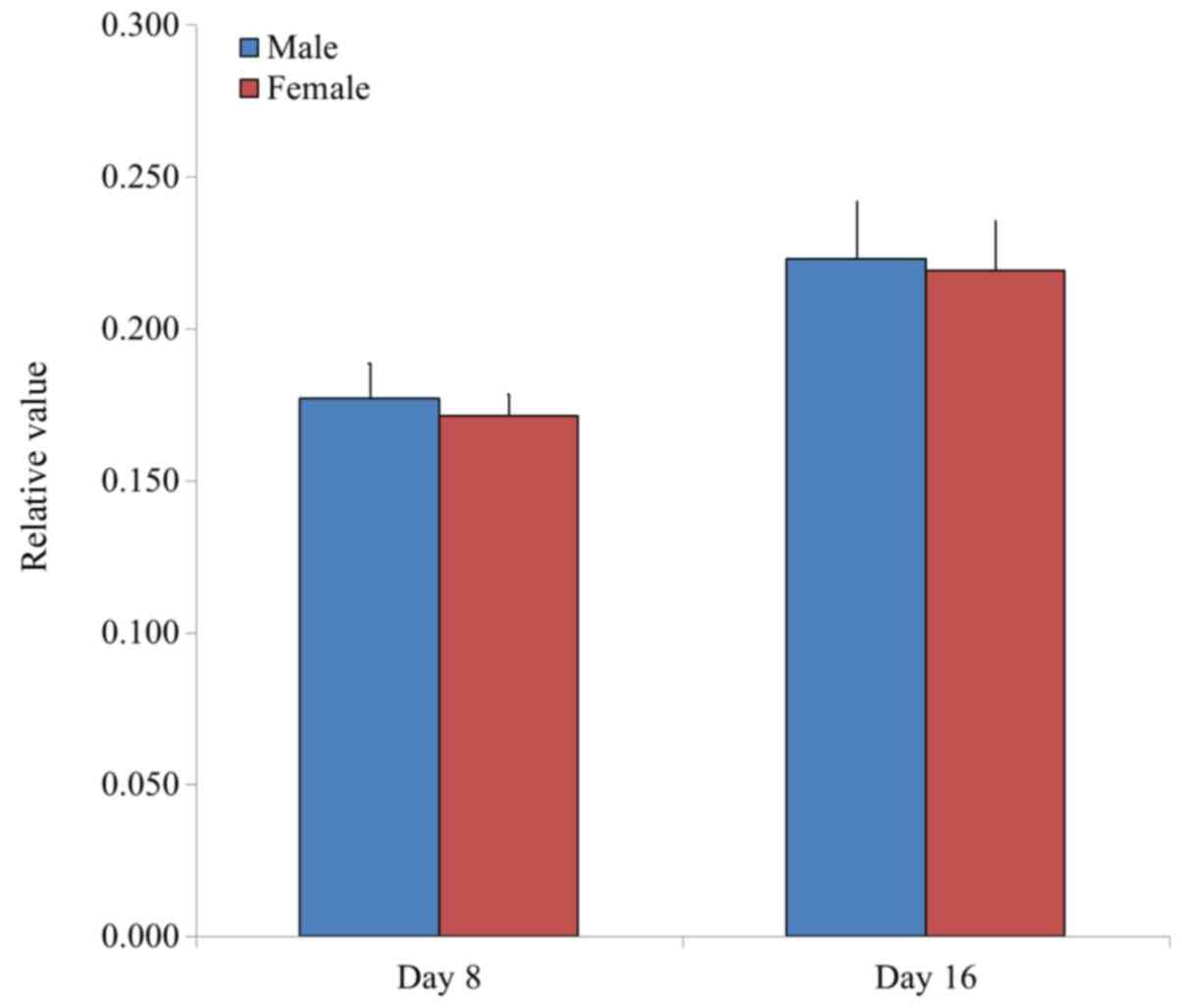
respectively (Fig. 3). Furthermore,
the relative values of adipogenesis were 0.177±0.011 and
0.171±0.007 for male and female groups on day 8, respectively,
whilst the relative values of adipogenesis were 0.223±0.019 and
0.219±0.016 for male and female groups on day 16, respectively
(Fig. 4).
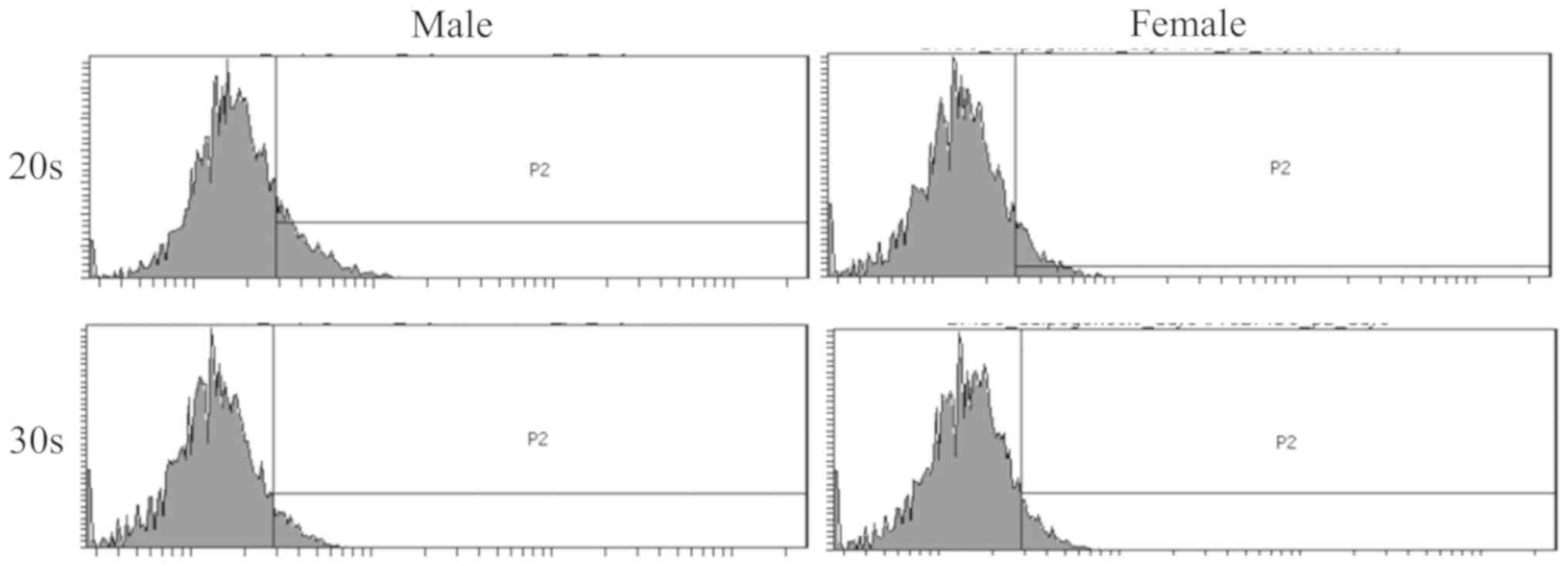
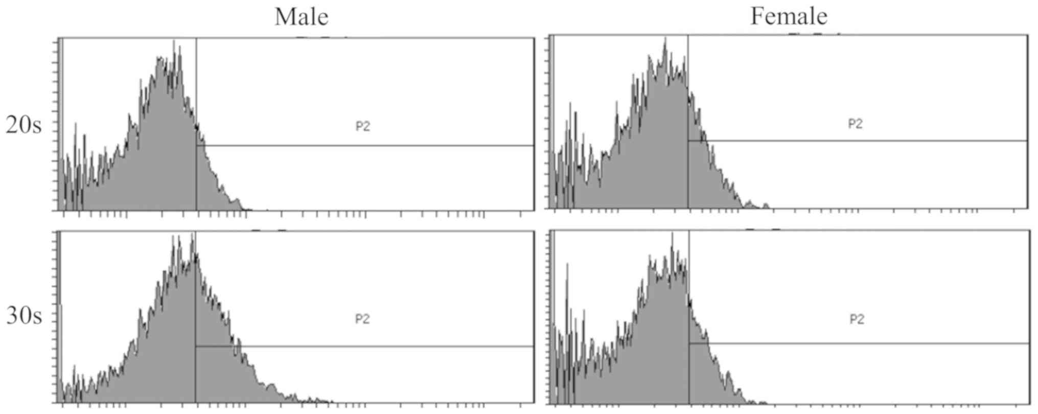
To determine the phenotype of isolated BMSCs
following growth in adipogenic media, expression of the CD44
surface marker was examined in BMSCs isolated from male and female
participants in their 20s and 30s. CD44 surface marker expression
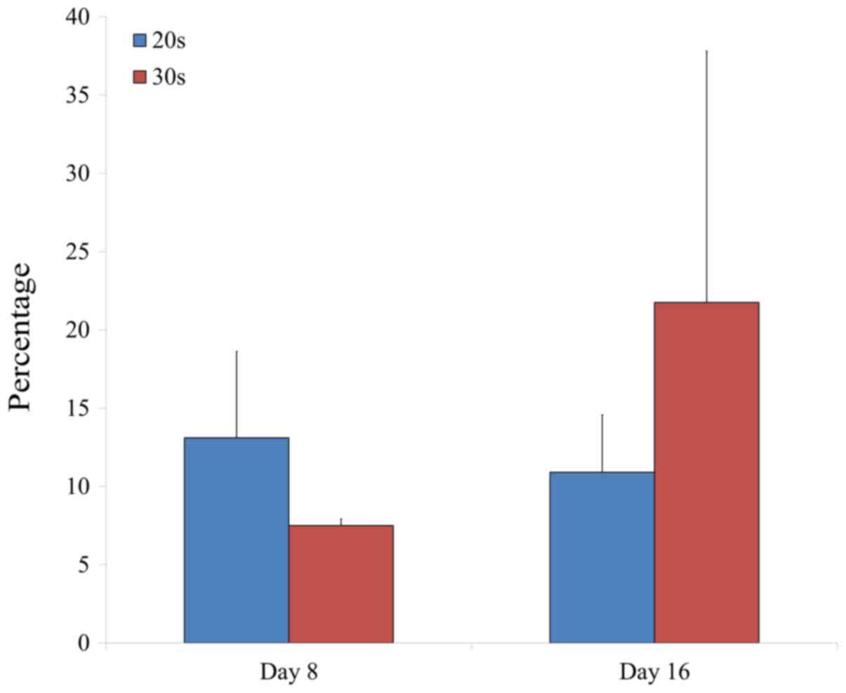
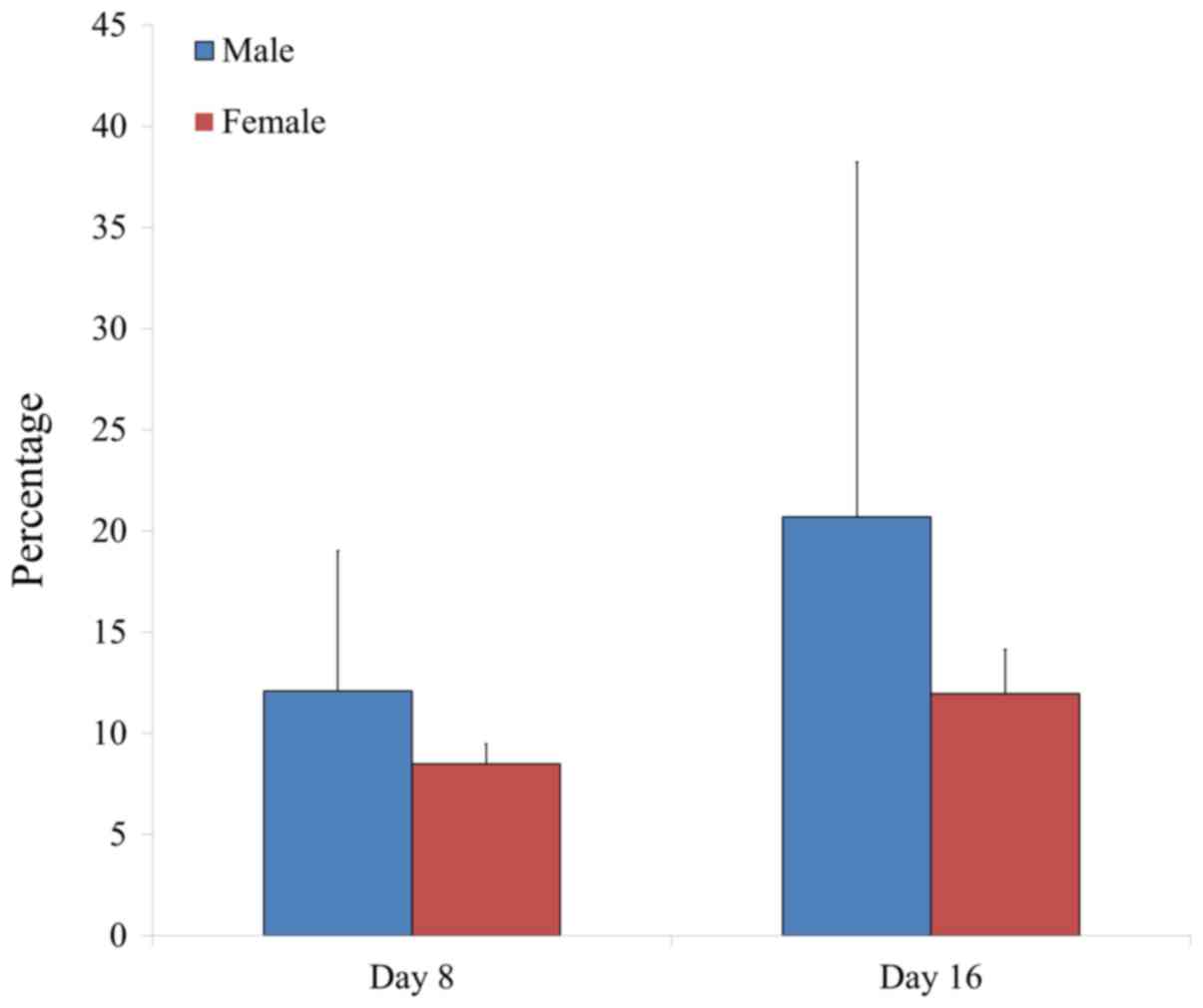
was analyzed on day 8 and 16 by flow cytometry (Figs. 5 and 6). The percentage of CD44 expression was
13.1±5.5 and 7.5±0.4% for the 20s and 30s age groups on day 8,
respectively, whilst the percentage of CD44 expression was 10.9±3.7
and 21.8±16.1% for the 20s and 30s age groups on day 16,
respectively (Fig. 7). Furthermore,
the percentage of CD44 expression was 12.1±6.9 and 8.5±1.0% for
male and female groups on day 8, respectively, whilst the
percentage of CD44 expression was 20.7±17.5 and 12.0±2.2% for male
and female groups on day 16, respectively (Fig. 8).
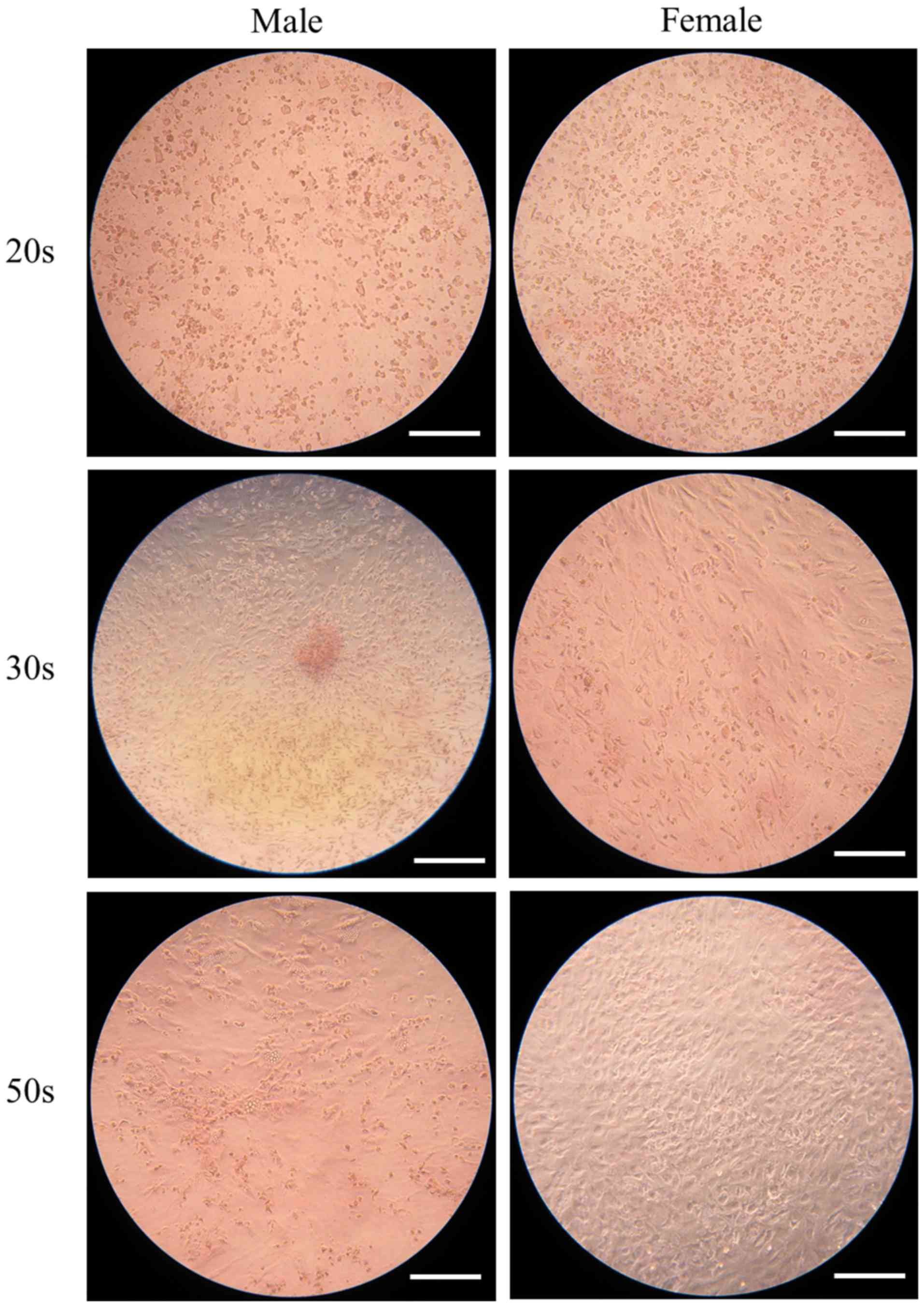
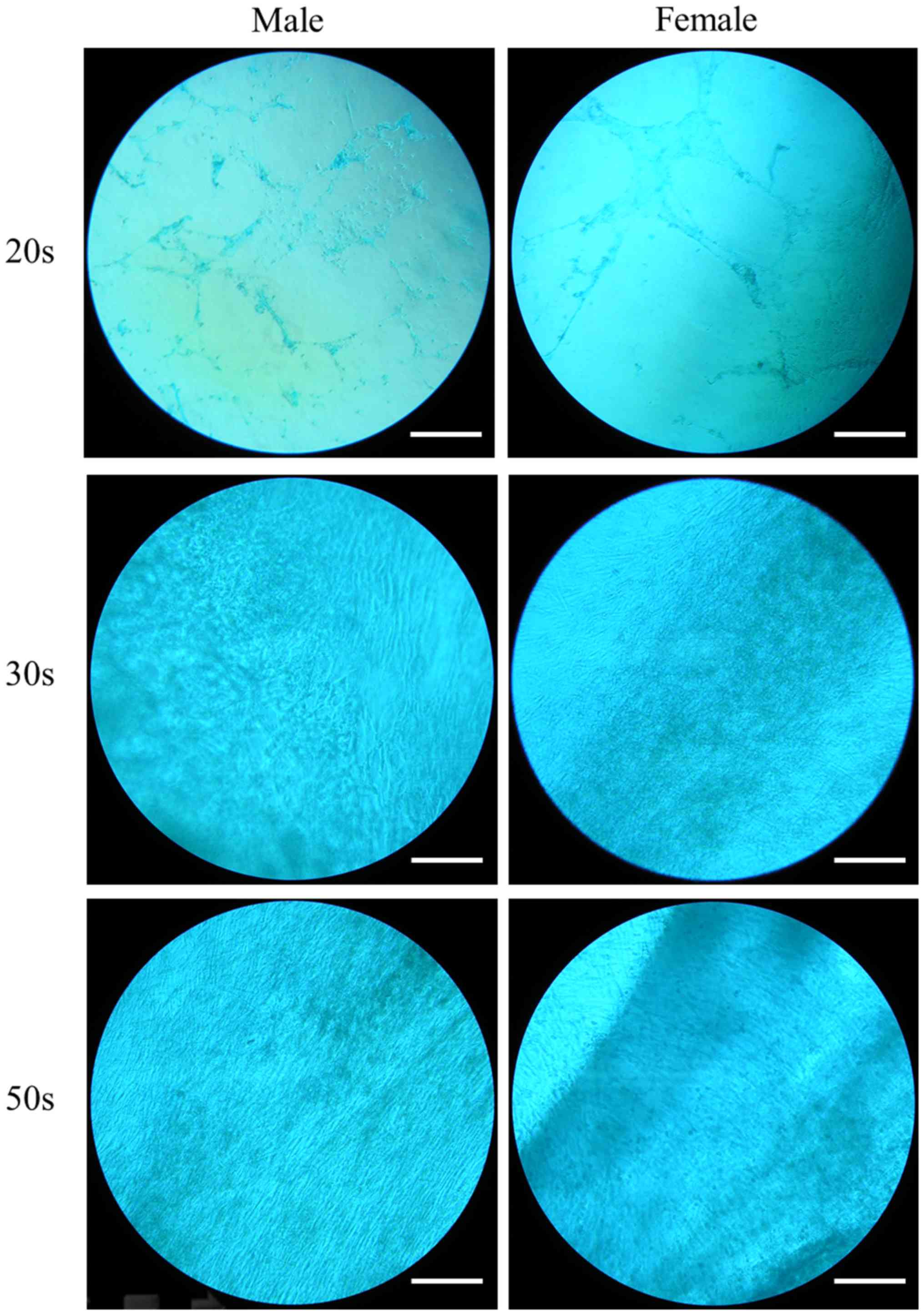
Morphologic evaluation of chondrogenic
differentiation
The ability of the isolated BMSCs to differentiate
into chondrocytes was examined in BMSCs isolated from male and
female participants in their 20s, 30s and 50s and grown in
chondrogenic media. Following eight days of growth in chondrogenic
media, observed changes in cell morphology were very similar among
the different age groups. In addition, similar changes in Alcian
blue staining intensity were also observed (Fig. 9). Following 16 days of growth in
chondrogenic media, observed changes in cell morphology were very
similar among the different age groups. In addition, similar
changes in Alcian blue staining intensity were also observed
(Fig. 10). In general, Alcian blue
staining intensity was increased at day 16 compared with day 8
(P<0.05; Fig. 11).
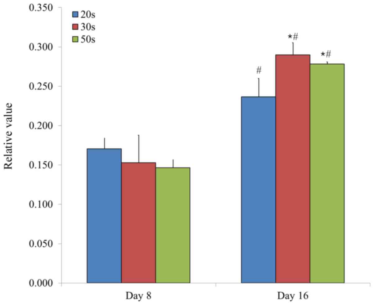
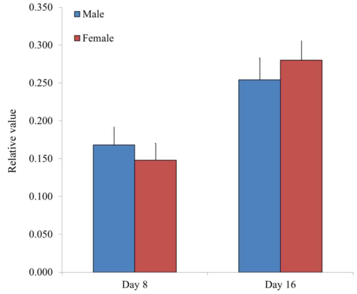
Chondrogenesis was evaluated by measuring the
relative intensity of Alcian blue staining in BMSCs. Relative
values of chondrogenesis were 0.171±0.013, 0.153±0.035 and
0.147±0.010 for the 20s, 30s and 50s age groups on day 8,
respectively, whilst the relative values of chondrogenesis were
0.237±0.023, 0.290±0.016 and 0.278±0.003, for the 20s, 30s and 50s
age groups on day 16, respectively (P<0.05, 50s and 30s vs. 20s
group at day 16; Fig. 11).
Furthermore, the relative values of chondrogenesis were 0.168±0.023
and 0.148±0.022 for male and female groups on day 8, respectively,
whilst the relative values of chondrogenesis were 0.254±0.029 and
0.280±0.026 for male and female groups on day 16, respectively
(Fig. 12).
Discussion
The current study investigated the effect of
demographic factors on adipogenic and chondrogenic differentiation
potential in BMSC spheroids. A previous study demonstrated that the
expression levels of adipocyte-associated genes significantly
increased in BMSCs isolated from female mice compared with BMSCs
isolated from male mice (7).
Similarly, gene expression analysis by reverse
transcription-quantitative polymerase chain reaction examining
aging and gender-associated effects on BMSC differentiation,
identified an increase in adipogenesis with aging (14). In addition, a previous study
demonstrated that adipogenesis is enhanced in BMSCs isolated from
older female mice (15).
Furthermore, a reduced chondrogenic and adipogenic differentiation
potential was observed in BMSCs isolated from patients with
advanced osteoarthritis (9).
However, a previous study demonstrated that there were no
age-associated changes in the adipocytic colony formation or the
steady-state level mRNA expression of adipogenic gene markers in
marrow stromal cells isolated from patients with osteoporosis
compared with age-matched healthy controls (5). In the current study, no significant
differences in the androgenic differentiation potential of BMSCs
isolated from participants in the 20s, 30s and 50s age groups were
observed. Similarly, no gender-associated effects were observed in
the androgenic differentiation potential of isolated BMSCs.
A previous study demonstrated histological,
immunohistochemical and molecular evidence for the in vitro
chondrogenic differentiation of bone marrow-derived mesenchymal
progenitor cells (16). In addition,
when bone marrow-derived cells were passaged in a monolayer culture
as many as 20 times, cells maintained the chondrogenic
differentiation potential after each passage (17). There was a significant reduction in
in vitro chondrogenic activity in cultures of
patient-derived mesenchymal stem cells (MSCs) from patients with
advanced osteoarthritis compared with normal cultures (9). A previous study demonstrated that the
chondrogenic potential of human adult MSCs is independent of age or
osteoarthritis etiology (18).
However, the chondrogenic potential of BMSCs decreased with age in
rat models (19). In the current
study, no significant differences in the chondrogenic
differentiation potential of BMSCs isolated from participants in
the 20s, 30s and 50s age groups were observed on day 8, however on
day 16 there was a significant difference in the chondrogenic
differentiation potential of BMSCs isolated from participants in
the 30s and 50s age groups, compared with those in the 20s age
group. In addition, no gender-associated effects were observed in
the chondrogenic differentiation potential of isolated BMSCs.
The effects of aging and gender on adipogenic and
chondrogenic differentiation potential is varied, and this may be
due to changes in the model system used, the passage and stage of
differentiation, and the culture period (20,21).
Investigating the effects of aging have proved contradictory due to
the relative narrow age range of participants in a number of the
studies (22).
The differentiation potential of MSCs varies
depending on their origin. In a previous study, adipose
tissue-derived MSCs isolated from young female participants were
revealed to be more resistant to senescence under in vitro
culture conditions when compared with those isolated from older
patients (23). The effect of gender
and anatomical region on induction of osteogenic differentiation of
human adipose-derived stem cells was examined, which revealed that
male-derived cells differentiated faster and more efficiently
compared with female-derived cells (24). In addition, the influence of gender
on the chondrogenic potential of muscle-derived stem cells (MDSCs)
was examined, which revealed that male MDSCs displayed a higher
chondrogenic differentiation with increased cartilage regeneration
potential (25). Furthermore, a
decrease in the chondrogenic potential of periosteum with aging was
observed in a rabbit model (26).
However, the chondrogenic potential of periosteum-derived stem
cells varied between donor sites, an effect potentially caused by
differences in total cell count (21).
The current study demonstrated no significant
differences in the adipogenic and chondrogenic differentiation
potential of BMSCs derived from healthy male donors compared with
healthy female donors. Similarly, there were no significant
differences in the adipogenic and chondrogenic differentiation
among the different age groups.
Acknowledgements
Not applicable.
Funding
The current study was partially supported by the
Research Fund of Seoul St. Mary's Hospital, The Catholic University
of Korea. The current study was also supported by the Basic Science
Research Program through the National Research Foundation of Korea,
funded by a grant from the Ministry of Science, Information and
Communication Technology & Future Planning (grant no.
NRF-2017R1A1A1A05001307).
Availability of data and materials
All datasets used and/or analyzed during the current
study are available from the corresponding author upon reasonable
request.
Authors' contributions
HL, SM and JP designed the study. HL, SM and JP
collected and analyzed data. HL, SM and JP performed the
experiments. HL, SM and JP wrote the manuscript. All authors read
and approved the manuscript.
Ethics approval and consent to
participate
The Catholic MASTER cells supplied by the Catholic
Institute of Cell Therapy (Seoul, Korea) were derived from human
bone marrow donated by healthy donors after informed consent. The
current study was approved by the Institutional Review Board of
Seoul St. Mary's Hospital (Seoul, Republic of Korea). Written
informed consent was obtained from the participants as specified in
the Declaration of Helsinki.
Patient consent for publication
Not applicable.
Competing interests
The authors confirm that they have no competing
interests.
References
|
1
|
Tögel F and Westenfelder C: Adult bone
marrow-derived stem cells for organ regeneration and repair. Dev
Dyn. 236:3321–3331. 2007. View Article : Google Scholar : PubMed/NCBI
|
|
2
|
Totey S, Totey S and Pal R and Pal R:
Adult stem cells: A clinical update. J Stem Cells. 4:105–121.
2009.PubMed/NCBI
|
|
3
|
Maria OM, Khosravi R, Mezey E and Tran SD:
Cells from bone marrow that evolve into oral tissues and their
clinical applications. Oral Dis. 13:11–16. 2007. View Article : Google Scholar : PubMed/NCBI
|
|
4
|
D'Ippolito G, Schiller PC, Ricordi C, Roos
BA and Howard GA: Age-related osteogenic potential of mesenchymal
stromal stem cells from human vertebral bone marrow. J Bone Miner
Res. 14:1115–1122. 1999. View Article : Google Scholar : PubMed/NCBI
|
|
5
|
Justesen J, Stenderup K, Eriksen EF and
Kassem M: Maintenance of osteoblastic and adipocytic
differentiation potential with age and osteoporosis in human marrow
stromal cell cultures. Calcif Tissue Int. 71:36–44. 2002.
View Article : Google Scholar : PubMed/NCBI
|
|
6
|
Stenderup K, Rosada C, Justesen J,
Al-Soubky T, Dagnaes-Hansen F and Kassem M: Aged human bone marrow
stromal cells maintaining bone forming capacity in vivo evaluated
using an improved method of visualization. Biogerontology.
5:107–118. 2004. View Article : Google Scholar : PubMed/NCBI
|
|
7
|
Bragdon B, Burns R, Baker AH, Belkina AC,
Morgan EF, Denis GV, Gerstenfeld LC and Schlezinger JJ: Intrinsic
Sex-linked variations in osteogenic and adipogenic differentiation
potential of bone marrow multipotent stromal cells. J Cell Physiol.
230:296–307. 2015. View Article : Google Scholar : PubMed/NCBI
|
|
8
|
Muschler GF, Nitto H, Boehm CA and Easley
KA: Age- and gender-related changes in the cellularity of human
bone marrow and the prevalence of osteoblastic progenitors. J
Orthop Res. 19:117–125. 2001. View Article : Google Scholar : PubMed/NCBI
|
|
9
|
Murphy JM, Dixon K, Beck S, Fabian D,
Feldman A and Barry F: Reduced chondrogenic and adipogenic activity
of mesenchymal stem cells from patients with advanced
osteoarthritis. Arthritis Rheum. 46:704–713. 2002. View Article : Google Scholar : PubMed/NCBI
|
|
10
|
Zheng YH, Xiong W, Su K, Kuang SJ and
Zhang ZG: Multilineage differentiation of human bone marrow
mesenchymal stem cells in vitro and in vivo. Exp Ther Med.
5:1576–1580. 2013. View Article : Google Scholar : PubMed/NCBI
|
|
11
|
Jeong CH, Kim SM, Lim JY, Ryu CH, Jun JA
and Jeun SS: Mesenchymal stem cells expressing brain-derived
neurotrophic factor enhance endogenous neurogenesis in an ischemic
stroke model. Biomed Res Int. 2014:1291452014. View Article : Google Scholar : PubMed/NCBI
|
|
12
|
Lee JE, Kim BB, Ko Y, Jeong SH and Park
JB: Effects of Cimicifugae Rhizoma on the osteogenic and adipogenic
differentiation of stem cells. Exp Ther Med. 13:443–448. 2017.
View Article : Google Scholar : PubMed/NCBI
|
|
13
|
Park JB, Bae SS, Lee PW, Lee W, Park YH,
Kim H, Lee K and Kim I: Comparison of stem cells derived from
periosteum and bone marrow of jaw bone and long bone in rabbit
models. Tissue Eng Regen Med. 9:224–230. 2012. View Article : Google Scholar
|
|
14
|
Jiang Y, Mishima H, Sakai S, Liu YK,
Ohyabu Y and Uemura T: Gene expression analysis of major
lineage-defining factors in human bone marrow cells: Effect of
aging, gender and age-related disorders. J Orthop Res. 26:910–917.
2008. View Article : Google Scholar : PubMed/NCBI
|
|
15
|
Bolt AM, Grant MP, Wu TH, Flores Molina M,
Plourde D, Kelly AD, Negro Silva LF, Lemaire M, Schlezinger JJ,
Mwale F and Mann KK: Tungsten promotes sex-specific adipogenesis in
the bone by altering differentiation of bone marrow-resident
mesenchymal stromal cells. Toxicol Sci. 150:333–346. 2016.
View Article : Google Scholar : PubMed/NCBI
|
|
16
|
Johnstone B, Hering TM, Caplan AI,
Goldberg VM and Yoo JU: In vitro chondrogenesis of bone
marrow-derived mesenchymal progenitor cells. Exp Cell Res.
238:265–272. 1998. View Article : Google Scholar : PubMed/NCBI
|
|
17
|
Yoo JU, Barthel TS, Nishimura K, Solchaga
L, Caplan AI, Goldberg VM and Johnstone B: The chondrogenic
potential of human bone-marrow-derived mesenchymal progenitor
cells. J Bone Joint Surg Am. 80:1745–1757. 1998. View Article : Google Scholar : PubMed/NCBI
|
|
18
|
Scharstuhl A, Schewe B, Benz K, Gaissmaier
C, Buhring HJ and Stoop R: Chondrogenic potential of human adult
mesenchymal stem cells is independent of age or osteoarthritis
etiology. Stem Cells. 25:3244–3251. 2007. View Article : Google Scholar : PubMed/NCBI
|
|
19
|
Zheng H, Martin JA, Duwayri Y, Falcon G
and Buckwalter JA: Impact of aging on rat bone marrow-derived stem
cell chondrogenesis. J Gerontol A Biol Sci Med Sci. 62:136–148.
2007. View Article : Google Scholar : PubMed/NCBI
|
|
20
|
Barbero A, Grogan S, Schäfer D, Heberer M,
Mainil-Varlet P and Martin I: Age related changes in human
articular chondrocyte yield, proliferation and post-expansion
chondrogenic capacity. Osteoarthritis Cartilage. 12:476–484. 2004.
View Article : Google Scholar : PubMed/NCBI
|
|
21
|
Gallay SH, Miura Y, Commisso CN,
Fitzsimmons JS and O'Driscoll SW: Relationship of donor site to
chondrogenic potential of periosteum in vitro. J Orthop Res.
12:515–525. 1994. View Article : Google Scholar : PubMed/NCBI
|
|
22
|
Stolzing A, Jones E, McGonagle D and Scutt
A: Age-related changes in human bone marrow-derived mesenchymal
stem cells: Consequences for cell therapies. Mech Ageing Dev.
129:163–173. 2008. View Article : Google Scholar : PubMed/NCBI
|
|
23
|
Ock SA, Lee YM, Park JS, Shivakumar SB,
Moon SW, Sung NJ, Lee WJ, Jang SJ, Park JM, Lee SC, et al:
Evaluation of phenotypic, functional and molecular characteristics
of porcine mesenchymal stromal/stem cells depending on donor age,
gender and tissue source. J Vet Med Sci. 78:987–995. 2016.
View Article : Google Scholar : PubMed/NCBI
|
|
24
|
Aksu AE, Rubin JP, Dudas JR and Marra KG:
Role of gender and anatomical region on induction of osteogenic
differentiation of human adipose-derived stem cells. Ann Plast
Surg. 60:306–322. 2008. View Article : Google Scholar : PubMed/NCBI
|
|
25
|
Matsumoto T, Kubo S, Meszaros LB, Corsi
KA, Cooper GM, Li G, Usas A, Osawa A, Fu FH and Huard J: The
influence of sex on the chondrogenic potential of muscle-derived
stem cells: Implications for cartilage regeneration and repair.
Arthritis Rheum. 58:3809–3819. 2008. View Article : Google Scholar : PubMed/NCBI
|
|
26
|
O'Driscoll SW, Saris DB, Ito Y and
Fitzimmons JS: The chondrogenic potential of periosteum decreases
with age. J Orthop Res. 19:95–103. 2001. View Article : Google Scholar : PubMed/NCBI
|