Introduction
Diabetic cataract is a diabetic complication caused
by poor glycemic control (1).
Cataract usually occurs in middle-aged and elderly people, but
experts say that diabetes can greatly accelerate its formation
(2). In addition, people live
increasingly unhealthy and irregular life, such as small daily
exercise intensity, unhealthy diet, and excessive alcoholism and
smoking, leading to more and more people with diabetes. Diabetic
cataract has become a common multiple eye disease in chronic
complications of diabetes patients (3,4), and its
incidence has shown a trend of younger age (5).
Pathological changes of Lens epithelial cells (LECs)
are the direct cause of diabetic cataract in diabetes patients, but
the specific mechanism is not very clear (6). A large number of studies have found
that transforming growth factor-β1 (TGF-β1) is closely related to
the differentiation and proliferation of cataract LECs (7,8). The
expression of TGF-β1 in cataract LECs is higher than that in normal
cataract LECs (9). Lens capsule
constitutes the extracellular matrix of LECs, and its changes have
a great influence on the differentiation and proliferation of LECs.
Matrix metalloproteinase (MMP) is an enzyme that decomposes the
extracellular matrix of LECs (10,11).
Studies have currently confirmed that MMP-9 is correlated with
diabetic cataract (12). However,
there are few studies on the correlation of TGF-β1 and MMP-9 with
diabetic cataract. In this study, the expression of TGF-β1 and
MMP-9 in LECs of diabetic cataract rats and its effect on the
occurrence and development of diabetic cataract were
investigated.
Materials and methods
Study objects
A total of 40 female Sprague-Dawley (SD) rats
(Guangdong Medical Laboratory Animal Center, Foshan, China) were
fed with LAD0011 feed (Nantong Teluofei Feed Technology Co., Ltd.,
Nantong, China). They were randomly divided into control and study
group, with 20 rats in each group. The average age of SD rats in
study group was 8.45±0.39 weeks with a body weight of 200–225 g,
and that in control group was 8.51±0.32 weeks with a body weight of
200–225 g. Indoor temperature 21.5±0.5°C and humidity 45–65%, with
fluorescent lighting, and unrestricted food and drink.
The study was approved by the Ethics Committee of
The Central Hospital of Wuhan, Tongji Medical College, Huazhong
University of Science and Technology (Wuhan, China). Patients who
participated in this research had complete clinical data. The
signed informed consents were obtained from the patients or the
guardians.
Establishment of diabetic cataract rat
models
Main reagents
Rabbit anti-rat TGF-β1 polyclonal antibody (cat. no.
ABP57257; Amyjet Scientific Co., Ltd., Shanghai, China), rabbit
anti-rat MMP-9 polyclonal antibody (cat. no. E-AB-31531;
Elabscience Biotechnology Co., Ltd., Wuhan, China), streptozotocin
(cat. no. K0050; Shanghai BaoMan Biotech. Co., Ltd., Shanghai,
China), DAB developing kit and SP immunohistochemistry kit (cat.
nos. DA1010 and SP0041) both from Beijing Solarbio Science &
Technology Co., Ltd., were used in this study.
Modeling methods
A total of 40 rats were randomly divided into study
and control group, with 20 rats in each group. Rats in control
group were intraperitoneally injected with the equal amount of
citrate buffer solution. Rats in study group were given a single
intraperitoneal injection of 2% streptozotocin (55 mg/kg), and the
blood glucose concentration of the tail venous blood of rats was
detected 3 days later. Lens changes of the two groups of rats after
modeling were observed under a slit lamp. The lens of rats in
control group was transparent; the blood glucose of rats in study
group reached 16.7 mmol/l, with flocculent turbidity and vacuoles
in the lens of rats, indicating successful diabetic cataract rat
modeling. At T1 and T2 after the start of the experiment, 12 rats
in study and control group were sacrificed by intraperitoneal
injection of pentobarbital solution (50 mg/kg) 3 min after the
inhalation of ether, and their eyeballs were removed. The stripped
lens was immediately fixed with neutral formalin solution with a
volume fraction of 10% to prepare slices. Immunohistochemistry was
used to detect TGF-β1 and MMP-9 in LECs, and phosphate-buffered
saline instead of primary antibody as a negative control, with a
working titer of primary TGF-β1 and MMP-9 antibody of 1:100.
Judgment of results
The results showed that brown granules in LECs
slurry were positive, while non-brown granules were negative. The
Image proplus image analysis system was used to perform
quantitative analysis on TGF-β1 and MMP-9 in study and control
groups.
Statistical analysis
SPSS v.17.0 (SPSS, Inc., Chicago, IL, USA) software
system was used for statistical analysis. Measurement data were
expressed as mean ± standard deviation. t-test was used for
comparison between the two groups, repeated measures analysis of
variance for comparison at different time points in the group, with
Least Significant Difference test, and Pearson analysis for
correlation analysis. P<0.05 was considered to indicate a
statistically significant difference.
Results
Comparison of general information
between two groups of rats
The length and age of rats in study group were
18.14±2.02 cm and 8.45±0.39 weeks, respectively. Those in control
group were 18.75±1.05 cm and 8.51±0.32 weeks, respectively. There
were no statistically significant differences in length and age
between the two groups of rats (P>0.05). Glucose concentrations
in the blood of rats in study group before and after modeling were
13.24±2.43 and 20.12±3.48 mmol/l, respectively. Those in control
group were 12.93±2.51 and 12.76±2.46 mmol/l, respectively. There
was no statistically significant difference in the glucose
concentration in the blood of rats between study group and control
group before modeling (P>0.05). The glucose concentration in the
blood of rats after modeling was significantly higher than that
before modeling in study group, with a statistically significant
difference (P<0.001). There was no statistically significant
difference in the blood glucose concentration between before and
after modeling in control group (P>0.05). The glucose
concentration in the blood of rats was significantly higher in
study group than that in control group after modeling, with a
statistically significant difference (P<0.001; Table I).
 | Table I.Comparison of general information
between study and control groups of rats. |
Table I.
Comparison of general information
between study and control groups of rats.
| Index | Study group
(n=20) | Control group
(n=20) | t | P-value |
|---|
| Length (cm) | 18.14±2.02 | 18.75±1.05 | 1.198 |
0.234 |
| Age (weeks) |
8.45±0.39 |
8.51±0.32 | 0.532 |
0.598 |
| Glucose (mmol/l) |
|
|
|
|
| Before
modeling | 13.24±2.43 | 12.93±2.51 | 0.397 |
0.694 |
| After
modeling |
20.12±3.48a | 12.76±2.46 | 7.723 | <0.001 |
Comparison of TGF-β1 and MMP-9
quantitative expression between two groups after successful
modeling
Comparison of TGF-β1 quantitative
expression between two groups
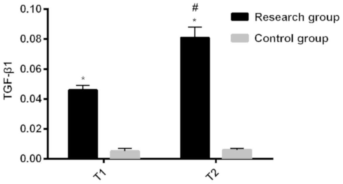
The expression of TGF-β1 protein in LECs of rats in
study group was 0.046±0.003 at T1 and 0.081±0.007 at T2 and in
control group was 0.005±0.002 at T1 and 0.006±0.001 at T2. The
expression of TGF-β1 protein in LECs of rats in study group at T2
was significantly higher than that at T1, with a statistically
significant difference (P<0.001). There was no statistically
significant difference in that at T1 and T2 in control group
(P>0.05). Expression in LECs of rats was significantly higher in
study than that in control group at T1 and T2, with a statistically
significant difference (P<0.001). It is indicated that the
expression of TGF-β1 protein in LECs of diabetic cataract rats was
higher in study group than that in normal control rats at the same
time points, which increased with time, showing an upward trend
(Table II and Fig. 1).
| Table II.Comparison of TGF-β1 quantitative
expression between study and control groups. |
Table II.
Comparison of TGF-β1 quantitative
expression between study and control groups.
| Index | Study group
(n=20) | Control group
(n=20) | t | P-value |
|---|
| T1 | 0.046±0.003 | 0.005±0.002 | 50.850 | <0.001 |
| T2 | 0.081±0.007 | 0.006±0.001 | 47.430 | <0.001 |
| t | 20.550 | 2.000 |
|
|
| P-value | <0.001 | 0.053 |
|
|
Comparison of MMP-9 quantitative
expression between two groups
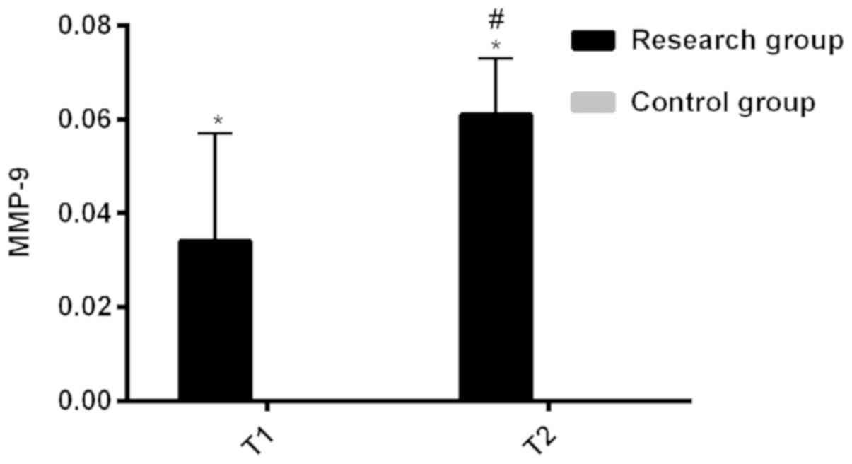
The expression of MMP-9 protein in LECs of rats in
study group was 0.034±0.023 at T1 and 0.061±0.012 at T2. That in
control group was 0.001±0.001 at T1 and T2. The expression of MMP-9
protein in LECs of rats in study group at T2 was significantly
higher than that at T1, with a statistically significant difference
(P<0.001). There was no statistically significant difference in
that at T1 and T2 in control group after successful modeling
(P>0.05). Expression in LECs of rats was significantly higher in
study group than that in control group at T1 and T2, with a
statistically significant difference (P<0.001). It is indicated
that the expression of MMP-9 protein in LECs of diabetic cataract
rats was higher in study group than that in normal control rats at
the same time points, which increased with time, showing an upward
trend (Table III and Fig. 2).
| Table III.Comparison of MMP-9 quantitative
expression between study and control groups. |
Table III.
Comparison of MMP-9 quantitative
expression between study and control groups.
| Index | Study group
(n=20) | Control group
(n=20) | t | P-value |
|---|
| T1 | 0.034±0.023 | 0.001±0.001 |
6.611 | <0.001 |
| T2 | 0.061±0.012 | 0.001±0.001 | 22.730 | <0.001 |
| t |
4.654 | – |
|
|
| P-value | <0.001 | – |
|
|
Correlation analysis of TGF-β1
expression with MMP-9 expression in LECs of diabetic cataract
rats
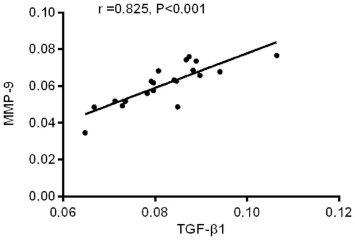
The TGF- 1 expression was positively correlated with
the MMP-9 expression in LECs of diabetic cataract rats (r=0.825,
P<001; Fig. 3).
Discussion
Posterior capsular turbidity is the most common in
lens turbidity types of diabetic cataract. This is due to
pathological changes of anterior and posterior lens capsule caused
by abnormal differentiation of LECs with rapid lesion progression
(13). MMPs, main mediators of
extracellular matrix degradation, are a class of proteolytic
enzymes. They are present in the form of zymogens, and their main
physiological role is to degrade extracellular matrix (14,15).
Gelatinase B (MMP-9) is a matrix hydrolase that degrades type IV
collagen (16). Luo et al
found that TGF-β1 promotes the expression of extracellular matrix
such as collagen and fibronectin, which is beneficial to cell
repair and embryo development. TGF-β1 is generally produced by cell
autocrine and paracrine (17).
TGF-β1 produced by normal cell autocrine or paracrine needs
activation to exert its effect, and most of TGF-β1 are present in a
potentially inactive form (18).
There are currently few studies on the correlation of TGF-β1 and
MMP-9 with diabetic cataract. In this study, the expression of
TGF-β1 and MMP-9 in LECs of diabetic cataract rats and its effect
on the occurrence and development of diabetic cataract were
investigated.
Experimental rats were randomly divided into study
group and control group. Rats in study group were successfully
modeled diabetic cataract rats, and rats in control group were
normal rats. First, the general information of the two groups of
rats was compared. The results showed that there were no
statistically significant differences in length and age between the
two groups of rats, demonstrating that the two groups of rats are
comparable. The glucose concentration in the blood of rats in study
group after modeling was significantly higher than that before
modeling, and that after modeling was significantly higher in study
group than that in control group, with statistically significant
differences. Long-term hyperglycemia in diabetes will lead to
various chronic complications in different tissues, such as chronic
damage and dysfunction in the heart, blood vessels and eyes. Among
them, cataract and retinopathy are the most common in eye
complications of diabetes (19,20).
Then, the quantitative expressions of TGF-β1 and MMP-9 between the
two groups were compared at T1 and T2 after modeling. The results
showed that the expression of TGF-β1 protein in LECs of rats in
study group at T2 was significantly higher than that at T1, and it
was significantly higher in study group than that in control group
at T1 and T2, with statistically significant differences.
Therefore, it is hypothesized that the expression of TGF-β1 protein
in LECs of diabetic cataract rats was higher in study group than
that in normal control rats at the same time points, which
increases with time, showing an upward trend. Studies have shown
that the expression level of TGF-β1 is significantly increased in
diabetes patients in the early stage, suggesting that high glucose
environment may be the main cause of TGF-β1 production and
activation (21,22). The expression of MMP-9 protein in
LECs of rats was significantly higher in study group than that in
control group at T1 and T2. Therefore, it is speculated that the
expression of MMP-9 protein in LECs of diabetic cataract rats was
higher in study group than that in normal control rats at the same
time points, which increases with time, showing an upward trend. In
recent years, a large number of studies on MMPS have shown that
MMP-9 is highly expressed in LECs of diabetic cataract, but less
expressed in LECs of non-diabetic cataract (11,23,24),
which is similar to findings in this study and supports our
results. Finally, Pearson analysis was used to perform correlation
analysis on the expression of TGF-β1 and MMP-9 in LECs of diabetic
cataract rats. The results showed that the TGF-β1 expression was
positively correlated with the MMP-9 expression in LECs of diabetic
cataract rats. Therefore, it is believed that the TGF-β1 expression
and the MMP-9 expression in LECs of diabetic cataract rats have a
certain mutual regulation effect. Xu et al (9) reported that the interaction between
TGF-β1-induced cells and extracellular matrix comes into play
through regulating the transcriptional activity of MMP-9 in
LECs.
In this study, due to the insufficient number of
rats enrolled, there may be some contingency in some results. In
order to improve study results, the number of experimental rats
will be increased later in a further study.
In summary, the expression of MMP-9 protein in LECs
of diabetic cataract rats is higher than that in normal rats. It is
believed that the specific mechanism of diabetic cataract may be
the that hyperglycemia causes TGF-β1 activation, resulting in the
increased expression of MMP-9, and the extracellular matrix
degradation in LECs of diabetic cataract leads to the abnormal
proliferation and differentiation of LECs, causing cataract. The
increased expression of TGF-β1 and MMP-2 proteins are correlated
with the occurrence and development of diabetic cataract.
Acknowledgements
Not applicable.
Funding
No funding was received.
Availability of data and materials
The datasets used and/or analyzed during the present
study are available from the corresponding author on reasonable
request.
Authors' contributions
KL and WA conceived and designed the study. WA
helped with establishment of diabetic cataract rat models, and KL
interpreted the data by using Pearson analysis. Both authors read
and approved the final manuscript.
Ethics approval and consent to
participate
The study was approved by the Ethics Committee of
The Central Hospital of Wuhan, Tongji Medical College, Huazhong
University of Science and Technology (Wuhan, China). Patients who
participated in this research had complete clinical data. The
signed informed consents were obtained from the patients or the
guardians.
Patient consent for publication
Not applicable.
Competing interests
The authors declare that they have no competing
interests.
References
|
1
|
Chang KC, Li L, Sanborn TM, Shieh B,
Lenhart P, Ammar D, LaBarbera DV and Petrash JM: Characterization
of Emodin as a therapeutic agent for diabetic cataract. J Nat Prod.
79:1439–1444. 2016. View Article : Google Scholar : PubMed/NCBI
|
|
2
|
Tan JS, Wang JJ and Mitchell P: Influence
of diabetes and cardiovascular disease on the long-term incidence
of cataract: The Blue Mountains eye study. Ophthalmic Epidemiol.
15:317–327. 2008. View Article : Google Scholar : PubMed/NCBI
|
|
3
|
Lu Q, Hao M, Wu W, Zhang N, Isaac AT, Yin
J, Zhu X, Du L and Yin X: Antidiabetic cataract effects of GbE,
rutin and quercetin are mediated by the inhibition of oxidative
stress and polyol pathway. Acta Biochim Pol. 65:35–41. 2018.
View Article : Google Scholar : PubMed/NCBI
|
|
4
|
Bhadada SV, Vyas VK and Goyal RK:
Protective effect of Tephrosia purpurea in diabetic cataract
through aldose reductase inhibitory activity. Biomed Pharmacother.
83:221–228. 2016. View Article : Google Scholar : PubMed/NCBI
|
|
5
|
Lee JY, Jeong HS, Lee DY, Sohn HJ and Nam
DH: Early postoperative intraocular pressure stability after
combined 23-gauge sutureless vitrectomy and cataract surgery in
patients with proliferative diabetic retinopathy. Retina.
32:1767–1774. 2012. View Article : Google Scholar : PubMed/NCBI
|
|
6
|
Takamura Y, Sugimoto Y, Kubo E, Takahashi
Y and Akagi Y: Immunohistochemical study of apoptosis of lens
epithelial cells in human and diabetic rat cataracts. Jpn J
Ophthalmol. 45:559–563. 2001. View Article : Google Scholar : PubMed/NCBI
|
|
7
|
de Iongh RU, Wederell E, Lovicu FJ and
McAvoy JW: Transforming growth factor-beta-induced
epithelial-mesenchymal transition in the lens: A model for cataract
formation. Cells Tissues Organs. 179:43–55. 2005. View Article : Google Scholar : PubMed/NCBI
|
|
8
|
Hales AM, Chamberlain CG, Murphy CR and
McAvoy JW: Estrogen protects lenses against cataract induced by
transforming growth factor-beta (TGFbeta). J Exp Med. 185:273–280.
1997. View Article : Google Scholar : PubMed/NCBI
|
|
9
|
Xu GX, Hu JZ, Zheng WD, Zhang S and Wang
TT: Expression and significance of transforming growth
factor-beta1, matrix metalloproteinase-2 and its inhibitor in lens
epithelial cells of diabetic cataract. Zhonghua Yan Ke Za Zhi.
39:411–414. 2003.(In Chinese). PubMed/NCBI
|
|
10
|
Nathu Z, Dwivedi DJ, Reddan JR, Sheardown
H, Margetts PJ and West-Mays JA: Temporal changes in MMP mRNA
expression in the lens epithelium during anterior subcapsular
cataract formation. Exp Eye Res. 88:323–330. 2009. View Article : Google Scholar : PubMed/NCBI
|
|
11
|
Kwon JW, Choi JA and Jee D: Matrix
metalloproteinase-1 and matrix metalloproteinase-9 in the aqueous
humor of diabetic macular edema patients. PLoS One.
11:e01597202016. View Article : Google Scholar : PubMed/NCBI
|
|
12
|
Dwivedi DJ, Pino G, Banh A, Nathu Z,
Howchin D, Margetts P, Sivak JG and West-Mays JA: Matrix
metalloproteinase inhibitors suppress transforming growth
factor-beta-induced subcapsular cataract formation. Am J Pathol.
168:69–79. 2006. View Article : Google Scholar : PubMed/NCBI
|
|
13
|
Boscia F, Grattagliano I, Vendemiale G,
Micelli-Ferrari T and Altomare E: Protein oxidation and lens
opacity in humans. Invest Ophthalmol Vis Sci. 41:2461–2465.
2000.PubMed/NCBI
|
|
14
|
Lindsey ML, Iyer RP, Jung M,
DeLeon-Pennell KY and Ma Y: Matrix metalloproteinases as input and
output signals for post-myocardial infarction remodeling. J Mol
Cell Cardiol. 91:134–140. 2016. View Article : Google Scholar : PubMed/NCBI
|
|
15
|
Backstrom JR and Tökés ZA: The 84-kDa form
of human matrix metalloproteinase-9 degrades substance P and
gelatin. J Neurochem. 64:1312–1318. 1995. View Article : Google Scholar : PubMed/NCBI
|
|
16
|
Inoue Y, Abe K, Obata K, Yoshioka T,
Ohmura G, Doh K, Yamamoto K, Hoshiai H and Noda K:
Immunohistochemical studies on matrix metalloproteinase-9 (MMP-9)
and type-IV collagen in endometrial carcinoma. J Obstet Gynaecol
Res. 23:139–145. 1997. View Article : Google Scholar : PubMed/NCBI
|
|
17
|
Luo D, Guan Q, Wang K, Nguan CYC and Du C:
TGF-β1 stimulates movement of renal proximal tubular epithelial
cells in a three-dimensional cell culture via an autocrine TGF-β2
production. Exp Cell Res. 350:132–139. 2017. View Article : Google Scholar : PubMed/NCBI
|
|
18
|
Kawarada Y, Inoue Y, Kawasaki F, Fukuura
K, Sato K, Tanaka T, Itoh Y and Hayashi H: TGF-β induces p53/Smads
complex formation in the PAI-1 promoter to activate transcription.
Sci Rep. 6:354832016. View Article : Google Scholar : PubMed/NCBI
|
|
19
|
Selim S: Frequency and pattern of chronic
complications of diabetes and their association with glycemic
control among adults with type 2 diabetes in Bangladesh. Diabetes
Metab Syndr. 11 (Suppl 1):S329–S332. 2017. View Article : Google Scholar : PubMed/NCBI
|
|
20
|
Akiyode O and Tran C: Overview of ocular
anti-vascular endothelial growth factor therapy in the management
of diabetic eye complications. Diabetes Spectr. 29:44–49. 2016.
View Article : Google Scholar : PubMed/NCBI
|
|
21
|
Voelker J, Berg PH, Sheetz M, Duffin K,
Shen T, Moser B, Greene T, Blumenthal SS, Rychlik I, Yagil Y, et
al: Anti-TGF-β1 antibody therapy in patients with diabetic
nephropathy. J Am Soc Nephrol. 28:953–962. 2017. View Article : Google Scholar : PubMed/NCBI
|
|
22
|
Xu MT, Sun S, Zhang L, Xu F, Du SL, Zhang
XD and Wang DW: Diabetes mellitus affects the biomechanical
function of the callus and the expression of TGF-beta1 and BMP2 in
an early stage of fracture healing. Braz J Med Biol Res.
49:e47362016.PubMed/NCBI
|
|
23
|
Alapure BV, Praveen MR, Gajjar DU,
Vasavada AR, Parmar TJ and Arora AI: Matrix metalloproteinase-2 and
−9 activities in the human lens epithelial cells and serum of
steroid induced posterior subcapsular cataracts. Mol Vis. 18:64–73.
2012.PubMed/NCBI
|
|
24
|
Dynlacht JR: The role of age, sex and
steroid sex hormones in radiation cataractogenesis. Radiat Res.
180:559–566. 2013. View Article : Google Scholar : PubMed/NCBI
|