Introduction
Severe acute pancreatitis (SAP) is characterized by
rapid progression, severe systemic complications, multiple organs
dysfunction, and high mortality (1).
The activation of pancreatic enzymes, release of inflammatory
factors, and subsequent systemic inflammatory response syndrome
(SIRS) and multiple organ dysfunction syndromes (MODS) are the main
pathological processes of SAP. The bile reflux deprived from
biliary obstruction or the pancreatic duct obstruction (or
increased pressure in the pancreatic duct) is considered as the
most important factor for developing SAP (2). Targeted treatment such as
sphincterotomy or nasobiliary drainage may prevent the progression
of acute pancreatitis (AP) or SAP (3,4), which
indicates that sustained bile drainage or biliary decompression
could decrease the organ injury in SAP and improve the
prognosis.
Exploration for the mechanisms of SAP progression
and the relevant treatment strategies are particularly important.
Inflammation-associated factors serve a vital role in the
progression of SAP. The imbalance between pro-inflammatory factors
including tumor necrosis factor (TNF)-α and high mobility group box
1 (HMGB1) and anti-inflammatory factors like hemeoxygenase-1 (HO-1)
is indispensable for SAP progression, which leads to local
pancreatic inflammation and systemic complications (5–7).
Previous studies have demonstrated that TNF-α and interleukin-6
(IL-6) activate the Kupffer cells in the liver and then produce
pro-inflammatory factors, thus inducing early liver injury, and
gut-derived cytokines reaching the lung, resulting in acute
respiratory distress syndrome (8–11). TNF-α
affects the kidney function directly or indirectly via cytotoxicity
or by inducing the release of endothelin, respectively (12).
A previous study has demonstrated that biliary tract
external drainage protected multiple organs against SAP associated
injuries via HO-1 upregulation (7).
HO-1 is also demonstrated to modulate some immunoinflammatory and
autoimmune diseases, such as multiple sclerosis, which may
represent a novel treatment approach for the above diseases
(13,14). Furthermore, the antioxidant factor
nuclear factor E2-related factor 2 could also promote anticancer
activity via the modulation of HO-1 (15). Nuclear transcription factor (NF)-κB
is an important pro-inflammatory factor and NF-κB-p65 or RelA is a
member of NF-κB family, which is associated with cell proliferation
and apoptosis (16,17). Based on the previous studies, the
present study aimed to further investigate the role of sustained
bile external drainage in decreasing the injury of SAP and the
association of sustained bile drainage group (BDG) on inflammatory
factors TNF-α, HMGB1, IL-10, NF-κB-p65, and HO-1 in SAP rats at
different periods (3, 6 and 12 h). The present study provided
evidence for the application of sustained BDG in the treatment of
SAP.
Materials and methods
Reagents
Sodium taurocholate was from Sigma-Aldrich; Merck
KGaA (purity >97%). TNF-α (JYM0635Ra), HO-1 (JYM0356Ra), IL-10
(JYM0651Ra), and HMGB1 (JYM0371Ra) ELISA kit were all purchased
from ColorfulGene Biological Technology, NF-κB-p65 mouse antibody
(8242S) was from Cell Signaling Technology, Inc.
Establishment of animal model
A total of 72 8-week-old female Sprague-Dawley (SD)
rats weighing 190–230 g were randomly divided into four groups
(n=18): Sham operation group (SOG), SOG + bile drainage group
(BDG), SAP group, and SAP + BDG. The animals (6 weeks of age) were
purchased from Jinan Peng Yue Laboratory Animal Breeding Company
and fed with food and water freely under the same environment at
22–26°C for 2 weeks with a 12 h light/dark cycle. The present study
was approved by the Ethics Committee of Shandong Provincial
Qianfoshan Hospital affiliated to Shandong University.
The SD rats received intraperitoneal anesthesia with
3% pentobarbital sodium (30 mg/kg body weight). The duodenum, bile
duct, and pancreas were separated, and 4% sodium taurocholate
solution was injected by retrograde puncture of biliopancreatic
duct through duodenum to set up the SAP model (1 mg/kg body
weight). The SOG group received operation only without any other
interventions. After the injection of sodium taurocholate, a
cannula of diameter 0.8 mm was inserted into the bile duct through
duodenum. The cannula was then fixed with the bile duct, duodenum
and abdominal wall separately when the drainage of the bile was
unobstructed to establish SAP + BDG model. The SOG + BDG group only
fixed a cannula into bile duct without the application of 4% sodium
taurocholate solution. Pentobarbital sodium (200 mg/kg body weight)
with an intraperitoneal injection was used to sacrifice the rats
prior to blood collection from the abdominal aorta and the tissue
samples were collected.
Analysis of serum amylase (AMY)
The blood samples (5 ml) were obtained at various
time points (t=3 h, 6 h, 12 h) from abdominal aorta. After standing
for 15 min at room temperature, the samples were centrifuged at
1,370 × g for 10 min at 4°C. The serum supernatant was collected.
The level of serum AMY was detected in the clinical laboratory of
Qianfoshan Hospital (7).
H&E staining
Tissue samples of pancreas, liver, and lung were
collected at various time points (t=3 h, 6, and 12 h) and immersed
in 10% paraformaldehyde and paraffin embedded. Thick tissue
sections (4-µm) were obtained. Subsequently, the sections were
dewaxed in xylene and dehydrated through a serial alcohol gradient
followed by washing with PBS for 5 min at room temperature. The
sections were stained with hematoxylin for 5 min and eosin for 30
sec at room temperature. Following the staining, sections were
dehydrated by increasing concentrations of ethanol and xylene. The
histomorphology was observed with an optical microscope
(magnification, ×200). The evaluation standard of histopathologic
scores of the pancreas were recorded in Table I (18). The current study selected three
sections and five fields (magnification, ×200) for every section
randomly. The scores were subsequently calculated in accordance
with the standard of table I for every field in four parts (edema,
inflammation, hemorrhage and necrosis). The average value for the
histopathologic scores of the pancreas were then calculated.
 | Table I.Histologic scoring for acute
hemorrhagic necrotizing pancreatitis. |
Table I.
Histologic scoring for acute
hemorrhagic necrotizing pancreatitis.
| Condition | Score | Description |
|---|
| Edema | 0 | Absent |
|
| 1 | Focally increased
between lobules |
|
| 2 | Diffusely increased
between lobules |
|
| 3 | Tense acini, widely
separated lobules |
|
| 4 | Gross lobular
separation |
| Inflammation | 0 | Absent |
|
| 1 | Around ductal
margins |
|
| 2 | In parenchyma
(<50% of lobules) |
|
| 3 | In parenchyma (51
to 75% of lobules) |
|
| 4 | Massive
collections, abscesses |
| Hemorrhage | 0 | Absent |
|
| 1 | Blood in parenchyma
(<25%) |
|
| 2 | Blood in parenchyma
(25 to 50%) |
|
| 3 | Blood in parenchyma
(50 to 75%) |
|
| 4 | Blood in 100% of
lobules |
| Necrosis | 0 | Absent |
|
| 1 | Periductal
parenchymal destruction |
|
| 2 | Focal parenchymal
necrosis (<20%) |
|
| 3 | Diffuse loss of
lobules (20 to 50%) |
|
| 4 | Severe loss of
lobules (>50%) |
ELISA
The SD rats were divided into four groups: SOG, SOG
+ BDG, SAP group, and SAP + BDG. Blood (5 ml) from the abdominal
aorta was collected at various time points (t=3 h, 6, and 12 h) in
every group. After 15 min, the blood was centrifuged at 1,370 × g
for 10 min at room temperature and serum supernatants were
collected. Measurement of TNF-α, HO-1, IL-10, and HMGB1 levels in
blood supernatants was performed using commercially available ELISA
kits (ColorfulGene) according to the manufacturer's protocol.
Optical density (OD) values were obtained at 450 nm using the
Spectra Max 190 (Molecular Devices, LLC).
Western blotting
The SD rats were divided into four groups as stated
above; the pancreas tissues were collected and lysed in cold radio
immunoprecipitation assay lysis buffer (Beyotime Institute of
Biotechnology) with 1 nM phenylmethylsufonyl fluoride for 30 min on
ice, followed by centrifugation at 16,000 × g for 15 min at 4°C.
Protein concentrations of cleared lysates were determined with a
bicinchoninic acid Protein Assay kit (Beyotime Institute of
Biotechnology). Total amount of proteins (40 µg) were separated on
10% SDS-polyacrylamide gel electrophoresis gels (Beyotime Institute
of Biotechnology) and then electrotransferred to a polyvinylidene
fluoride (PVDF) membrane. PVDF membranes were blocked with 5%
skimmed milk and incubated overnight at 4°C with the NF-κB-p65
primary antibody (cat. no. 8242S; 1:1,000; Cell Signaling
Technology, Inc.) and GADPH primary antibody (cat. no. ab37168;
1:1,000; Abcam) in PBS-Tween. Following three washes with PBS
containing 0.1% Tween-20 for 15 min each, the membranes were
incubated with horse radish peroxidase-conjugated anti-rabbit IgG
(cat. no. A0280; 1:1,000; Beyotime Institute of Biotechnology) at
37°C for 1 h and then washed three times with PBS containing 0.1%
Tween-20. A TANON-4500SF chemiluminescence system (Tanon Science
and Technology Co., Ltd, Shanghai, China) was used to detect the
target proteins.
Statistics
All the data are expressed as the mean ± standard
deviation. Multiple group comparisons of the means were carried out
by one-way analysis of variance followed by the least significant
difference test as the post hoc test. Statistical analyses were
performed with SPSS software (version 17.0, SPSS China). P<0.05
was considered to indicate a statistically significant
difference.
Results
Effects of sustained BDG on serum
levels of TNF-α, HO-1, IL-10, and HMGB1
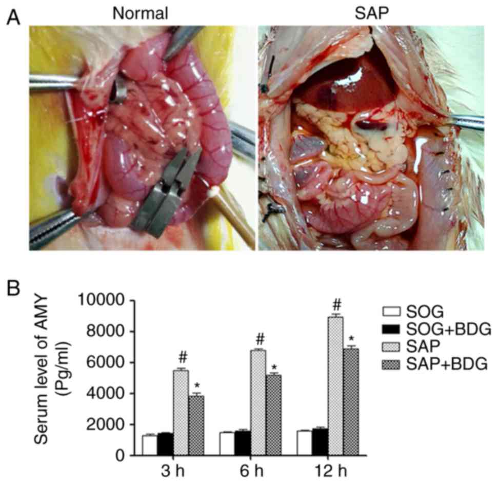
To explore the effect of BDG on pancreas in SAP, the
SAP rat models were induced by 4% sodium taurocholate solution via
injection of retrograde puncture of biliopancreatic duct through
duodenum, and the pancreas and intestine were obviously edematous
while ascites existed in the SAP rats (Fig. 1A, right panel). The serum levels of
AMY in the SAP group and SAP + BDG group were higher compared with
the SOG group and SOG + BDG group, while BDG could decrease the
level of AMY in SAP (Fig. 1B). SIRS
is the main reason for high mortality at the early stage of SAP,
the inflammatory cytokines induce the cascade effect, even the MODS
and death (19). The
pro-inflammatory factors TNF-α and HMGB1 were upregulated in SAP
rats, however sustained BDG could decrease the level of TNF-α in
SAP significantly at different times, while HMGB1 was only
downregulated at the 6 h of SAP by BDG, and no significant
differences were observed at 3 and 12 h (Fig. 2A and C). There was no marked
difference in the serum level of IL-10 in the SAP and SAP + BDG
groups (Fig. 2D). The
anti-inflammatory factor HO-1 was increased in SAP groups at early
stages (3 and 6 h), but decreased at 12 h, and then sustained BDG
upregulated the serum level of HO-1 compared with the SAP group at
different time check-points of SAP (Fig.
2B).
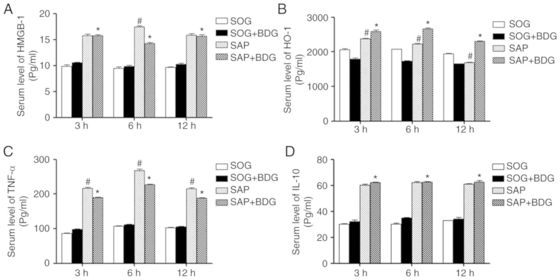 | Figure 2.Effect of sustained bile drainage on
serum expression of inflammatory factors. The Sprague-Dawley rats
were divided into four groups: SOG, SOG + BDG, SAP, and SAP + BDG.
The serum levels of (A) HMGB1, (B) HO-1, (C) TNF-α and (D) IL-10
were detected by ELISA assay at 3, 6, and 12 h (n=6).
#P<0.05 vs. SAP + BDG; *P<0.05 vs. SOG + BDG. BDG,
bile drainage group; HMGB1, high mobility group box 1; HO-1, heme
oxygenase-1; IL-10, interleukin-10; SAP, severe acute pancreatitis;
SOG, sham operation group; TNF-α, tumor necrosis factor-α. |
The effect of sustained BDG on the
multiple organ injury of SAP
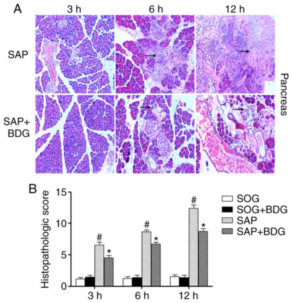
To evaluate the effect of BDG, the histopathologic
changes of pancreas, liver, and lung were observed. No significant
histopathologic changes in the SOG and SAP+SOG groups were observed
(Fig. S1). The inflammatory cells
infiltration, cell necrosis, the damage of pancreatic lobule was
higher in the SAP group, and obvious hemorrhage and tissue necrosis
in a time-dependent manner was observed, while BDG significantly
mitigated the abovementioned phenomena (Fig. 3A). The histopathologic scores of
pancreas in SAP were higher than the SOG groups, while BDG
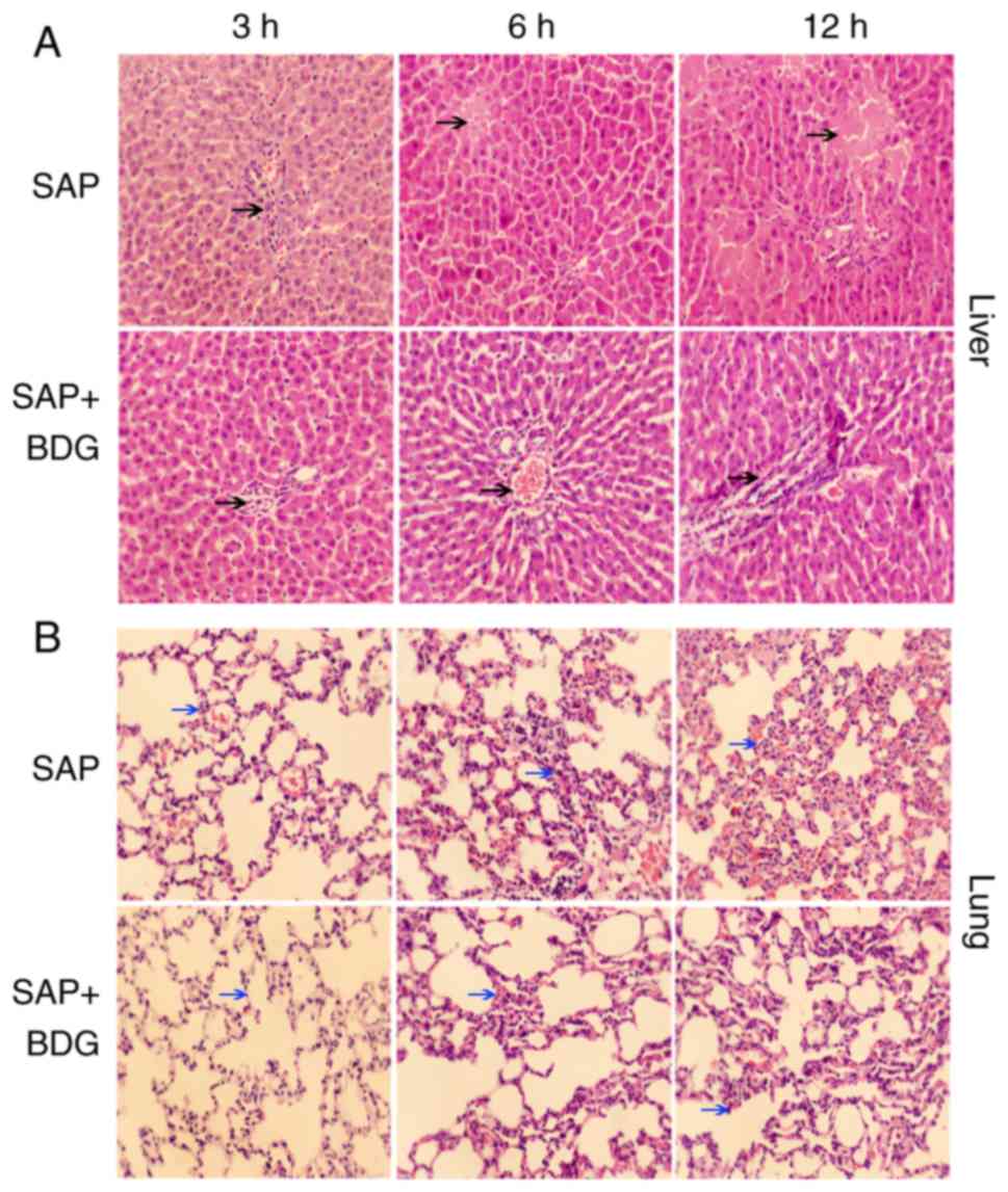
decreased the histopathologic scores of SAP significantly (Fig. 3B). Furthermore, compared with the SAP
+ BDG group the liver cells edema, necrosis, hepatic cord disorder,
and inflammatory cells infiltration were more obvious in SAP at all
time points (Fig. 4A). Finally, the
edema and hyperemia of lung tissue, the red blood cells and
inflammatory cells infiltration in alveolar cavity and interstitial
tissue were more serious than the SAP + BDG group (Fig. 4B).
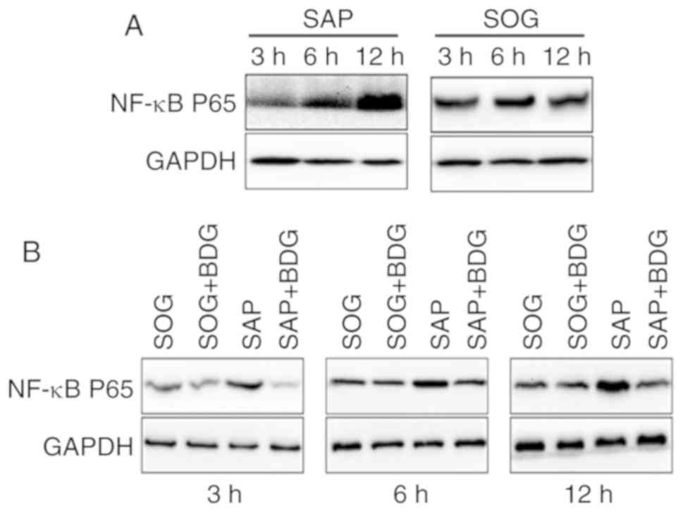
The modulation by BDG on the
expression level of NF-κB-p65 in the pancreas
During subsequent experiments, the rat pancreas
tissues at different time points was collected. The protein level
of NF-κB-p65 was upregulated significantly in the SAP group in a
time-dependent manner, while no marked difference was found in the
SOG group (Fig. 5A). To further
explore the role of BDG, the expression level of NF-κB-p65 at every
time point was monitored. The results indicated that the NF-κB-p65
expression levels of the SOG and SOG + BDG groups were similar and
those of SAP were the highest, among all groups tested. BDG
downregulated its expression compared with the SAP group
significantly at different time points (Fig. 5B).
Discussion
Severe acute pancreatitis (SAP) is a type of severe
digestive disease associated with rapid progression, severe
complications, and high morality. Blockage of the biliopancreatic
duct, alcohol consumption, infection, and medical injury are the
common reasons for SAP (20). The
mechanisms of SAP are complex. The activation of pancreatic
enzymes, the release of inflammatory factors, oxidative stress
reaction, translocation of gut microflora, and effect of nitric
oxide are responsible for the progress of SAP (21–23). The
imbalance between pro-inflammatory factors (TNF-α, IL-6, and HMGB1)
and anti-inflammatory factors (IL-2 and IL-10) results in the
cascade effect, and finally leads to multiple organ injury and poor
prognosis (24,25).
Bile reflux activates pancreatic enzymes, and the
degree of bile duct blockage is positively associated with the
degree of AP. Previous studies have demonstrated that removal of
bile or pancreatic duct obstruction effectively alleviated the
progress of SAP (4,7,26). In
animal experiments, bile drainage prolonged the survival and
relieved the pathological damage to the intestine (27). Hypoperfusion induced the liver to
release inflammatory factors, TNF-α, HMGB1, and IL-6 into the bile,
and further aggravates the injury of intestinal mucosa (28). Previous studies demonstrated that
bile external drainage protected the intestine and organ functions
under shock via blocking the ‘gut-liver-lung’ cytokines axis, and
decreased the injury to liver and lungs (7,29), which
is consistent with the present study. Meanwhile, biliary drainage
is not indicated for chronic non-progressive elevation of SAP, but
for a progressive increase in SAP or persistent jaundice (30). In the present study, based on the
H&E staining, it was found that the inflammation, cell necrosis
and organs structure damage were aggravated with the progress of
SAP in a time-dependent manner, and sustained BDG improved above
phenomena significantly and exhibited multiple organs protective
effects. The present study indicated that bile drainage or biliary
decompression could alleviate the progress of SAP and protect the
organs, and decrease the possibility of SIRS and MODS, which could
be considered as a potential treatment choice for SAP. Future
studies exploring the underlying mechanisms or signaling pathways
are required to provide more evidence for the clinical application
of sustained BDG.
A previous study reported that ascites induced the
secretion of TNF-α in pancreatic acinar cells (31). TNF-α mRNA was overexpressed in
pancreas tissues of SAP rat models, and the phenomenon was also
observed in the liver and lungs along with the progress of SAP
(32). Another pro-inflammatory
factor, HMGB1, is also crucial for the pathogenesis of SAP. HMGB1
could activate toll-like receptors 4 (TLR4) signaling pathway,
which is indispensable for the inflammatory process, and the
specific inhibitors of HMGB1 or TLR4 (such as eritoran and
[(S,R)-3-phenyl-4,5-dihydro-5-isoxasole acetic acid)] could
alleviate the inflammation and improve the prognosis (33–35). The
present study revealed that TNF-α and HMGB1 were upregulated in the
SAP and SAP + BDG groups compared with the control groups, but BDG
decreased the levels of TNF-α significantly and HMGB1 at 6 h. HO-1
is considered as an important anti-inflammatory factor, which is
responsible for cytothesis and tissue protection (36). HO-1 could exert anti-inflammatory
effects via the production of carbon monoxide (CO), which may be a
possible factor of early treatment of SAP, while CO has been proven
effective in some immuno-inflammatory and autoimmune diseases
(37,38). Under stress, HO-1 significantly
inhibits apoptosis of the islet cells and protects the pancreatic
β-cells (39,40). It was demonstrated that HO-1 levels
were increased in both SAP groups, and BDG upregulated the serum
level of HO-1 compared with the SAP group. The present study
demonstrated that sustained BDG could decrease the inflammation
process by modulating the expressions of TNF-α, HO-1 and HMGB1, and
alleviate the degree of SIRS and SAP model organ injury. However,
SOG+BDG group could not return SAP levels to normal compared with
SOG group, while BDG is considered as an effective method to slow
the progress of SAP. Furthermore, other clinical strategies,
including the inhibition of pancreatin, nutritional support and
fasting, are also indispensible (1,4).
Meanwhile, the associated signaling pathways involved in the above
inflammatory factors still remain to be explored. NF-κB is crucial
for the inflammatory activities via pro-inflammatory cytokines
release. Picroside II is demonstrated to reduce the level of NF-κB
and autophagy, and improves the antioxidant and anti-inflammatory
activities of SAP models (41). The
present study demonstrated that the NF-κB-p65 level of SAP was
upregulated, and BDG downregulated the expression of NF-κB-p65
significantly. According to the modulation of NF-κB-p65, bile
drainage could decrease the release of pro-inflammatory cytokines.
However, whether TNF-α and HMGB1 are the downstream factors still
remains unclear. The activation of NF-κB signaling pathway is
important for the progress of SAP, and it is indicated that
sustained BDG could decrease the activity of NF-κB signaling and
further improve the prognosis of SAP.
In conclusion, the present study demonstrated that
BDG decreased the progression of SAP effectively. The
histopathologic changes of pancreas, liver, and lung were
significantly attenuated by BDG. It was highlighted that the
underlying mechanisms were the downregulation of TNF-α, NF-κB p65
and HMGB1 and upregulation of HO-1 modulated by BDG.
Supplementary Material
Supporting Data
Acknowledgements
Not applicable.
Funding
The present study was supported by grants from
Science and Technology Development Plan of Jinan City (grant no.
20140821), Key Research and Development Plan of Shandong Province
(grant no. 2016GSF201108), and Science and Technology Development
Plan of Shandong Province (grant no. 2011YD18017).
Availability of data and materials
The analyzed data sets generated during the study
are available from the corresponding author on reasonable
request.
Authors' contributions
HT and JL conceived, designed and supervised the
present study. JC and ZF developed the methodology. FW, QW, SZ and
FL performed the study and acquired the data. FW and QW reviewed
the manuscript and analyzed/interpreted the data.
Ethics approval
The present study was approved by the Ethics
Committee of Shandong Provincial Qianfoshan Hospital affiliated to
Shandong University.
Patient consent for publication
Not applicable.
Competing interests
The authors declare that they have no competing
interests.
References
|
1
|
Bai Y, Liu Y, Jia L, Jiang H, Ji M, Lv N,
Huang K, Zou X, Li Y, Tang C, et al: Severe acute pancreatitis in
China: Etiology and mortality in 1976 patients. Pancreas.
35:232–237. 2007. View Article : Google Scholar : PubMed/NCBI
|
|
2
|
Alexakis N, Lombard M, Raraty M, Ghaneh P,
Smart HL, Gilmore I, Evans J, Hughes M, Garvey C, Sutton R and
Neoptolemos JP: When is pancreatitis considered to be of biliary
origin and what are the implications for management? Pancreatology.
7:131–141. 2007. View Article : Google Scholar : PubMed/NCBI
|
|
3
|
Youn YH, Lim HC, Jahng JH, Jang SI, You
JH, Park JS, Lee SJ and Lee DK: The Increase in balloon size to
over 15 mm does not affect the development of pancreatitis after
endoscopic papillary large balloon dilatation for bile duct stone
removal. Dig Dis Sci. 56:1572–1577. 2011. View Article : Google Scholar : PubMed/NCBI
|
|
4
|
Siqin D, Wang C, Zhou Z and Li Y: The key
event of acute pancreatitis: Pancreatic duct obstruction and bile
reflux, not a single one can be omitted. Med Hypothes. 72:589–591.
2009. View Article : Google Scholar
|
|
5
|
Neyrinck AM, Margagliotti S, Gomez C and
Delzenne NM: Kupffer cell-derived prostaglandin E2 is involved in
regulation of lipid synthesis in rat liver tissue. Cell Biochem
Funct. 22:327–332. 2004. View
Article : Google Scholar : PubMed/NCBI
|
|
6
|
Mäck C, Jungermann K, Götze O and
Schieferdecker HL: Anaphylatoxin C5a actions in rat liver:
Synergistic enhancement by C5a of lipopolysaccharide-dependent
alpha(2)-macroglobulin gene expression in hepatocytes via IL-6
release from Kupffer cells. J Immunol. 167:3972–3979. 2001.
View Article : Google Scholar : PubMed/NCBI
|
|
7
|
Wang JL, Chen Y, Song XQ, Lu ML, Zhao B,
Ma L, Chen EZ and Mao EQ: Biliary tract external drainage protects
against multiple organs injuries of severe acute pancreatitis rats
via hemeoxygenas-1 upregulation. Pancreatology. 17:219–227. 2017.
View Article : Google Scholar : PubMed/NCBI
|
|
8
|
Gonzalez RJ, Moore EE, Ciesla DJ, Biffl
WL, Johnson JL and Silliman CC: Mesenteric lymph is responsible for
post-hemorrhagic shock systemic neutrophil priming. J Trauma.
51:1069–1072. 2001.PubMed/NCBI
|
|
9
|
Davidson MT, Deitch EA, Lu Q, Osband A,
Feketeova E, Németh ZH, Haskó G and Xu DZ: A study of the biologic
activity of trauma-hemorrhagic shock mesenteric lymph over time and
the relative role of cytokines. Surgery. 136:32–41. 2004.
View Article : Google Scholar : PubMed/NCBI
|
|
10
|
Fanous MY, Phillips AJ and Windsor JA:
Mesenteric lymph: The bridge to future management of critical
illness. JOP. 8:374–399. 2007.PubMed/NCBI
|
|
11
|
Akbarshahi H, Sam A, Chen C, Rosendahl AH
and Andersson R: Early activation of pulmonary TGF-β1/Smad2
signaling in mice with acute pancreatitis-associated acute lung
injury. Mediators Inflamm. 2014:1480292014. View Article : Google Scholar : PubMed/NCBI
|
|
12
|
Wang Z, Cheng Z, Cristofaro V, Li J, Xiao
X, Gomez P, Ge R, Gong E, Strle K, Sullivan MP, et al: Inhibition
of TNF-α improves the bladder dysfunction that is associated with
type 2 diabetes. Diabetes. 61:2134–2145. 2012. View Article : Google Scholar : PubMed/NCBI
|
|
13
|
Fagone P, Mangano K, Di Marco R,
Touil-Boukoffa C, Chikovan T, Signorelli S, Lombardo GA, Patti F,
Mammana S and Nicoletti F: Expression of DNA methylation genes in
secondary progressive multiple sclerosis. J Neuroimmunol.
290:66–69. 2016. View Article : Google Scholar : PubMed/NCBI
|
|
14
|
Li BZ, Guo B, Zhang HY, Liu J, Tao SS, Pan
HF and Ye DQ: Therapeutic potential of HO-1 in autoimmune diseases.
Inflammation. 37:1779–1788. 2014. View Article : Google Scholar : PubMed/NCBI
|
|
15
|
Liu R and Yan X: Sulforaphane protects
rabbit corneas against oxidative stress injury in keratoconus
through activation of the Nrf-2/HO-1 antioxidant pathway. Int J Mol
Med. 42:2315–2328. 2018.PubMed/NCBI
|
|
16
|
Altavilla D, Famulari C, Passaniti M,
Galeano M, Macrì A, Seminara P, Minutoli L, Marini H, Calò M,
Venuti FS, et al: Attenuated cerulein-induced pancreatitis in
nuclear factor-kappaB-deficient mice. Lab Invest. 83:1723–1732.
2003. View Article : Google Scholar : PubMed/NCBI
|
|
17
|
Ijaz T, Sun H, Pinchuk IV, Milewicz DM,
Tilton RG and Brasier AR: Deletion of NF-κB/RelA in angiotensin
II-sensitive mesenchymal cells blocks aortic vascular inflammation
and abdominal aortic aneurysm formation. Arterioscler Thromb Vasc
Biol. 37:1881–1890. 2017. View Article : Google Scholar : PubMed/NCBI
|
|
18
|
Kusske AM, Rongione AJ, Ashley SW,
McFadden DW and Reber HA: Interleukin-10 prevents death in lethal
necrotizing pancreatitis in mice. Surgery. 120:284–289. 1996.
View Article : Google Scholar : PubMed/NCBI
|
|
19
|
Gunjaca I, Zunic J, Gunjaca M and Kovac Z:
Circulating cytokine levels in acute pancreatitis-model of
SIRS/CARS can help in the clinical assessment of disease severity.
Inflammation. 35:758–763. 2012. View Article : Google Scholar : PubMed/NCBI
|
|
20
|
Frossard JL, Steer ML and Pastor CM: Acute
pancreatitis. Lancet. 371:143–152. 2008. View Article : Google Scholar : PubMed/NCBI
|
|
21
|
Petersen OH: Ca2+ signaling in pancreatic
acinar cells: Physiology and pathophysiology. Braz J Med Biol Res.
42:9–16. 2009. View Article : Google Scholar : PubMed/NCBI
|
|
22
|
Pandol SJ, Saluja AK, Imrie CW and Banks
PA: Acute pancreatitis: Bench to the bedside. Gastroenterology.
132:1127–1151. 2007. View Article : Google Scholar : PubMed/NCBI
|
|
23
|
Leaphart CL and Tepas JJ III: The gut is a
motor of organ system dysfunction. Surgery. 141:563–569. 2007.
View Article : Google Scholar : PubMed/NCBI
|
|
24
|
Sharma D, Jakkampudi A, Reddy R, Reddy PB,
Patil A, Murthy HVV, Rao GV, Reddy DN and Talukdar R: Association
of systemic inflammatory and anti-inflammatory responses with
adverse outcomes in acute pancreatitis: Preliminary results of an
ongoing study. Dig Dis Sci. 62:3468–3478. 2017. View Article : Google Scholar : PubMed/NCBI
|
|
25
|
Petrov MS, Shanbhag S, Chakraborty M,
Phillips AR and Windsor JA: Organ failure and infection of
pancreatic necrosis as determinants of mortality in patients with
acute pancreatitis. Gastroenterology. 139:813–820. 2010. View Article : Google Scholar : PubMed/NCBI
|
|
26
|
Pellegrini CA: Surgery for gallstone
pancreatitis. Am J Surg. 165:515–518. 1993. View Article : Google Scholar : PubMed/NCBI
|
|
27
|
Jackson GD, Dai Y and Sewell WA: Bile
mediates intestinal pathology in endotoxemia in rats. Infect Immun.
68:4714–4719. 2000. View Article : Google Scholar : PubMed/NCBI
|
|
28
|
Landahl P, Ansari D and Andersson R:
Severe acute pancreatitis: Gut barrier failure, systemic
inflammatory response, acute lung injury, and the role of the
mesenteric lymph. Surg Infect (Larchmt). 16:651–656. 2015.
View Article : Google Scholar : PubMed/NCBI
|
|
29
|
Yang R, Miki K, Oksala N, Nakao A,
Lindgren L, Killeen ME, Mennander A, Fink MP and Tenhunen J: Bile
high-mobility group box 1 contributes to gut barrier dysfunction in
experimental endotoxemia. Am J Physiol Regul Integr Comp Physiol.
297:R362–R369. 2009. View Article : Google Scholar : PubMed/NCBI
|
|
30
|
Saluja SS, Kalayarasan R, Mishra PK,
Srivastava S, Chandrasekar S and Godhi S: Chronic pancreatitis with
benign biliary obstruction: Management issues. World J Surg.
38:2455–2459. 2014. View Article : Google Scholar : PubMed/NCBI
|
|
31
|
Ramudo L, Manso MA and De Dios I: Biliary
pancreatitis-associated ascitic fluid activates the production of
tumor necrosis factor-alpha in acinar cells. Crit Care Med.
33:143–148; discussion 248. 2005. View Article : Google Scholar : PubMed/NCBI
|
|
32
|
Norman JG, Fink GW and Franz MG: Acute
pancreatitis induces intrapancreatic tumor necrosis factor gene
expression. Arch Surg. 130:966–970. 1995. View Article : Google Scholar : PubMed/NCBI
|
|
33
|
Yang ZY, Ling Y, Yin T, Tao J, Xiong JX,
Wu HS and Wang CY: Delayed ethyl pyruvate therapy attenuates
experimental severe acute pancreatitis via reduced serum high
mobility group box 1 levels in rats. World J Gastroenterol.
14:4546–4550. 2008. View Article : Google Scholar : PubMed/NCBI
|
|
34
|
Musumeci D, Roviello GN and Montesarchio
D: An overview on HMGB1 inhibitors as potential therapeutic agents
in HMGB1-related pathologies. Pharmacol Ther. 141:347–357. 2014.
View Article : Google Scholar : PubMed/NCBI
|
|
35
|
Lee JC, Menacherry S, Diehl MC, Giffear
MD, White CJ, Juba R, Bagarazzi ML, Muthumani K, Boyer J, Agarwal
V, et al: Safety, bioavailability, and pharmacokinetics of
VGX-1027-A novel oral anti-inflammatory drug in healthy human
subjects. Clin Pharmacol Drug Dev. 5:91–101. 2016. View Article : Google Scholar : PubMed/NCBI
|
|
36
|
Aziz NM, Kamel MY and Rifaai RA: Effects
of hemin, a heme oxygenase-1 inducer in L-arginine-induced acute
pancreatitis and associated lung injury in adult male albino rats.
Endocr Regul. 51:20–30. 2017. View Article : Google Scholar : PubMed/NCBI
|
|
37
|
Song L, Li J, Yuan X, Liu W, Chen Z, Guo
D, Yang F, Guo Q and Song H: Carbon monoxide-releasing molecule
suppresses inflammatory and osteoclastogenic cytokines in nicotine-
and lipopolysaccharide-stimulated human periodontal ligament cells
via the heme oxygenase-1 pathway. Int J Mol Med. 40:1591–1601.
2017. View Article : Google Scholar : PubMed/NCBI
|
|
38
|
Mangano K, Cavalli E, Mammana S, Basile
MS, Caltabiano R, Pesce A, Puleo S, Atanasov AG, Magro G, Nicoletti
F and Fagone P: Involvement of the Nrf2/HO-1/CO axis and
therapeutic intervention with the CO-releasing molecule CORM-A1, in
a murine model of autoimmune hepatitis. J Cell Physiol.
233:4156–4165. 2018. View Article : Google Scholar : PubMed/NCBI
|
|
39
|
Tobiasch E, Günther L and Bach FH: Heme
oxygenase-1 protects pancreatic beta cells from apoptosis caused by
various stimuli. J Investig Med. 49:566–571. 2001. View Article : Google Scholar : PubMed/NCBI
|
|
40
|
Ribeiro MM, Klein D, Pileggi A, Molano RD,
Fraker C, Ricordi C, Inverardi L and Pastori RL: Heme oxygenase-1
fused to a TAT peptide transduces and protects pancreatic
beta-cells. Biochem Biophys Res Commun. 305:876–881. 2003.
View Article : Google Scholar : PubMed/NCBI
|
|
41
|
Piao X, Liu B, Guo L, Meng F and Gao L:
Picroside II shows protective function for severe acute
pancreatitis in rats by preventing NF-κB-dependent autophagy. Oxid
Med Cell Longev. 2017:70857092017. View Article : Google Scholar : PubMed/NCBI
|