Introduction
Myocardial ischemia has a high incidence of
morbidity and mortality worldwide (1). It has been shown that acute myocardial
ischemia is a leading cause of death (2). In addition to sudden death, myocardial
ischemia also leads to various clinical manifestations such as
cardiac myocyte hypertrophy (3–5).
Therapeutic interventions in myocardial ischemia have been explored
extensively and many treatment options have been published.
Myocardial ischemia-reperfusion is an effective treatment method
for acute myocardial ischemia, which can reduce myocardial
infarction size and relieve symptoms of heart failure (6). However, myocardial ischemia-reperfusion
injury exists and remains a complex problem. Dysregulation of many
signaling pathways have been suggested to be associated with heart
damage caused by myocardial ischemia, especially the dysregulation
of intracellular signaling pathways. Signaling disorders leading to
cell proliferation, differentiation, autophagy and apoptosis under
physiological or pathological conditions may be closely related to
cardiac damage (7).
As a traditional Chinese herbal medicine, rhizome
gastrodiae has been used in China for thousands of years. According
to previous studies, rhizome gastrodiae has nootropic, analgesic
and anti-inflammatory effects and can improve microcirculation and
general circulatory functions (8,9).
Gastrodin is the major active ingredient in rhizome gastrodiae,
which demonstrates a high application value (10). It was reported that gastrodin has
neuroprotective effects on cultured cortical neurons (11). Gastrodin can inhibit expression of
pro-inflammatory cytokines and cyclooxygenase-2 (12). Gastrodin can regulate signaling
pathways involved in inflammatory responses. Studies have suggested
that the beneficial effects of gastrodin may be derived from its
antioxidant properties (13). GAS
can protect primary cultured rat hippocampal neurons from beta
amyloid-induced neurotoxicity (14).
In this study, the protective effect of gastrodin on myocardial
ischemia-reperfusion injury and the possible molecular mechanism
were explored, aiming to provide a reference for the treatment of
this disease.
Materials and methods
Experimental animals and grouping
Male 7–8-week-old Sprague Dawley (SD) rats, weighing
210–250 g, were raised in a cage, with free access to food and
water, on a 12:12-h light/dark cycle for a week. The temperature
was 22±2°C, and the relative humidity was 60±5%. The rats were
randomly divided into the sham operation group, the myocardial
ischemia-reperfusion model group and the gastrodin group (500
mg/kg), with equal number of 20 rats per group. The rats in the
gastrodin group were administered gastrodin twice daily by gavage
for 7 days before model establishment and 3 days after model
establishment. The rats in the sham operation and the myocardial
ischemia-reperfusion model groups were given an equal volume of
saline during the same time period. The rats used in this study
were provided by the Experimental Animal Center of Nantong
University with a permission number SCXX (Su) 2002–0019. All
experimental procedures were performed in accordance with the Guide
for the Care and Use of Laboratory Animals published by National
Institutes of Health (Bethesda, MD, USA). The protocols were
approved by the Animal Ethics Committee of the Affiliated Hospital
of Nantong University (Nantong, China).
Establishment of myocardial ischemia
model
To induce myocardial ischemia, the rats were
anesthetized and subjected to open-chest surgery to expose the
heart. Ligation of the left descending coronary artery was
performed 2 mm below the border between the left atrium and the
ventricle using 5/0 nylon suture. Myocardial reperfusion induced by
ligature removal was performed in the myocardial
ischemia-reperfusion model group. Those who only threaded but not
ligated were the sham operation model group. Ischemia was confirmed
by visually observed cyanosis and electrocardiogram demonstrating
ST segment elevation and QRS widening. Successful establishment of
the ischemia and reperfusion models was judged according to the
criteria. Myocardial reperfusion induced by ligature removal was
performed in the myocardial ischemia-reperfusion model group. Those
who only threaded but not ligated were the sham operation model
group. All rats were euthanized on the 8th day after myocardial
ischemia surgery, and the heart tissue was harvested.
Western blot analysis
Lysis of heart tissues was performed using the RIPA
lysis buffer to extract proteins, followed by concentration
measurement of the protein extracts and the BCA protein
quantification kit was purchased from Thermo Fisher Scientific,
Inc. (Waltham, MA, USA). A total of 5 µl of protein was loaded per
lane. The proteins in the sample were separated using SDS-PAGE gel
electrophoresis and transferred to the membrane (NC membrane;
MilliporeSigma, Burlington, MA, USA) using a wet system. The
primary antibodies [monoclonal Bax antibody (1:1,500; cat. no.
YS-28034R) and monoclonal Bcl-2 antibody (1:1,000; cat. no.
YSm-10846M) purchased from Santa Cruz Biotechnology, Inc., Dallas,
TX, USA, and monoclonal caspase-3 antibody (1:500; cat. no.
ab131000) purchased from Abcam, Cambridge, UK] were added after the
membrane was blocked with 5% BSA, followed by incubation at 37°C
overnight. Then, HRP-labeled goat anti-rabbit secondary antibody
(1:1,000; cat. no. A0208, Beyotime Institute of Biotechnology,
Shanghai, China) was added, and color was developed at 37°C. The
percentages of separation and stacking gels were 10 and 5%,
respectively.
Fluorescence-based reverse
transcription-quantitative polymerase chain reaction (RT-qPCR)
Total RNA was extracted from the obtained
cardiomyocytes in acccordance with the instructions of TRIzol kit
(Invitrogen; Thermo Fisher Scientific, Inc.). The complementary DNA
(cDNA) was synthesized via reverse transcription of the single
stranded RNA in acccordance with the instructions of cDNA kit
(Toyobo Co., Ltd., Osaka, Japan). SYBR-Green PCR kit (Toyobo Co.,
Ltd.) was used. Amplification of DNA was carried out, followed by
analysis. The thermocycling conditions were: 42°C × 15 min, 92°C ×
5 min, (95°C × 20 sec → 55°C × 15 sec → 72°C × 20 sec) × 30 sec,
96°C × 30 sec, 55°C × 30 sec, 95°C × 30 sec. All expression levels
were calculated using the 2−ΔΔCq method (15). The primers used in RT-qPCR are shown
in Table I.
 | Table I.Primers used in RT-qPCR assay. |
Table I.
Primers used in RT-qPCR assay.
| Genes | Primer sequences |
|---|
| GAPDH | F:
5′-GACAACTITGGCATCGTGGA-3′ |
|
| R:
5′-ATGCAGGGATGATGTTCTGG-3 |
| Bax | F:
5′-AGACACCTGAGCTGACCTTGGAG-3′ |
|
| R:
5′-GTTGAAGTTGCCATCAGCAAACA-3′ |
| Bcl-2 | F:
5′-TGAACCGGCATCTGCACAC-3′ |
|
| R:
5′-CGTCTTCAGAGACAGCCAGGAG-3′ |
| Caspase-3 | F:
5′-CATACAGCGGAACTGTCGAT-3′ |
|
| R:
5′-GTTCAGCAAGGCGCATAGTG-3′ |
Terminal deoxynucleotidyl
transferase-mediated dUTP
As previously described (16), apoptosis was assessed using TUNEL
assay (Roche Molecular Diagnostics, Pleasanton, CA, USA). Frozen
sections (6 mm thickness) or cells were stained with DAPI (Thermo
Fisher Scientific, Inc.) at room temperature for 5 min, allowing
for visualization of the nucleus. Monoclonal anti-α-actinin
antibody (1:1,000; cat. no. sc-17829; Santa Cruz Biotechnology,
Inc.) were added, followed by incubation at 4°C overnight. The
sections were then incubated with goat anti-rat α-actinin antibody
at 37°C for 2 h. Positive cells were counted using a fluorescence
microscopy.
HE staining
All specimens were fixed by formaldehyde, after
which they were embedded in paraffin and routinely sectioned. The
samples were then stained, followed by pathological examination
under light microscope (Olympus Corp., Tokyo, Japan).
ELISA
Serum levels of IL-6, IL-10, TNF-α and IL-1β were
measured using ELISA kits according to the instructions of the kits
(Abcam).
Statistical analysis
GraphPad Prism Software (GraphPad Software, Inc., La
Jolla, CA, USA) was used for statistical analysis. The count data
were analyzed by χ2 test. One-way analysis of variance
was used for comparison for multiple groups, and Fishers Least
Significant Difference test was the post-hoc test used after
one-way analysis. All data were expressed as (mean ± standard
deviation). P<0.05 was considered to indicate a statistically
significant difference.
Results
Alleviation of myocardial
ischemia-reperfusion injury by gastrodin
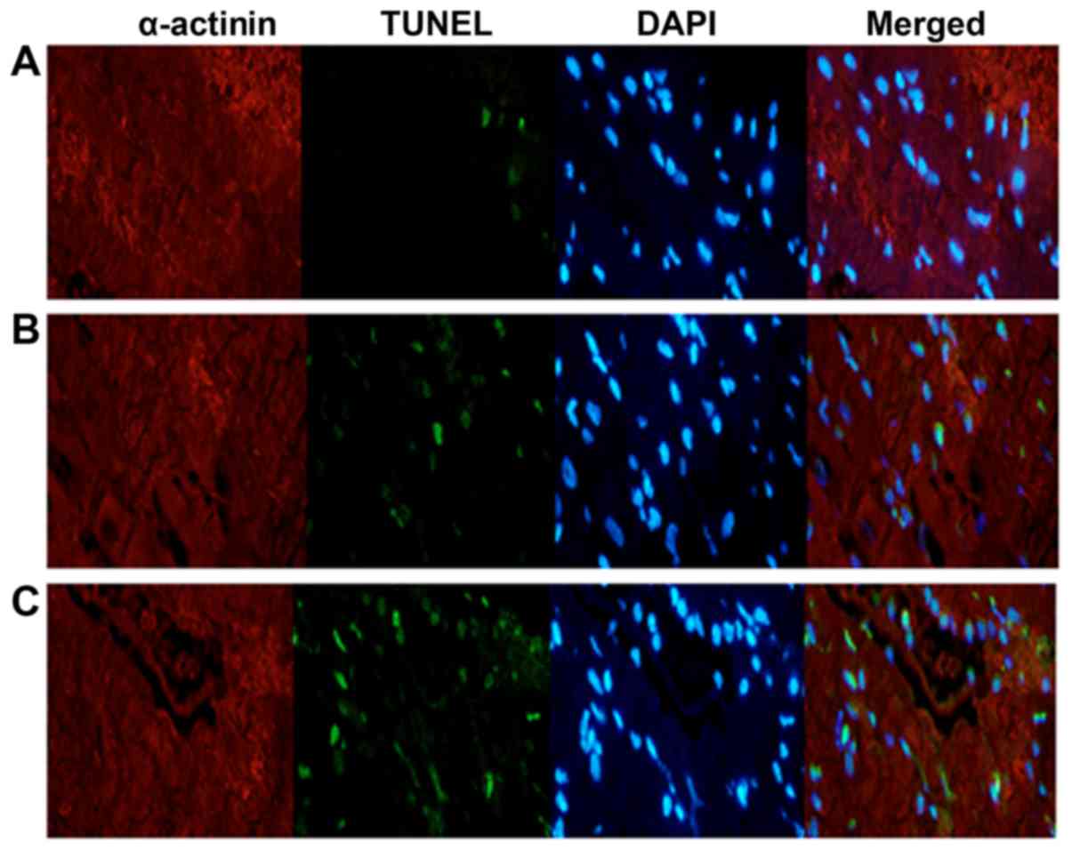
As shown in the TUNEL assay results (Table II and Fig. 1), the cardiomyocyte apoptotic rates
in the sham operation, myocardial ischemia-reperfusion model and
gastrodin groups were 2.1±0.8, 24.2±8.5, and 12.1±3.7%,
respectively. Compared with the other two groups, the atrial
apoptosis rate of the gastrodin treatment group was higher than
that of the sham operation group, but the myocardial apoptosis rate
of the gastrodin treatment group was significantly lower than that
of the myocardial ischemia-reperfusion model group (P<0.05).
| Table II.Cardiomyocyte apoptotic rate in the
three groups (%). |
Table II.
Cardiomyocyte apoptotic rate in the
three groups (%).
| Groups | Apoptotic rate
(%) |
|---|
| Sham operation
(n=20) |
2.1±0.8 |
| Gastrodin (n=20) | 12.1±3.7a |
| Myocardial
ischemia-reperfusion model (n=20) |
24.2±8.5a,b |
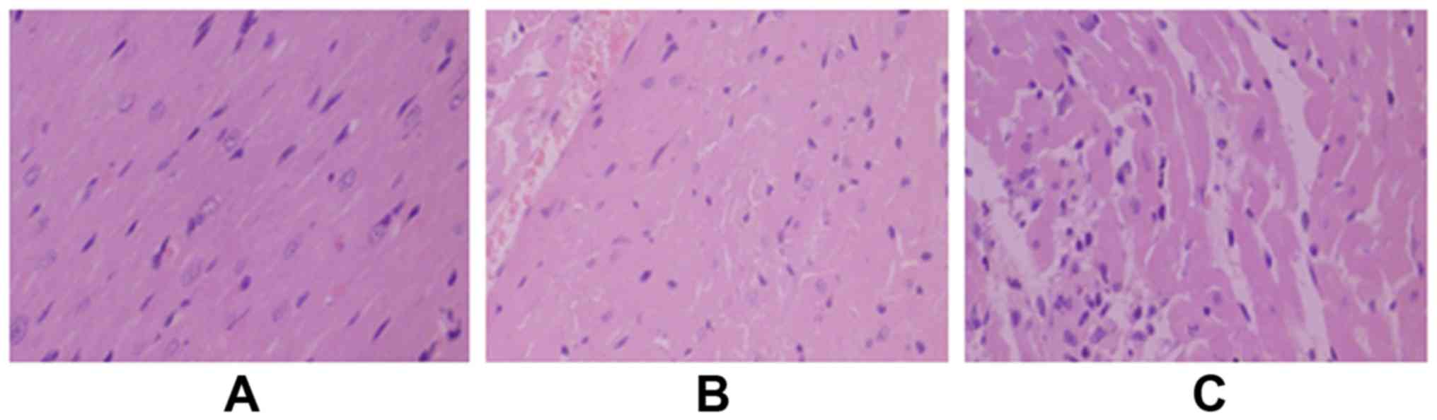
Morphological changes of myocardial tissues were
examined by HE staining, and the results are shown in Fig. 2. Cardiomyocytes in the sham operation
group were neatly arranged. In the myocardial ischemia-reperfusion
model group, the cardiomyocytes were in disordered arrangements.
Widening gaps and inflammatory factor exudates were observed. The
degree of cardiomyocyte damage in the gastrodin group was
significantly less than that in the myocardial ischemia-reperfusion
model group.
Expression levels of proteins involved
in cardiomyocyte pathways
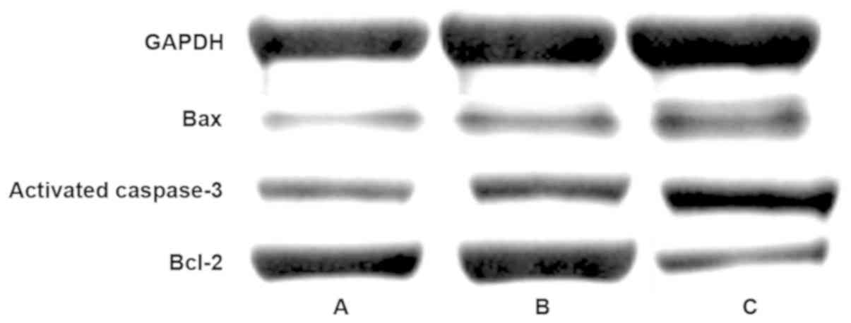
As shown in the results of western blot analysis
(Table III and Fig. 3), the expression levels of Bax and
activated caspase-3 were 0.38±0.16 and 0.43±0.21, respectively, in
the gastrodin group, as well as 0.87±0.36 and 0.93±0.36
respectively, in the sham operation group as well as 1.42±0.28 and
1.67±0.37 respectively, in the myocardial ischemia-reperfusion
model group. The differences between the two groups were
statistically significant (P<0.05). The expressions of Bax and
activated caspase-3 protein in the gastrodin, sham operation and
myocardial ischemia-reperfusion groups showed an increasing trend,
and the differences between the groups were statistically
significant (P<0.05). The expression level of Bcl-2 in the
gastrodin group increased to 0.59±0.24.
| Table III.Expression levels of proteins involved
in cardiomyocyte pathways (mean ± standard deviation). |
Table III.
Expression levels of proteins involved
in cardiomyocyte pathways (mean ± standard deviation).
| Groups | Bax | Bcl-2 | Bcl-2/Bax | Activated
caspase-3 |
|---|
| Sham operation
(n=20) | 0.87±0.36 | 0.92±0.25 | 1.06±0.23 | 0.93±0.36 |
| Gastrodin (n=20) |
0.38±0.16a |
0.59±0.24a | 1.55±0.34 |
0.43±0.21a |
| Myocardial
ischemia-reperfusion model (n=20) |
1.42±0.28a,b |
0.34±0.27a,b | 0.31±0.14 |
1.67±0.37a,b |
mRNA expression levels of genes
related to cardiomyocyte pathways
As shown in the RT-qPCR results (Table IV), the mRNA expression levels of
pro-apoptotic genes Bax and activated caspase-3 were 0.98±0.31 and
0.99±0.40, respectively, in the sham operation group, as well as
0.46±0.27 and 0.55±0.19, respectively, in the gastrodin group, and
1.24±0.45 and 0.98±0.42, respectively, in the cardiac
ischemia-reperfusion group. The expression levels of Bax and
activated caspase-3 mRNA in the gastrodin, sham operation and
myocardial ischemia-reperfusion groups showed an increasing trend,
and the differences between the groups were statistically
significant (P<0.05). The expression levels of the
anti-apoptotic gene Bcl-2 in the sham operation, gastrodin, and
myocardial ischemia-reperfusion model groups were 0.97±0.38,
0.89±0.36 and 0.32±0.15, respectively, (P<0.05).
| Table IV.mRNA expression levels of genes
related to cardiomyocyte pathways (mean ± standard deviation). |
Table IV.
mRNA expression levels of genes
related to cardiomyocyte pathways (mean ± standard deviation).
| Groups | Bax | Bcl-2 | Activated
caspase-3 |
|---|
| Sham operation
(n=20) | 0.98±0.31 | 0.97±0.38 | 0.99±0.40 |
| Gastrodin
(n=20) |
0.46±0.27a |
0.89±0.36a |
0.55±0.19a |
| Myocardial
ischemia-reperfusion model (n=20) |
1.24±0.45a,b |
0.32±0.15a,b |
0.98±0.42b |
Serum levels of inflammatory
factors
As shown in the ELISA results (Table V), the serum levels of IL-6, TNF-α
and IL-1β were 41±16, 26±9 and 37±14, respectively, in the sham
operation group, as well as 138±42, 126±35 and 145±42,
respectively, in the myocardial ischemia-reperfusion model group
and 59±23, 58±17 and 49±15 in the gastrodin group, respectively.
The differences between the two groups were statistically
significant (P<0.05). The serum levels of IL-10 were 88±17 and
167±45, respectively, in the myocardial ischemia-reperfusion model
and gastrodin groups, and the difference was statistically
significant. In the gastrodin group, the higher level of IL-10 was
clearly opposite to the lower levels of IL-6, TNF-α and IL-1β.
| Table V.Serum levels of inflammatory factors
(mean ± standard deviation). |
Table V.
Serum levels of inflammatory factors
(mean ± standard deviation).
| Groups | IL-6 (ng/l) | IL-10 (ng/l) | TNF-α (ng/l) | IL-1β (ng/l) |
|---|
| Sham operation
(n=20) | 41±16 |
45±18 | 26±9 | 37±14 |
| Gastrodin
(n=20) |
59±23a | 167±45a |
58±17a | 49±15a |
| Myocardial
ischemia-reperfusion model (n=20) |
138±42a,b |
88±17a,b |
126±35a,b |
145±42a,b |
Discussion
Studies have shown that defects in many signaling
pathways are associated with myocardial ischemia-reperfusion injury
(17,18). For example, the interaction between
activated PI3K/AKT pathway and downstream target proteins,
including Bcl-2 and Bax, promotes cell survival (19). If the PI3K/AKT pathway is
dysregulated, alteration of cell signaling such as Bcl-2 can occur,
eventually leading to apoptosis. Ke et al reported that
activation of the PI3K/AKT pathway protects against
isoproterenol-induced myocardial ischemic injury (17). In another study, it was reported that
physiological heart growth was induced by activating the PI3K/AKT
pathway (20).
Some studies suggested that myocardial
ischemia-reperfusion may aggravate cardiac dysfunction and
myocardial cell damage, leading to inflammation and partial
apoptosis. It is important to explore the underlying mechanism in
order to prevent myocardial ischemia-reperfusion injury. According
to literature, multiple factors such as calcium overload,
neutrophil aggregation and cardiomyocyte apoptosis are involved in
myocardial ischemia-reperfusion injury (21,22).
Gastrodin is an effective monomer composition
extracted from rhizome gastrodiae, which demonstrates various
pharmacological effects. It not only improves microcirculation and
general circulatory functions but also possesses neuroprotective
effects and suppresses inflammatory factors. Myocardial ischemia is
a serious condition that normally leads to devastating consequences
such as necrosis and apoptosis (23). In this study, myocardial
ischemia-reperfusion injury was investigated, aimed at providing
more treatment information for this disease. Results from this
study showed that the cardiomyocyte apoptotic rate in the
myocardial ischemia-reperfusion model group increased
substantially, compared with that in the sham operation group.
Gastrodin significantly reduced cardiomyocyte apoptosis as
suggested by the lower apoptotic rate in the gastrodin group,
compared with the myocardial ischemia-reperfusion model group.
In this study, the effect of gastrodin on expression
of activated caspase-3, Bax and Bcl-2 was also explored. Caspase-3
is capable of digesting specific protein substrates, thereby
inhibiting DNA repair and leading to chromosomal DNA fragmentation
and cell death. Members of the Bcl-2 family proteins play a
critical role in regulating the autonomous apoptotic pathway. It is
known that Bax and Bcl-2 can migrate to different parts of the cell
to exert a role of signal transduction, thereby regulating cell
survival or apoptosis (24). The
results showed that gastrodin significantly downregulated
expression of Bax and activated caspase-3 but upregulated Bcl-2
expression in the gastrodin group, compared with the myocardial
ischemia-reperfusion model group. These findings suggested that
gastrodin has significant protective effects in myocardial
ischemia-reperfusion injury.
Recent studies demonstrated that inflammatory
lesions also played an important role in myocardial
ischemia-reperfusion injury (25,26).
IL-6 is a cytokine that is mainly secreted from monocytes,
macrophages and T/B lymphocytes. It can induce the expression of
ICAM-1 gene in cardiomyocytes and promote the entry of
granulocytes into the ischemic region. The overexpression of ICAM-1
mRNA participates in LFA-1 signaling transduction on the surface of
neutrophil leading to adhesion of neutrophils and endothelial
cells. It also mediates neutrophil infiltration and exacerbates
inflammatory responses by increasing pro-inflammatory factors
(27,28). Pro-inflammatory cytokines TNF-α and
IL-1β also play an important role in apoptotic processes. To assess
anti-inflammatory effects of gastrodin, the expression levels of
IL-6, IL-10, TNF-α and IL-1β were measured. The results showed that
there were statistically significant differences in the expression
of TNF-α, IL-6, IL-10 and IL-1β between the groups. IL-1β, TNF-α
and IL-6 were all upregulated in the myocardial
ischemia-reperfusion model group, suggesting that myocardial
ischemia-reperfusion may increase myocardial injury through
inflammatory responses. Compared with the myocardial
ischemia-reperfusion model group, gastrodin significantly reduced
the expression of these cytokines in the gastrodin group. IL-10, as
a multifunctional negative regulator, is involved in regulation of
a variety of biological processes and plays a protective role. In
this study, the expression level of IL-10 increased substantially
in the gastrodin group, compared with the myocardial
ischemia-reperfusion model group. It may play a role in protecting
ischemic cardiomyocytes from apoptosis by suppressing inflammation.
Findings in this study suggested that gastrodin may reduce
inflammatory damage by suppressing inflammatory factors, thereby
protecting the myocardial tissue.
In this study, it was revealed that gastrodin can
decrease the release of inflammatory factors while increase the
expression of protective cytokines. It reduced cardiomyocyte
apoptosis by inhibiting apoptotic signaling, thereby alleviating
myocardial ischemia-reperfusion injury. Therefore, findings in this
study suggested that gastrodin can provide protection against
myocardial ischemia-reperfusion injury, which may serve as a
reference for better treatment of myocardial ischemia-reperfusion
injury.
Acknowledgements
Not applicable.
Funding
No funding was received.
Availability of data and materials
The datasets used and/or analyzed during the current
study are available from the corresponding author on reasonable
request.
Authors' contributions
XH drafted the manuscript. XH, HS and KL conceived
and designed the study. LZ, FW and QY performed PCR. KL and LZ were
responsible for animal model construction. All authors read and
approved the final manuscript.
Ethics approval and consent to
participate
This study was approved by Affiliated Hospital of
Nantong University (Nantong, China).
Patient consent for publication
Not applicable.
Competing interests
The authors declare that they have no competing
interests.
References
|
1
|
Steg PG, Greenlaw N, Tendera M, Tardif JC,
Ferrari R, Al Zaibag M, Dorian P, Hu D, Shalnova S, Sokn FJ, et al
Prospective Observational Longitudinal Registry of Patients with
Stable Coronary Artery Disease (CLARIFY) Investigators, :
Prevalence of anginal symptoms and myocardial ischemia and their
effect on clinical outcomes in outpatients with stable coronary
artery disease: data from the international observational CLARIFY
registry. JAMA Intern Med. 174:1651–1659. 2014. View Article : Google Scholar : PubMed/NCBI
|
|
2
|
Cheng D, Zhu C, Cao J and Jiang W: The
protective effects of polyphenols from jujube peel (Ziziphus Jujube
Mill) on isoproterenol-induced myocardial ischemia and
aluminum-induced oxidative damage in rats. Food Chem Toxicol.
50:1302–1308. 2012. View Article : Google Scholar : PubMed/NCBI
|
|
3
|
Misumida N, Kobayashi A, Saeed M, Fox JT
and Kanei Y: Electrocardiographic left ventricular hypertrophy as a
predictor for nonsignificant coronary artery disease in patients
with non ST segment elevation myocardial infarction. Angiology.
67:27–33. 2016. View Article : Google Scholar : PubMed/NCBI
|
|
4
|
Sung HK, Chan YK, Han M, Jahng JWS, Song
E, Danielson E, Berger T, Mak TW and Sweeney G: Lipocalin-2 (NGAL)
attenuates autophagy to exacerbate cardiac apoptosis induced by
myocardial ischemia. J Cell Physiol. 232:2125–2134. 2017.
View Article : Google Scholar : PubMed/NCBI
|
|
5
|
Panza JA, Holly TA, Asch FM, She L,
Pellikka PA, Velazquez EJ, Lee KL, Borges-Neto S, Farsky PS, Jones
RH, et al: Inducible myocardial ischemia and outcomes in patients
with coronary artery disease and left ventricular dysfunction. J Am
Coll Cardiol. 61:1860–1870. 2013. View Article : Google Scholar : PubMed/NCBI
|
|
6
|
Patel RD and Saver JL: Evolution of
reperfusion therapies for acute brain and acute myocardial
ischemia: a systematic, comparative analysis. Stroke. 44:94–98.
2013. View Article : Google Scholar : PubMed/NCBI
|
|
7
|
Zhang J, Yu XH, Yan YG, Wang C and Wang
WJ: PI3K/Akt signaling in osteosarcoma. Clin Chim Acta.
444:182–192. 2015. View Article : Google Scholar : PubMed/NCBI
|
|
8
|
Kim HJ, Moon KD, Lee DS and Lee SH: Ethyl
ether fraction of Gastrodia elata Blume protects amyloid beta
peptide-induced cell death. J Ethnopharmacol. 84:95–98. 2003.
View Article : Google Scholar : PubMed/NCBI
|
|
9
|
Kim HJ, Moon KD, Oh SY, Kim SP and Lee SR:
Ether fraction of methanol extracts of Gastrodia elata, a
traditional medicinal herb, protects against kainic acid-induced
neuronal damage in the mouse hip-pocampus. Neurosci Lett.
314:65–68. 2001. View Article : Google Scholar : PubMed/NCBI
|
|
10
|
Park S, da Kim S and Kang S: Gastrodia
elata Blume water extracts improve insulin resistance by decreasing
body fat in dietinduced obese rats: vanillin and
4-hydroxybenzaldehyde are the bioactive candidates. Eur J Nutr.
50:107–118. 2011. View Article : Google Scholar : PubMed/NCBI
|
|
11
|
Xu X, Lu Y and Bie X: Protective effects
of gastrodin on hypoxia-induced toxicity in primary cultures of rat
cortical neurons. Planta Med. 73:650–654. 2007. View Article : Google Scholar : PubMed/NCBI
|
|
12
|
Dai JN, Zong Y, Zhong LM, Li YM, Zhang W,
Bian LG, Ai QL, Liu YD, Sun J and Lu D: Gastrodin inhibits
expression of inducible NO synthase, cyclooxygenase-2 and
proinflammatory cytokines in cultured LPS-stimulated microglia via
MAPK pathways. PLoS One. 6:e218912011. View Article : Google Scholar : PubMed/NCBI
|
|
13
|
Shu C, Chen C, Zhang DP, Guo H, Zhou H,
Zong J, Bian Z, Dong X, Dai J, Zhang Y and Tang Q: Gastrodin
protects against cardiac hypertrophy and fibrosis. Mol Cell
Biochem. 359:9–16. 2012. View Article : Google Scholar : PubMed/NCBI
|
|
14
|
Zhao X, Zou Y, Xu H, Fan L, Guo H, Li X,
Li G, Zhang X and Dong M: Gastrodin protect primary cultured rat
hippocampal neurons against amyloid-beta peptide-induced
neurotoxicity via ERK1/2-Nrf2 pathway. Brain Res. 1482:13–21. 2012.
View Article : Google Scholar : PubMed/NCBI
|
|
15
|
Livak KJ and Schmittgen TD: Analysis of
relative gene expression data using real time quantitative PCR and
the 2(-Delta Delta C(T)) method. Methods. 25:402–408. 2001.
View Article : Google Scholar : PubMed/NCBI
|
|
16
|
Abbate A, Salloum FN, Van Tassell BW,
Vecile E, Toldo S, Seropian I, Mezzaroma E and Dobrina A:
Alterations in the interleukin-1/interleukin-1 receptor antagonist
balance modulate cardiac remodeling following myocardial infarction
in the mouse. PLoS One. 6:e279232011. View Article : Google Scholar : PubMed/NCBI
|
|
17
|
Ke Z, Wang G, Yang L, Qiu H, Wu H, Du M,
Chen J, Song J, Jia X and Feng L: Crude terpene glycoside component
from Radix paeoniae rubra protects against isoproterenol-induced
myocardial ischemic injury via activation of the PI3K/AKT/mTOR
signaling pathway. J Ethnopharmacol. 206:160–169. 2017. View Article : Google Scholar : PubMed/NCBI
|
|
18
|
Cui G, Shan L, Hung M, Lei S, Choi I,
Zhang Z, Yu P, Hoi P, Wang Y and Lee SM: A novel Danshensu
derivative confers cardioprotection via PI3K/Akt and Nrf2 pathways.
Int J Cardiol. 168:1349–1359. 2013. View Article : Google Scholar : PubMed/NCBI
|
|
19
|
Ouyang ZH, Wang WJ, Yan YG, Wang B and Lv
GH: The PI3K/Akt pathway: A critical player in intervertebral disc
degeneration. Oncotarget. 8:57870–57881. 2017. View Article : Google Scholar : PubMed/NCBI
|
|
20
|
De Los Santos S, García-Pérez V,
Hernández-Reséndiz S, Palma-Flores C, González-Gutiérrez CJ,
Zazueta C, Canto P and Coral-Vázquez RM: (−)-Epicatechin induces
physiological cardiac growth by activation of the PI3K/Akt pathway
in mice. Mol Nutr Food Res. 61:612017. View Article : Google Scholar
|
|
21
|
Correa F, Buelna-Chontal M, Chagoya V,
García-Rivas G, Vigueras RM, Pedraza-Chaverri J, García-Niño WR,
Hernández-Pando R, León-Contreras JC and Zazueta C: Inhibition of
the nitric oxide/cyclic guanosine monophosphate pathway limited the
cardioprotective effect of post-conditioning in hearts with apical
myocardial infarction. Eur J Pharmacol. 765:472–481. 2015.
View Article : Google Scholar : PubMed/NCBI
|
|
22
|
Kambara T, Shibata R, Ohashi K, Matsuo K,
Hiramatsu-Ito M, Enomoto T, Yuasa D, Ito M, Hayakawa S, Ogawa H, et
al: C1q/tumor necrosis factor-related protein 9 protects against
acute myocardial injury through an adiponectin receptor
I-AMPK-dependent mechanism. Mol Cell Biol. 35:2173–2185. 2015.
View Article : Google Scholar : PubMed/NCBI
|
|
23
|
Broughton BR, Reutens DC and Sobey CG:
Apoptotic mechanisms after cerebral ischemia. Stroke. 40:e331–e339.
2009. View Article : Google Scholar : PubMed/NCBI
|
|
24
|
Pape M, Engelhard K, Eberspächer E,
Hollweck R, Kellermann K, Zintner S, Hutzler P and Werner C: The
long-term effect of sevoflurane on neuronal cell damage and
expression of apoptotic factors after cerebral ischemia and
reperfusion in rats. Anesth Analg. 103:173–179. 2006. View Article : Google Scholar : PubMed/NCBI
|
|
25
|
Smith IC, Vigna C, Levy AS, Denniss SG,
Rush JW and Tupling AR: The effects of buthionine sulfoximine
treatment on diaphragm contractility and SERCA pump function in
adult and middle aged rats. Physiol Rep. 3:e125472015. View Article : Google Scholar : PubMed/NCBI
|
|
26
|
Komin N, Moein M, Ellisman MH and Skupin
A: Multiscale modeling indicates that temperature dependent
[Ca2+]i spiking in astrocytes is quantitatively
consistent with modulated SERCA activity. Neural Plast.
2015:6834902015. View Article : Google Scholar : PubMed/NCBI
|
|
27
|
Yan J, Schmid E, Hosseinzadeh Z, Honisch
S, Shumilina E, Fuchs J and Lang F: Impact of Janus kinase 3 on
cellular Ca release, store operated Ca(2+) entry and Na(+)/Ca(2+)
exchanger activity in dendritic cells. Cell Physiol Biochem.
36:2287–2298. 2015. View Article : Google Scholar : PubMed/NCBI
|
|
28
|
Wiernicki B, Rozwadowska N, Malcher A,
Kolanowski T, Zimna A, Rugowska A and Kurpisz M: Human myoblast
transplantation in mice infarcted heart alters the expression
profile of cardiac genes associated with left ventricle remodeling.
Int J Cardiol. 202:710–721. 2016. View Article : Google Scholar : PubMed/NCBI
|