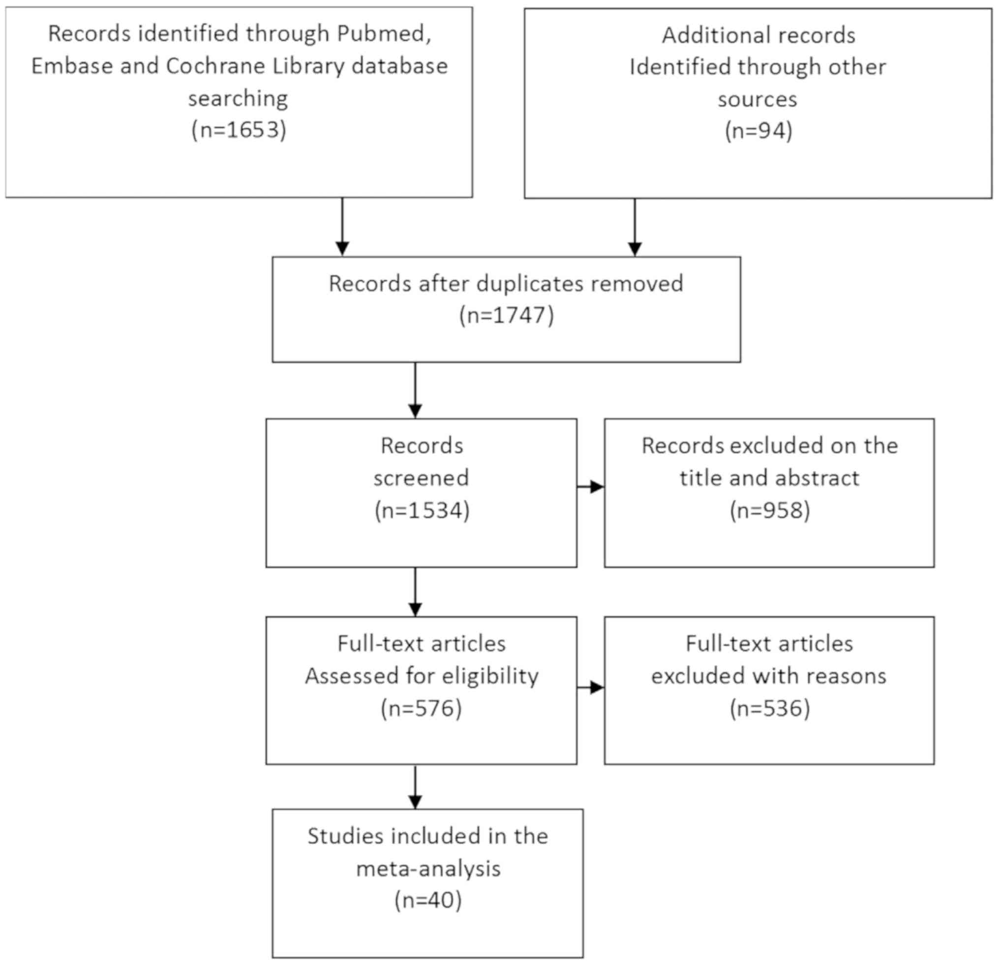
|
1
|
Kahi CJ, Anderson JC, Waxman I, Kessler
WR, Imperiale TF, Li X and Rex DK: High-definition
chromocolonoscopy vs. high-definition white light colonoscopy for
average-risk colorectal cancer screening. Am J Gastroenterol.
105:1301–1307. 2010. View Article : Google Scholar : PubMed/NCBI
|
|
2
|
Muto T, Bussey HJ and Morson BC: The
evolution of cancer of the colon and rectum. Cancer. 36:2251–2270.
1975. View Article : Google Scholar : PubMed/NCBI
|
|
3
|
Winawer SJ, Zauber AG, Ho MN, O'Brien MJ,
Gottlieb LS, Sternberg SS, Waye JD, Schapiro M, Bond JH, Panish JF,
et al: Prevention of colorectal cancer by colonoscopic polypectomy.
The National Polyp Study Workgroup. N Engl J Med. 329:1977–1981.
1993. View Article : Google Scholar : PubMed/NCBI
|
|
4
|
Kahi CJ, Imperiale TF, Juliar BE and Rex
DK: Effect of screening colonoscopy on colorectal cancer incidence
and mortality. Clin Gastroenterol Hepatol. 7:770–775; quiz 711.
2009. View Article : Google Scholar : PubMed/NCBI
|
|
5
|
Zauber AG, Winawer SJ, O'Brien MJ,
Lansdorp-Vogelaar I, van Ballegooijen M, Hankey BF, Shi W, Bond JH,
Schapiro M, Panish JF, et al: Colonoscopic polypectomy and
long-term prevention of colorectal-cancer deaths. N Engl J Med.
366:687–696. 2012. View Article : Google Scholar : PubMed/NCBI
|
|
6
|
Matsuda T, Ono A, Sekiguchi M, Fujii T and
Saito Y: Advances in image enhancement in colonoscopy for detection
of adenomas. Nat Rev Gastroenterol Hepatol. 14:305–314. 2017.
View Article : Google Scholar : PubMed/NCBI
|
|
7
|
Jin XF, Chai TH, Shi JW, Yang XC and Sun
QY: Meta-analysis for evaluating the accuracy of endoscopy with
narrow band imaging in detecting colorectal adenomas. J
Gastroenterol Hepatol. 27:882–887. 2012. View Article : Google Scholar : PubMed/NCBI
|
|
8
|
Omata F, Ohde S, Deshpande GA, Kobayashi
D, Masuda K and Fukui T: Image-enhanced, chromo, and cap-assisted
colonoscopy for improving adenoma/neoplasia detection rate: A
systematic review and meta-analysis. Scand J Gastroenterol.
49:222–237. 2014. View Article : Google Scholar : PubMed/NCBI
|
|
9
|
Whiting P, Rutjes AW, Reitsma JB, Bossuyt
PM and Kleijnen J: The development of QUADAS: A tool for the
quality assessment of studies of diagnostic accuracy included in
systematic reviews. BMC Med Res Methodol. 3:252003. View Article : Google Scholar : PubMed/NCBI
|
|
10
|
Dias S, Welton NJ, Sutton AJ and Ades AE:
A generalised linear modelling framework for pairwise and network
meta-analysis of randomised controlled trials. NICE DSU Technical
Support Document. 2:2011.
|
|
11
|
Sturtz S, Ligges U and Gelman A:
R2WinBUGS: A package for running WinBUGS from R. J Stat Softw.
12:1–16. 2005. View Article : Google Scholar
|
|
12
|
Aminalai A, Rösch T, Aschenbeck J, Mayr M,
Drossel R, Schröder A, Scheel M, Treytnar D, Gauger U, Stange G, et
al: Live image processing does not increase adenoma detection rate
during colonoscopy: A randomized comparison between FICE and
conventional imaging (Berlin Colonoscopy Project 5, BECOP-5). Am J
Gastroenterol. 105:2383–2388. 2010. View Article : Google Scholar : PubMed/NCBI
|
|
13
|
Bisschops R, Tejpar S, Willekens H, De
Hertogh G and Van Cutsem E: Su1432 I-SCAN detects more polyps in
lynch syndrome (HNPCC) patients: A prospective controlled
randomized back-to-back study. Gastrointest Endosc. 75:AB3302012.
View Article : Google Scholar
|
|
14
|
Boparai KS, van den Broek FJ, van Eeden S,
Fockens P and Dekker E: Increased polyp detection using narrow band
imaging compared with high resolution endoscopy in patients with
hyperplastic polyposis syndrome. Endoscopy. 43:676–682. 2011.
View Article : Google Scholar : PubMed/NCBI
|
|
15
|
Brooker JC, Saunders BP, Shah SG, Thapar
CJ, Thomas HJ, Atkin WS, Cardwell CR and Williams CB: Total colonic
dye-spray increases the detection of diminutive adenomas during
routine colonoscopy: A randomized controlled trial. Gastrointest
Endosc. 56:333–338. 2002. View Article : Google Scholar : PubMed/NCBI
|
|
16
|
Cha JM, Lee JI, Joo KR, Jung SW and Shin
HP: A prospective randomized study on computed virtual
chromoendoscopy versus conventional colonoscopy for the detection
of small colorectal adenomas. Dig Dis Sci. 55:2357–2364. 2010.
View Article : Google Scholar : PubMed/NCBI
|
|
17
|
Chung SJ, Kim D, Song JH, Kang HY, Chung
GE, Choi J, Kim YS, Park MJ and Kim JS: Comparison of detection and
miss rates of narrow band imaging, flexible spectral imaging
chromoendoscopy and white light at screening colonoscopy: A
randomised controlled back-to-back study. Gut. 63:785–791. 2014.
View Article : Google Scholar : PubMed/NCBI
|
|
18
|
Chung SJ, Kim D, Song JH, Park MJ, Kim YS,
Kim JS, Jung HC and Song IS: Efficacy of computed virtual
chromoendoscopy on colorectal cancer screening: A prospective,
randomized, back-to-back trial of Fuji Intelligent Color
Enhancement versus conventional colonoscopy to compare adenoma miss
rates. Gastrointest Endosc. 72:136–142. 2010. View Article : Google Scholar : PubMed/NCBI
|
|
19
|
East JE, Ignjatovic A, Suzuki N, Guenther
T, Bassett P, Tekkis PP and Saunders BP: A randomized, controlled
trial of narrow-band imaging vs. high-definition white light for
adenoma detection in patients at high risk of adenomas. Colorectal
Dis. 14:e771–e778. 2012. View Article : Google Scholar : PubMed/NCBI
|
|
20
|
East JE, Stavrindis M, Thomas-Gibson S,
Guenther T, Tekkis PP and Saunders BP: A comparative study of
standard vs. high definition colonoscopy for adenoma and
hyperplastic polyp detection with optimized withdrawal technique.
Aliment Pharmacol Ther. 28:768–776. 2008. View Article : Google Scholar : PubMed/NCBI
|
|
21
|
Glenn T, Hoffman BJ, Romagnuolo J and
Hawes RH: Does narrow band imaging (NBI) enhance colon polyp
detection? Gastrointest Endosc. 61:AB2772005. View Article : Google Scholar
|
|
22
|
Gross SA, Buchner AM, Crook JE, Cangemi
JR, Picco MF, Wolfsen HC, DeVault KR, Loeb DS, Raimondo M, Woodward
TA and Wallace MB: A comparison of high definition-image enhanced
colonoscopy and standard white-light colonoscopy for colorectal
polyp detection. Endoscopy. 43:1045–1051. 2011. View Article : Google Scholar : PubMed/NCBI
|
|
23
|
Helbig CD and Rex DK: Narrow band imaging
(NBI) versus white-light (WL) for colon polyp detection using high
definition (HD) colonoscopes. Gastrointest Endosc. 63:AB2132006.
View Article : Google Scholar
|
|
24
|
Hong SN, Choe WH, Lee JH, Kim SI, Kim JH,
Lee TY, Kim JH, Lee SY, Cheon YK, Sung IK, et al: Prospective,
randomized, back-to-back trial evaluating the usefulness of i-SCAN
in screening colonoscopy. Gastrointest Endosc. 75:1011–1021.e2.
2012. View Article : Google Scholar : PubMed/NCBI
|
|
25
|
Hüneburg R, Lammert F, Rabe C, Rahner N,
Kahl P, Büttner R, Propping P, Sauerbruch T and Lamberti C:
Chromocolonoscopy detects more adenomas than white light
colonoscopy or narrow band imaging colonoscopy in hereditary
nonpolyposis colorectal cancer screening. Endoscopy. 41:316–322.
2009. View Article : Google Scholar : PubMed/NCBI
|
|
26
|
Hurlstone DP, Cross SS, Slater R, Sanders
DS and Brown S: Detecting diminutive colorectal lesions at
colonoscopy: A randomised controlled trial of pan-colonic versus
targeted chromoscopy. Gut. 53:376–380. 2004. View Article : Google Scholar : PubMed/NCBI
|
|
27
|
Ikematsu H, Saito Y, Tanaka S, Uraoka T,
Sano Y, Horimatsu T, Matsuda T, Oka S, Higashi R, Ishikawa H and
Kaneko K: The impact of narrow band imaging for colon polyp
detection: A multicenter randomized controlled trial by tandem
colonoscopy. J Gastroenterol. 47:1099–1107. 2012. View Article : Google Scholar : PubMed/NCBI
|
|
28
|
Inoue T, Murano M, Murano N, Kuramoto T,
Kawakami K, Abe Y, Morita E, Toshina K, Hoshiro H, Egashira Y, et
al: Comparative study of conventional colonoscopy and pan-colonic
narrow-band imaging system in the detection of neoplastic colonic
polyps: A randomized, controlled trial. J Gastroenterol. 43:45–50.
2008. View Article : Google Scholar : PubMed/NCBI
|
|
29
|
Kaltenbach T, Friedland S and Soetikno R:
A randomised tandem colonoscopy trial of narrow band imaging versus
white light examination to compare neoplasia miss rates. Gut.
57:1406–1412. 2008. View Article : Google Scholar : PubMed/NCBI
|
|
30
|
Kiriyama S, Matsuda T, Nakajima T,
Sakamoto T, Saito Y and Kuwano H: Detectability of colon polyp
using computed virtual chromoendoscopy with flexible spectral
imaging color enhancement. Diagn Ther Endosc. 2012:5963032012.
View Article : Google Scholar : PubMed/NCBI
|
|
31
|
Kuiper T, van den Broek FJ, Naber AH, Van
Soest EJ, Scholten P, Mallant-Hent RCh, van den Brande J, Jansen
JM, van Oijen AH, Marsman WA, et al: Endoscopic trimodal imaging
detects colonic neoplasia as well as standard video endoscopy.
Gastroenterology. 140:1887–1894. 2011. View Article : Google Scholar : PubMed/NCBI
|
|
32
|
Lapalus MG, Helbert T, Napoleon B, Rey JF,
Houcke P and Ponchon T; Société Française d'Endoscopie Digestive, :
Does chromoendoscopy with structure enhancement improve the
colonoscopic adenoma detection rate? Endoscopy. 38:444–448. 2006.
View Article : Google Scholar : PubMed/NCBI
|
|
33
|
Le Rhun M, Coron E, Parlier D, Nguyen JM,
Canard JM, Alamdari A, Sautereau D, Chaussade S and Galmiche JP:
High resolution colonoscopy with chromoscopy versus standard
colonoscopy for the detection of colonic neoplasia: A randomized
study. Clin Gastroenterol Hepatol. 4:349–354. 2006. View Article : Google Scholar
|
|
34
|
Matsuda T, Saito Y, Fu KI, Uraoka T,
Kobayashi N, Nakajima T, Ikehara H, Mashimo Y, Shimoda T, Murakami
Y, et al: Does autofluorescence imaging videoendoscopy system
improve the colonoscopic polyp detection rate?-A pilot study. Am J
Gastroenterol. 103:1926–1932. 2008. View Article : Google Scholar : PubMed/NCBI
|
|
35
|
Moriichi K, Fujiya M, Sato R, Watari J,
Nomura Y, Nata T, Ueno N, Maeda S, Kashima S, Itabashi K, et al:
Back-to-back comparison of auto-fluorescence imaging (AFI) versus
high resolution white light colonoscopy for adenoma detection. BMC
Gastroenterol. 12:752012. View Article : Google Scholar : PubMed/NCBI
|
|
36
|
Paggi S, Radaelli F, Amato A, Meucci G,
Mandelli G, Imperiali G, Spinzi G, Terreni N, Lenoci N and Terruzzi
V: The impact of narrow band imaging in screening colonoscopy: A
randomized controlled trial. Clin Gastroenterol Hepatol.
7:1049–1054. 2009. View Article : Google Scholar : PubMed/NCBI
|
|
37
|
Pellisé M, Fernández-Esparrach G, Cárdenas
A, Sendino O, Ricart E, Vaquero E, Gimeno-García AZ, de Miguel CR,
Zabalza M, Ginès A, et al: Impact of wide-angle, high-definition
endoscopy in the diagnosis of colorectal neoplasia: A randomized
controlled trial. Gastroenterology. 135:1062–1068. 2008. View Article : Google Scholar : PubMed/NCBI
|
|
38
|
Pohl J, Lotterer E, Balzer C, Sackmann M,
Schmidt KD, Gossner L, Schaab C, Frieling T, Medve M, Mayer G, et
al: Computed virtual chromoendoscopy versus standard colonoscopy
with targeted indigocarmine chromoscopy: A randomised multicentre
trial. Gut. 58:73–78. 2009. View Article : Google Scholar : PubMed/NCBI
|
|
39
|
Ramsoekh D, Haringsma J, Poley JW, van
Putten P, van Dekken H, Steyerberg EW, van Leerdam ME and Kuipers
EJ: A back-to-back comparison of white light video endoscopy with
autofluorescence endoscopy for adenoma detection in high-risk
subjects. Gut. 59:785–793. 2010. View Article : Google Scholar : PubMed/NCBI
|
|
40
|
Rastogi A, Early DS, Gupta N, Bansal A,
Singh V, Ansstas M, Jonnalagadda SS, Hovis CE, Gaddam S, Wani SB,
et al: Randomized, controlled trial of standard-definition
white-light, high-definition white-light, and narrow-band imaging
colonoscopy for the detection of colon polyps and prediction of
polyp histology. Gastrointest Endosc. 74:593–602. 2011. View Article : Google Scholar : PubMed/NCBI
|
|
41
|
Rex DK and Helbig CC: High yields of small
and flat adenomas with high-definition colonoscopes using either
white light or narrow band imaging. Gastroenterology. 133:42–47.
2007. View Article : Google Scholar : PubMed/NCBI
|
|
42
|
Rotondano G, Bianco MA, Sansone S, Prisco
A, Meucci C, Garofano ML and Cipolletta L: Trimodal endoscopic
imaging for the detection and differentiation of colorectal
adenomas: A prospective single-centre clinical evaluation. Int J
Colorectal Dis. 27:331–336. 2012. View Article : Google Scholar : PubMed/NCBI
|
|
43
|
Sabbagh LC, Reveiz L, Aponte D and De
Aguiar S: Narrow-band imaging does not improve detection of
colorectal polyps when compared to conventional colonoscopy: A
randomized controlled trial and meta-analysis of published studies.
BMC Gastroenterol. 11:1002011. View Article : Google Scholar : PubMed/NCBI
|
|
44
|
Stoffel EM, Turgeon DK, Stockwell DH,
Normolle DP, Tuck MK, Marcon NE, Baron JA, Bresalier RS, Arber N,
Ruffin MT, et al: Chromoendoscopy detects more adenomas than
colonoscopy using intensive inspection without dye spraying. Cancer
Prev Res (Phila). 1:507–513. 2008. View Article : Google Scholar : PubMed/NCBI
|
|
45
|
Togashi K, Hewett DG, Radford-Smith GL,
Francis L, Leggett BA and Appleyard MN: The use of indigocarmine
spray increases the colonoscopic detection rate of adenomas. J
Gastroenterol. 44:826–833. 2009. View Article : Google Scholar : PubMed/NCBI
|
|
46
|
Tribonias G, Theodoropoulou A,
Konstantinidis K, Vardas E, Karmiris K, Chroniaris N, Chlouverakis
G and Paspatis GA: Comparison of standard vs high-definition,
wide-angle colonoscopy for polyp detection: A randomized controlled
trial. Colorectal Dis 12 (10 Online). e260–e266. 2010. View Article : Google Scholar
|
|
47
|
Van den Broek FJ, Fockens P, Van Eeden S,
Kara MA, Hardwick JC, Reitsma JB and Dekker E: Clinical evaluation
of endoscopic trimodal imaging for the detection and
differentiation of colonic polyps. Clin Gastroenterol Hepatol.
7:288–295. 2009. View Article : Google Scholar
|
|
48
|
Park SY, Lee SK, Kim BC, Han J, Kim JH,
Cheon JH, Kim TI and Kim WH: Efficacy of chromoendoscopy with
indigocarmine for the detection of ascending colon and cecum
lesions. Scand J Gastroenterol. 43:878–885. 2008. View Article : Google Scholar : PubMed/NCBI
|
|
49
|
Adler A, Aschenbeck J, Yenerim T, Mayr M,
Aminalai A, Drossel R, Schröder A, Scheel M, Wiedenmann B and Rösch
T: Narrow-band versus white-light high definition television
endoscopic imaging for screening colonoscopy: A prospective
randomized trial. Gastroenterology. 136:410–416.e1; quiz 715. 2009.
View Article : Google Scholar : PubMed/NCBI
|
|
50
|
Adler A, Pohl H, Papanikolaou IS,
Abou-Rebyeh H, Schachschal G, Veltzke-Schlieker W, Khalifa AC,
Setka E, Koch M, Wiedenmann B and Rösch T: A prospective randomised
study on narrow-band imaging versus conventional colonoscopy for
adenoma detection: Does narrow-band imaging induce a learning
effect? Gut. 57:59–64. 2008. View Article : Google Scholar : PubMed/NCBI
|
|
51
|
Ikematsu H, Sakamoto T, Togashi K, Yoshida
N, Hisabe T, Kiriyama S, Matsuda K, Hayashi Y, Matsuda T, Osera S,
et al: Detectability of colorectal neoplastic lesions using a novel
endoscopic system with blue laser imaging: A multicenter randomized
controlled trial. Gastrointest Endosc. 86:386–394. 2017. View Article : Google Scholar : PubMed/NCBI
|
|
52
|
Suzuki T, Hara T, Kitagawa Y, Takashiro H,
Nankinzan R, Sugita O and Yamaguchi T: Linked-color imaging
improves endoscopic visibility of colorectal nongranular flat
lesions. Gastrointest Endosc. 86:692–697. 2017. View Article : Google Scholar : PubMed/NCBI
|
|
53
|
Dos Santos CE, Malaman D, Lopes CV,
Pereira-Lima JC and Parada AA: Digital chromoendoscopy for
diagnosis of diminutive colorectal lesions. Diagn Ther Endosc.
2012:2795212012. View Article : Google Scholar
|
|
54
|
Leung FW: PDR or ADR as a quality
indicator for colonoscopy. Am J Gastroenterol. 108:1000–1002. 2013.
View Article : Google Scholar : PubMed/NCBI
|
|
55
|
Lau PC and Sung JJ: Flat adenoma in colon:
Two decades of debate. J Dig Dis. 11:201–207. 2010.PubMed/NCBI
|
|
56
|
Ogiso K, Yoshida N, Siah KT, Kitae H,
Murakami T, Hirose R, Inada Y, Dohi O, Okayama T, Kamada K, et al:
New-generation narrow band imaging improves visibility of polyps: A
colonoscopy video evaluation study. J Gastroenterol. 51:883–890.
2016. View Article : Google Scholar : PubMed/NCBI
|


