Introduction
Hypertensive disorders of pregnancy are unique.
Hypertension, proteinuria, and edema are the main clinical symptoms
that disappear after delivery. It is the second cause of maternal
death (1). The incidence rate is
9.4–10.4% in China (2), and 7–12%
worldwide (3). The basic
pathophysiological changes are systemic small arterial spasm,
endothelial damage and ischemia, and blood flow perfusion in
various organs and systems of the whole body is reduced, leading to
multiple organ damage (4). Its
aetiology remains unclear (5). For
severely ill patients, only the termination of pregnancy can
prevent the condition from worsening. The incidence of latent
preterm birth is increased (6).
Recent studies have found that the occurrence of PIH is associated
with dystrophic cell invasion into the uterus (7). The most prominent manifestation is
abnormal changes in the hemodynamics of the placenta bed. It is
characterized by large uterine spiral arterial resistance, narrow
tube diameter, and greatly reduced blood perfusion (8). The degradation of extracellular matrix
is the rate-limiting step of nourishing cell erosion. Matrix
metalloproteinase-9 (MMP-9) is the only effective hydrolase
secreted by trophoblast cells that can digest extracellular matrix
(ECM). This study detected the expression and characteristics
activity of MMP-9 and TIMP metallopeptidase inhibitor-1 (TIMP-1)
PIH placenta, in Uygur to analyze its correlation with PIH, with
the aim to provide theoretical basis for clinical work.
Patients and methods
Collection of materials
Ninety cases of placental tissue specimens of Uygur
patients who were hospitalized from December 2014 to September 2016
in the Department of Obstetrics of People's Hospital of Xinjiang
Uygur (Urumqi, China). Clinical diagnosis was 30 cases of severe
preeclampsia (severe group), and the maternal age was 22–35 years,
with an average of 30.9±4.5 years, 36.1 (32.3–38.5) weeks of
gestational age; 30 cases with mild preeclampsia (mild group),
22–35 years, average 29.0±4.0 years, 38.5 (37.9–39.4) weeks of
gestational age; 30 cases of normal pregnancy, no medical and
obstetric complications were the normal group, maternal gestational
age 39.0 (38.2–40.2) weeks, age 22 to 35 years, average 29.7±4.0
years. There was no significant difference in the number of births
(P>0.05). None of the patients received blood transfusion or
immunotherapy, and were excluded from chronic nephritis, essential
hypertension, epilepsy, cerebral hemorrhage, diabetic ketoacidosis,
hyperosmolar coma, hypoglycemia coma, malignant tumors and other
internal surgical diseases. Inclusion criteria: i) According to the
‘Obstetrics and Gynecology’ diagnostic criteria for early severe
preeclampsia, all patients were women with initial pregnancy, ii)
signed an informed consent, and iii) with the permission of the
Εthics Committee of the hospital, no drug treatment was
received.
This study was approved by the Ethics Committee of
People's Hospital of Xinjiang Uygur. Patients who participated in
this research had complete clinical data. The signed informed
consents were obtained from the patients or the guardians.
Specimen preparation
After the placenta was delivered, the placenta
tissue of 2 cm × 2 cm × 0.5 cm was taken from the maternal surface
to the fetal surface in the central place of the placenta, and
fixed in 10% neutral formalin for 24 h at 22°C, embedded in low
temperature paraffin. The thicknes of sections was 4 μm. The
microscope slides were washed and dried, and then immersed in a
polylysine solution diluted in deionized water (1:10) for 5 min,
and baked in a 600°C oven for 1 h for anti-stripping treatment. In
1 ml of reagent 2 (DAB substrate solution), 1 drop (about 50
microliters) of reagent 1 (DAB concentrate) was added and mixed,
then DAB solution was made. Dip 3 times in PBS, 5 min every time at
room temperature. The expression levels of MMP-9 and TIMP-1 were
determined by immunohistochemical staining. Immunohistochemical
antibodies were provided by ZS-BIO (Beijing, China), MMP-9 (cat.
no. ZA-0562; dil 1:100) and TIMP-1 (cat. no. ZA-0429; dil 1:200)
and performed strictly in accordance with the instructions. The
microscope OLYMPUS-BX51 (Olympus Corp., Tokyo, Japan) was used.
Evaluating the dyeing results
Under the microscope, the cytoplasm showed
brown-yellow granular or flaky staining as positive cells, and the
following evaluation methods were established according to the cell
staining intensity and coloring density. Five high power fields
were counted and at least 1,000 cells were evaluated per slice.
Coloring intensity: light coloring 1 point, moderate coloring 2
points, and strong coloring 3 points. Positive cells <10% 1
point, 10–49% 2 points, 50–79% 3 points, and ≥80% 4 points. The two
items were multiplied to obtain the total score of each case
stained. One to 3 were negative (−), 4 to 6 were weakly positive
(+), 7 to 9 were positive (++), and 10 to 12 were strongly positive
(+++).
Statistical analysis
SPSS 13.0 (SPSS, Inc., Chicago, IL, USA) was used to
analyze the data, and the measurement data are represented by (mean
± SD). The Chi-square tests or Fisher's exact probability method
was used for enumeration data.
Results
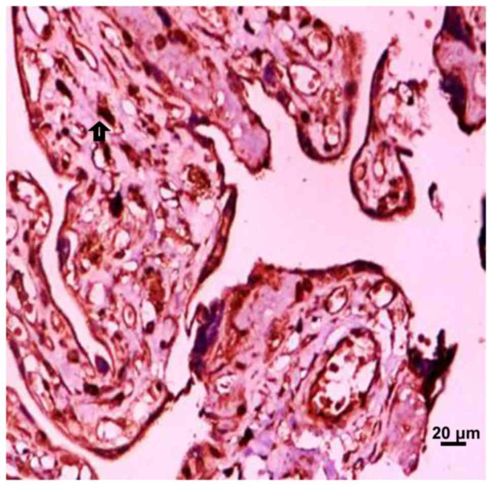
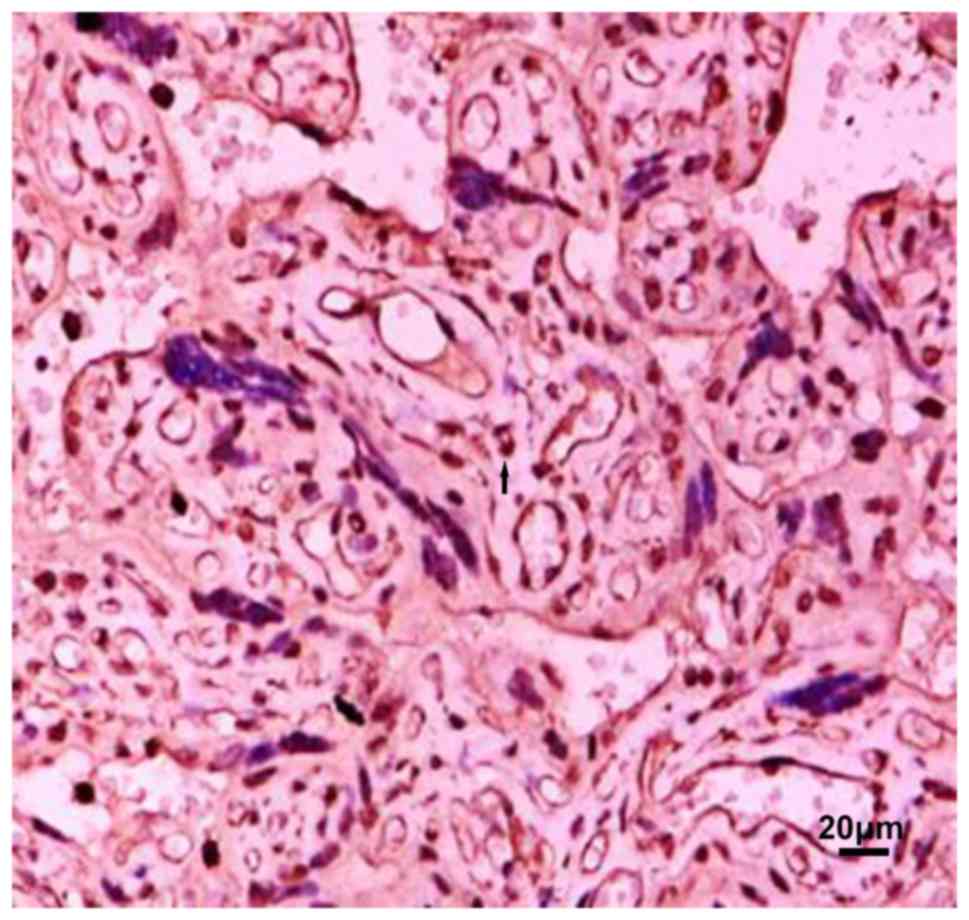
Localization and positive distribution
of MMP-9 in placental tissue
MMP-9 is mainly distributed in the cytoplasm of
trophoblast cells, vascular endothelial cells and villous
mesenchymal cells, but the expression intensity is different in
each group. The location of MMP-9 in the placenta tissue of the
preeclampsia group was the same as that in the normal late
pregnancy group. Among the four groups of placental grade (−), (+),
(++), (+++), the difference in the number of people in normal, mild
and severe group was statistically significant
(χ2=25.28, P<0.001). In the placenta grade (−), the
maximum number of people in the severe group was 6; in the placenta
grade (+). Most of the positive group was concentrated in the
severe group, and the least positive group was in the mild group;
the placenta grade (++). Most positive patients were concentrated
in mild group, and least number of positive patients were in severe
group. Number of positive patients with placenta grade (+++) was
concentrated in mild group, and least positive group was the severe
group. Distribution of MMP-9 positive particles in the placenta
tissue of severe preeclampsia group was significantly reduced
(Figs. 1–3 and Table
I). Pairwise comparison was carried out between mild and
severe, mild and normal, severe and normal groups. There was no
significant difference between mild and normal group. The
difference in MMP-9 between the other two groups was statistically
significant, that is, MMP-9 was expressed differently in the three
groups. With the aggravation of PIH, the distribution of positive
particles of MMP-9 was gradually reduced.
 | Table I.Expression of MMP-9 in placental
tissues in each group (n=30). |
Table I.
Expression of MMP-9 in placental
tissues in each group (n=30).
|
| Grading |
|---|
|
|
|
|---|
| Groups | − | + | ++ | +++ |
|---|
| Normal | 3 | 10 | 8 | 9 |
| Mild | 3 | 2 | 14 | 11 |
| Severe | 6 | 18 | 5 | 1 |
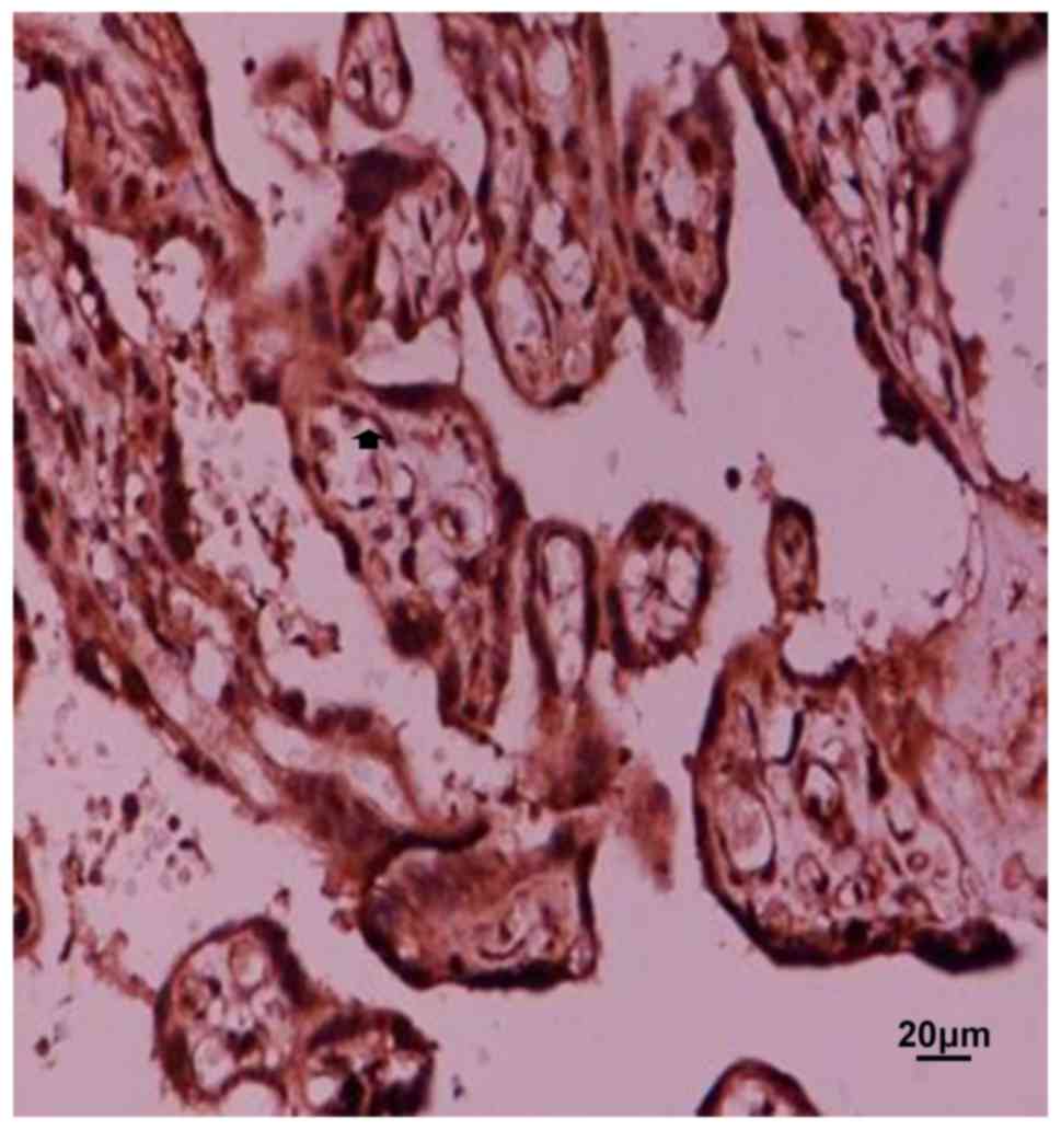
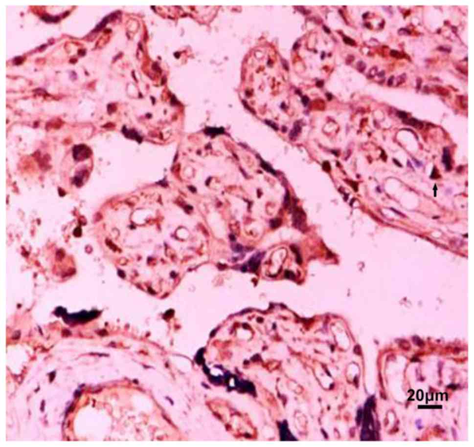
Location and positive expression of
TIMP-1 in placenta tissue
TIMP-1 is mainly distributed in the cytoplasm of
trophoblast cells, vascular endothelial cells and villous
mesenchymal cells. Pairwise comparison was carried out between mild
and severe, mild and normal, severe and normal groups. There was no
significant difference in the four groups of placental grades (−),
(+), (++), and (+++) (χ2=0.817, P>0.05), number of
positive in normal, mild and severe group was 0 in placenta grade
(−); most of the positive in the placenta grade (+), concentrated
in normal group, the least number of positives were in mild group,
the highest number of positive placenta (++) was concentrated in
normal group. The positive number of placenta in the (+++) group
was concentrated in mild group, and least positive group was normal
group. Number of positives in the three groups is basically
balanced (Figs. 4–6, Table
II).
| Table II.Expression of TIMP-1 in placental
tissues in each group (n=30). |
Table II.
Expression of TIMP-1 in placental
tissues in each group (n=30).
|
| Grading |
|---|
|
|
|
|---|
| Groups | − | + | ++ | +++ |
|---|
| Normal | 0 | 10 | 16 | 4 |
| Mild | 0 | 6 | 12 | 12 |
| Severe | 0 | 8 | 13 | 9 |
Positive expression ratio of MMP-9,
TIMP-1, MMP-9/TIMP-1 in placenta in each group
MMP-9 was expressed differently in the three groups
of placental tissues. With the aggravation of PIH, the distribution
of positive particles of MMP-9 was gradually reduced. MMP-9
positive particles in the placenta tissue of the severe
preeclampsia group were significantly reduced. TIMP-1 was expressed
in each group, and the difference was not statistically
significant. The positive expression ratio of MMP-9/TIMP-1 in the
severe group was lower than that in the normal pregnancy group and
the mild group, and the positive expression ratio of the two groups
gradually decreased as the condition worsened.
Discussion
Matrix metalloproteinases (MMPs) are a class of
proteolytic enzymes with similar structures and complex functions.
It is the most important enzyme in matrix degradation. It is named
after Zn2+ and Ca2+ and belongs to the family
of Zn2+-dependent enzymes. The interaction of MMPs with
the basement membrane (BM) is an important starting signal for
tumor invasion and metastasis (9).
MMP-9 is the only effective hydrolase secreted by trophoblast cells
that can digest ECM. It has been reported to be involved in
extracellular matrix (ECM) remodeling and processes of placental
angiogenesis with structural spiral arteries transformation, which
precedes proper trophoblastic invasion (10). TIMP-1 is a tissue inhibitory factor
corresponding to MMP-9 and is normally secreted by the same kind of
cells that secrete MMP-9. Usually combined with the MMP-9 zymogen
form into a 1:1 complex, which is linked by non-covalent bonds,
thereby preventing the activation of the zymogen and inhibiting the
activity of MMP-9 to degrade collagen. Even when activated MMP-9
acts on the basement membrane, it depends on the balance of MMP-9
and its inhibitory factor (TIMP-1), only when the balance is broken
(i.e. 30 MMP-9/TIMP-1 at >1:1), biological activity is produced
(11). Therefore, the balanced
expression of MMP-9 and TIMP-1 has profound implications for
regulating the depth of trophoblast invasion into the uterus.
Previous studies on serum MMP-9 and TIMP-1 in
patients with pregnancy-induced hypertension showed that serum
MMP-9 levels in severe group were significantly lower than those in
normal group and mild group (P<0.05), but there was no
significant difference between mild group and control group
(P>0.05), without statistical significance. There was no
significant difference in serum TIMP-1 between severe, mild and
normal groups (P>0.05), and the concentration of TIMP-1 in the
three groups did not change much (12), which is consistent with this
study.
Laird (13) found
that the placenta implantation process is strictly limited by the
time and space of MMPs and TIMPs, thus controlling the depth of
trophoblastic infiltration. The physiological changes in most
patients with PIH are limited to the aponeurotic segment of the
uterine artery. This study found that MMP-9 in placenta tissue is
mainly distributed in the cytoplasm of trophoblast cells, vascular
endothelial cells and villous mesenchymal cells, but the expression
intensity is different in each group, and MMP-9 is positive as the
severity of PIH is aggravated. The expression of the particles is
gradually decreasing. TIMP-1 and MMP-9 were consistently
distributed in each group, and the difference was not statistically
significant. The positive expression of MMP-9/TIMP-1 in the severe
group was lower than that in the normal pregnancy group and the
mild group, and the disease was aggravated. The positive expression
ratio of the two gradually decreased. It was observed that the
expression of MMP-9 decreased in the severe group, but the specific
inhibitor TIMP-1 had no corresponding change, and the equilibrium
state between the two changed. The expression of MMP-9 and TIMP-1
is related to the occurrence of preeclampsia and the development of
the disease. The level of MMP-9 in the placental tissue decreased,
and the balance between MMP-9 and TIMP-1 changed. The erosive cell
function was affected, which can lead to shallow implantation of
the placenta, resulting in ischemia and hypoxia of the placenta,
which eventually leads to maternal systemic vascular endothelial
damage, thus showing the clinical symptoms of PIH. Recent studies
have also shown that the extent of injury or disease can be
estimated by measuring MMP-9 concentration, and the prognostic
effect of treatment can be preliminarily predicted (7).
Moreover, in vitro experiments have shown
that the trophoblast infiltration ability is significantly reduced
because the expression of MMP-9 is lower than the normal level
required for infiltration (8). The
above suggests that MMP-9 and TIMP-1 act as a pair of mutually
restrictive factors playing an important role in the pathogenesis
of PIH.
Acknowledgements
Not applicable.
Funding
No funding was received.
Availability of data and materials
The datasets used and/or analyzed during the current
study are available from the corresponding author on reasonable
request.
Authors' contributions
YuyiZ wrote the manuscript. YuyiZ and YuboZ were
responsible for immunohistochemical staining. PL and YG evaluated
the results of dyeing. XL and YuyiZ worked on specimen preparation.
All the authors read and approved the final manuscript.
Ethics approval and consent to
participate
This study was approved by the Ethics Committee of
People's Hospital of Xinjiang Uygur (Urumqi, China). Patients who
participated in this research had complete clinical data. The
signed informed consents were obtained from the patients or the
guardians.
Patient consent for publication
Not applicable.
Competing interests
The authors declare that they have no competing
interests.
References
|
1
|
Cerdeira AS and Karumanchi SA: Angiogenic
factors in preeclampsia and related disorders. Cold Spring Harb
Perspect Med. 2:0065852012. View Article : Google Scholar
|
|
2
|
Vadhera RB and Simon M: Hypertensive
emergencies in pregnancy. Clin Obstet Gynec. 57:797–805. 2014.
View Article : Google Scholar : PubMed/NCBI
|
|
3
|
Winn VD, Gormley M and Fisher SJ: The
impact of preeclampsia on gene expression at the maternal-fetal
interface. Pregnancy Hypertens. 1:100–108. 2011. View Article : Google Scholar : PubMed/NCBI
|
|
4
|
Kleinrouweler CE, Wiegerinck MM,
Ris-Stalpers C, Bossuyt PM, van der Post JA, von Dadelszen P, Mol
BW and Pajkrt E; EBM CONNECT Collaboration, : Accuracy of
circulating placental growth factor, vascular endothelial growth
factor, soluble fms-like tyrosine kinase 1 and soluble endoglin in
the prediction of pre-eclampsia: A systematic review and
meta-analysis. BJOG. 119:778–787. 2012. View Article : Google Scholar : PubMed/NCBI
|
|
5
|
Chen J, Yu YH, Wang ZJ, Qin W and Zhang Q:
Relationship between preeclampsia umbilical blood flow and
perinatal outcomes. Nan Fang Yi Ke Da Xue Xue Bao. 29:745–746.
2009.(In Chinese). PubMed/NCBI
|
|
6
|
Zhu J, Zhong M, Pang Z and Yu Y:
Dysregulated expression of matrix metalloproteinases and their
inhibitors may participate in the pathogenesis of pre-eclampsia and
fetal growth restriction. Early Hum Dev. 90:657–664. 2014.
View Article : Google Scholar : PubMed/NCBI
|
|
7
|
Shi H, Liu L, Liu LM, Geng J and Chen L:
Inhibition of tumor growth by β-elemene through downregulation of
the expression of uPA, uPAR, MMP-2, and MMP-9 in a murine
intraocular melanoma model. Melanoma Res. 25:15–21. 2015.
View Article : Google Scholar : PubMed/NCBI
|
|
8
|
McCool WF, Durain D and Davis M: Overview
of latest evidence on uterine fibroids. Nurs Womens Health.
18:314–332. 2014. View Article : Google Scholar : PubMed/NCBI
|
|
9
|
Jaiswar SP, Gupta A, Sanchan R, Natu SN
and Shaili M: Latic dehydrogenase: A biochemical marker for
preeclampsia-eclampsia. J Obstet Gynaecol India. 61:645–648. 2011.
View Article : Google Scholar : PubMed/NCBI
|
|
10
|
Maged AM, Aid G, Bassiouny N, Eldin DS,
Dahab S and Ghamry NK: Association of biochemical markers with the
severity of pre-eclampsia. Int J Gynaecol Obstet. 136:138–144.
2017. View Article : Google Scholar : PubMed/NCBI
|
|
11
|
Sun C, Zhang Q, Hu B and Zhang K:
Investigation of the association between matrix metalloproteinase-9
genetic polymorphisms and development of pre-eclampsia in Chinese
pregnant women. Genet Mol Res. 15:1–6. 2016. View Article : Google Scholar
|
|
12
|
Zhang YY, Ren HY, Wang YT, Deng J, Liu XW
and Li XY: Expression of MMP-9 and TIMP-1 in placental tissue of
pregnancy-induced hypertension in Uigur women in Xinjiang. Progress
Modern Biomed. 19:3710–3712. 2009.(In Chinese).
|
|
13
|
Laird SM: Metalloproteinases and tissue
inhibitor ormetalloproteinase-1 (TIMP-1) in endometrial flushings
from pre-and post-menopausal women and from women with endometrial
adenocarcinoma. J Reprod Fertil. 115:225–232. 1999. View Article : Google Scholar : PubMed/NCBI
|