Introduction
Debris generated by prosthetic wear after artificial
total hip arthroplasty and the osteolysis caused by inflammation
cause symptoms such as pain, movement disorder and bone defect in
the hip joint. At this time, acetabular prosthesis needs to be
repaired to improve the therapeutic effect of hip replacement and
prolong the service life of the prosthesis (1,2).
At present, there are many repair methods for
acetabular prostheses, such as titanium mesh reconstruction,
structural bone graft reconstruction and acetabular plate
readdition. There are also many types of materials needed to repair
the prosthesis, such as hydroxyapatite, Cage and Bio-type cups, the
various prosthetic materials have their own advantages and
disadvantages, but the common purpose is to repair and reconstruct
the appropriate acetabulum to achieve good stability of the cup
prosthesis (3,4). In recent years, 3D printing technology
has developed rapidly and a variety of 3D technologies have been
applied in the medical field (5). 3D
printed titanium alloy is widely used in the repair and
re-treatment of various types of bone and joint injuries. Compared
with traditional bone and joint defects repair materials, 3D
printed titanium alloy has the advantages of strong forming ability
and short processing cycle, and is tailor-made according to the
patient.
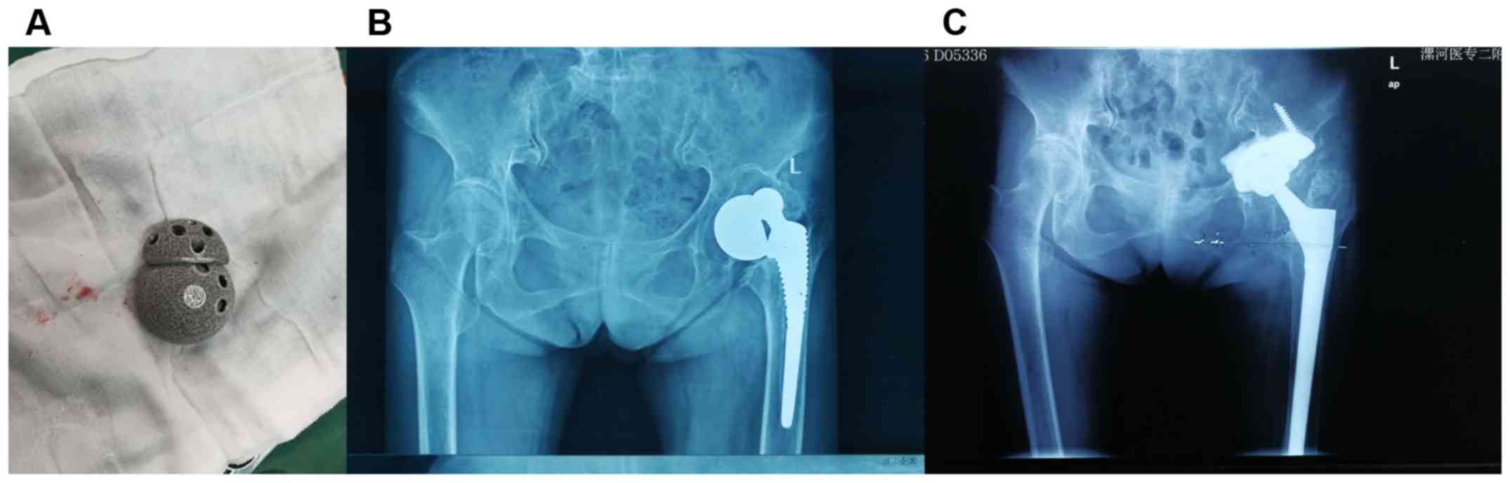
In this study, 3D printed titanium alloy trabecular
cup and titanium alloy block were used to repair the acetabular
defect after hip replacement. The curative effect and prognosis
were satisfactory.
Patients and methods
Research subjects
Forty-two patients who underwent acetabular repair
in the Second Affiliated Hospital of Luohe Medical College (Luohe,
China) from July 2014 to June 2017 were enrolled in observation and
control groups according to acetabular revision methods and
materials. There were 22 patients in the observation group,
including 12 males and 10 females, aged 22–58 years, with a mean
age of 35.8±6.7 years. There were 20 patients in the control group,
including 11 males and 9 females, aged 23–57 years, with a mean age
of 34.9±5.9 years.
This study was approved by Ethics Committee of the
Second Affiliated Hospital of Luohe Medical College. Patients who
participated in this study, signed the informed consent and had
complete clinical data.
Inclusion and exclusion criteria
Inclusion criteria were: i) patients who had
undergone total hip arthroplasty with only one side; ii) volumetric
or structural bone defects, pain and mobility disorders in
acetabular weight-bearing area, anterior-posterior column and inner
wall; iii) image examination showed that the displacement of the
cup in the vertical and horizontal directions exceeded 2.0 mm; iv)
rotation of the cup exceeded 5.0°; and v) acetabulum was rotated
and the screw was broken. Exclusion criteria were: i) trauma caused
by periprosthetic fracture; ii) first total hip arthroplasty; iii)
infection around the prosthesis; and iv) patients or family members
did not agree to participate in the study.
Renovation method
The patients were placed in lateral position.
General anesthesia was performed. Through posterior lateral
approach, incision of the sac, exposing the hip prosthesis, fully
revealing the acetabular rim, removing the prosthesis, removing
scars, bone cement and conjunctiva around the acetabulum were
performed. Bone was polished and the defect area of the acetabulum
and the extent of bone defect were evaluated. After treatment and
evaluation, patients in control group were equipped with non-3D
printed titanium alloy revision artificial hip joint for revision
in the acetabular defect. Patients in the observation group were
fitted with 3D printed pad at the acetabular defect to restore
acetabular edge of defect. A small amount of allogeneic or
autologous bone was placed to fill the bone defect cavity and
reconstruct the acetabulum. After that, installation of 3D printed
titanium trabecular cup was performed. If the femoral head
prosthesis was severely rubbed, femoral head prosthesis was
replaced intraoperatively. After the revision of the prosthesis,
saline was used to wash the cavity and wound was sutured.
Postoperative routine anti-infective treatment and other basic
treatments were performed. Patients were asked to wear
anti-spinning shoes to prevent hemorrhoids and ankle joint
contraction movement was performed within 6 weeks after surgery.
After 6 weeks, weight-bearing exercise was started according to the
bone growth of the interface.
Follow-up and efficacy evaluation
Patients were followed up at 3, 6 and 12 months
after surgery. Radiographic assessment and scale assessment were
performed during follow-up. Visual analog scale (VAS score) was
used to evaluate the preoperative and postoperative pain level
(6). Harris score was used to
evaluate the preoperative and postoperative hip function (7). Quality of life before and after surgery
was assessed using the Health Survey Scale (SF-36) (8). Assessment of acetabular prosthesis
position was performed using the DeLee and Charnley zoning method
(9): acetabular component is
unstable: a displacement of the cup or an outer acetabulum, and a
translucent line of at least 1.0 mm in the lower rim of the
acetabulum were considered as unstable acetabular component. Loose
acetabular prosthesis: acetabular abduction angle varies by
>10°, or a displacement of >6.0 mm occurs vertically or
horizontally. Evaluation of the ingrowth of the prosthesis was
performed using the bone growth evaluation criteria of the Anderson
Orthopaedic Institute (10): i) the
bright line disappeared; ii) the outer upper part of the acetabulum
and the inner lower acetabular bone edge were enhanced; iii) stress
shielding on the inner wall of the acetabulum; and iv) radial
trabecular bones are arranged perpendicular to the acetabular outer
upper part and the inner wall of the acetabulum and the acetabular
surface.
Statistical analysis
Statistical analysis was performed on the data in
this study using SPSS 19.0 statistical software (SPSS, Inc.,
Chicago, IL, USA). Measurement data were expressed as mean ±
standard deviation. Difference of measurement data between groups
were statistically analyzed by t-test. P<0.05 was considered to
indicate a statistically significant difference.
Results
Comparison of preoperative and
postoperative patient hip function Harris scores between two
groups
Hip function Harris scores were compared between the
groups before and 3, 6 and 12 months after surgery. There was no
significant difference in Harris score between the two groups
before operation (P>0.05). At 3, 6 and 12 months after
operation, Harris scores of the observation group were
significantly higher than those of the control group (P<0.05;
Table I and Fig. 1).
 | Table I.Comparison of preoperative and
postoperative patient hip function Harris scores between the two
groups. |
Table I.
Comparison of preoperative and
postoperative patient hip function Harris scores between the two
groups.
| Groups | Cases | Before treatment | 3 months after
treatment | 6 months after
treatment | 12 months after
treatment |
|---|
| Control group | 20 | 44.53±8.83 | 53.52±9.11 | 67.62±8.20 | 72.15±8.30 |
| Observation
group | 22 |
45.11±8.93a |
68.35±9.57a |
82.52±9.01a |
88.57±9.25a |
Comparison of preoperative and
postoperative pain VAS scores between the groups
Pain VAS scores of the two groups were compared
before and 3, 6 and 12 months after surgery. There was no
significant difference in pain VAS score between the groups before
operation (P>0.05). Pain VAS scores of the observation group
were significantly lower than those of the control group at 3, 6
and 12 months after operation (P<0.05; Table II).
| Table II.Comparison of preoperative and
postoperative pain VAS scores between the two groups (points). |
Table II.
Comparison of preoperative and
postoperative pain VAS scores between the two groups (points).
| Groups | Cases | Before treatment | 3 months after
treatment | 6 months after
treatment | 12 months after
treatment |
|---|
| Control group | 20 | 5.88±1.12 | 3.89±0.87 | 2.66±0.54 | 2.34±0.49 |
| Observation
group | 22 |
5.79±1.14a |
2.79±0.69a |
1.04±0.48a |
0.85±0.36a |
Comparison of preoperative and
postoperative quality of life SF-36 scores between the groups
Quality of life SF-36 scores were compared between
the groups before and 3, 6 and 12 months after surgery. There was
no significant difference in the quality of life SF-36 scores
between the two groups before treatment (P>0.05). The quality of
life SF-36 scores of the observation group were significantly
higher than those of the control group at 3, 6 and 12 months after
treatment (P<0.05; Table
III).
| Table III.Comparison of preoperative and
postoperative quality of life SF-36 scores between the two groups
(points). |
Table III.
Comparison of preoperative and
postoperative quality of life SF-36 scores between the two groups
(points).
| Groups | Cases | Before treatment | 3 months after
treatment | 6 months after
treatment | 12 months after
treatment |
|---|
| Control group | 20 | 330.13±90.34 | 501.25±86.36 | 620.41±90.44 | 650.62±90.11 |
| Observation
group | 22 |
334.57±91.31a |
589.32±83.16a |
735.67±92.46a |
752.41±90.56a |
Comparison of postoperative acetabular
component position and bone ingrowth between the groups of
patients
There was no change in displacement and abduction
angle in the observation group. In 20 patients, the cup was in
close contact with the bone surface 1 week after surgery and there
was no bright line under the X-ray. Two cases had bright lines 1
week after surgery, but the bright lines disappeared 6 months after
operation. None of the patients showed a bright line at the last
follow up. None of the patients had loosening of the prosthesis 6
months after surgery. There was continuous trabecular passage at
the junction of all the patients' prosthesis and host bone. In the
control group, 15 patients had tight contact with bone surface at 1
week after surgery but no bright lines were observed under X-ray
and 4 patients had bright lines at 1 week after surgery, but lines
disappeared at 6 months after surgery. None of the patients showed
bright line at the last follow-up. Revision failed in one patient,
18 patients had no loosening at 6 months after surgery and 18
patients had continuous trabecular passage at the junction of
prosthesis and host bone.
Discussion
In recent years, the number of patients undergoing
total hip arthroplasty increased year by year and the patients are
becoming younger and younger. Artificial total hip joints are worn
away during daily work and life activities and they need to be
refurbished after a long period of time. The difficulty of revision
surgery is greater than that of primary hip replacement (11). A large number of clinical
applications have found some methods and material design of hip
replacement in the past, such as placement of cups in high-rotation
centers, structural bone grafting of large bones, rotating
reinforcing rings, titanium mesh combined with particle bone
crushing and bone grafting, which have their own problems, such as
complicated operation procedures, long duration of surgery, large
amount of allogeneic bone, high replacement cost, easy infection,
poor biological stability and insufficient biomechanical efficacy
(12–14). Therefore, how to safely and
effectively repair the artificial hip joint has become a difficult
problem in orthopedics.
Electron beam melting technology is an important
part of 3D printing technology. Ideal bone growth interface can be
obtained by electron beam melting technology. Metal solid layer and
the surface porous layer of the printed prosthesis are formed once.
Preparation of titanium alloy trabecular cup used in hip repair in
this study is completed in a vacuum environment, so contamination
of the cup by external environment was avoid. In addition, it is
done at a constant temperature, resulting in good shape stability
and low residual stress in the printed cups (2). At present, non-cemented prosthesis is
often used in clinical revision of the hip joint. However,
prosthesis of these materials has a low porosity and a non-uniform
aperture. After revision, bone is difficult to grow into the
prosthesis, resulting in an unsatisfactory revision effect. 3D
printed titanium alloy trabecular cup and the pad have good
bio-identity and the cells are easy to attach and grow on the
surface of the prosthesis, which is beneficial to the osteogenic
differentiation of stem cells. In this study, 3D printed titanium
alloy trabecular metal cups and pads were used to repair the hip
joint. Postoperative hip function Harris scores, pain VAS scores
and quality of life SF-36 scores were improved compared with non-3D
printed titanium alloy trabecular metal cups and pads. In addition,
the stability and bone stenosis of hip prosthesis were better than
non-3D printed titanium trabecular metal cups and pads. Therefore,
use of 3D printed titanium trabecular metal cups and pads to repair
the hip joint can better restore patients' hip function, reduce
pain and improve the quality of life of patients. Improved clinical
outcomes were achieved due to sufficient porosity, uniform pore
size and excellent surface friction coefficient of 3D printed
titanium alloy trabecular metal cups and pads.
It was found that when using 3D printed titanium
alloy trabecular metal cups and pads to repair the hip joint, there
are a few points to note: i) posterior lateral surgical approach
can fully reveal the femoral shaft and posterior column of
acetabulum, but it also increases the risk of dislocation; ii) when
the acetabular prosthesis is removed with a thin bone knife, it can
be operated between the cup and the bone cement. Operation between
the acetabular wall and the bone cement can damage the acetabulum
and increase the risk of fracture; iii) when the acetabular
component is dislocated to acetabulum, care should be taken to
avoid damage to the blood vessel during revision surgery; iv) when
removing the bone cement at the bottom of the acetabulum, breaking
the acetabular humerus into the pelvic cavity must be avoided; and
v) when removing the bio-fixed cup prosthesis, a special acetabular
neutral locator should be used to avoid damage to the acetabular
wall.
Clinical effect of revision of the hip prosthesis
was satisfactory with 3D printed titanium trabecular metal cups and
pads. However, the long-term efficacy is uncertain because
follow-up was only performed for 1 year after surgery. Further
research is needed to monitor long-term efficacy.
Acknowledgements
Not applicable.
Funding
No funding was received.
Availability of data and materials
The datasets used and/or analyzed during the present
study are available from the corresponding author on reasonable
request.
Authors' contributions
LW and GW worked on the renovation method. PC and KL
collected and analyzed general data of patients. JL and SZ were
responsible for follow-up analysis. LW wrote the manuscript. All
authors read and approved the final manuscript.
Ethics approval and consent to
participate
The study was approved by the Ethics Committee of
the Second Affiliated Hospital of Luohe Medical College (Luohe,
China). Patients who participated in this study, signed the
informed consent and had complete clinical data.
Patient consent for publication
Not applicable.
Competing interests
The authors declare that they have no competing
interests.
References
|
1
|
Gladnick BP, Fehring KA, Odum SM, Christie
MJ, DeBoer DK and Fehring TK: Midterm survivorship after revision
total hip arthroplasty with a custom triflange acetabular
component. J Arthroplasty. 33:500–504. 2018. View Article : Google Scholar : PubMed/NCBI
|
|
2
|
Nakashima Y, Mashima N, Imai H, Mitsugi N,
Taki N, Mochida Y, Owan I, Arakaki K, Yamamoto T, Mawatari T, et
al: Clinical and radiographic evaluation of total hip
arthroplasties using porous tantalum modular acetabular components:
5-year follow-up of clinical trial. Mod Rheumatol. 23:112–118.
2013. View Article : Google Scholar : PubMed/NCBI
|
|
3
|
Pierannunzii L, Mambretti A and
D'Imporzano M: Trabecular metal cup without augments for acetabular
revision in case of extensive bone loss and low bone-prosthesis
contact. Int J Immunopathol Pharmacol. 24 (Suppl 2):133–137. 2011.
View Article : Google Scholar : PubMed/NCBI
|
|
4
|
Sadhu A, Nam D, Coobs BR, Barrack TN,
Nunley RM and Barrack RL: Acetabular component position and the
risk of dislocation following primary and revision total hip
arthroplasty: a matched cohort analysis. J Arthroplasty.
32:987–991. 2017. View Article : Google Scholar : PubMed/NCBI
|
|
5
|
Wang B, Hao Y, Pu F, Jiang W and Shao Z:
Computer-aided designed, three dimensional-printed hemipelvic
prosthesis for peri-acetabular malignant bone tumour. Int Orthop.
42:687–694. 2018. View Article : Google Scholar : PubMed/NCBI
|
|
6
|
Dimitroulis G, Austin S, Sin Lee PV and
Ackland D: A new three-dimensional, print-on-demand
temporomandibular prosthetic total joint replacement system:
Preliminary outcomes. J Craniomaxillofac Surg. 46:1192–1198. 2018.
View Article : Google Scholar : PubMed/NCBI
|
|
7
|
Gustke KA, Levering MF and Miranda MA: Use
of jumbo cups for revision of acetabulae with large bony defects. J
Arthroplasty. 29:199–203. 2014. View Article : Google Scholar : PubMed/NCBI
|
|
8
|
Adelani MA, Crook K, Barrack RL, Maloney
WJ and Clohisy JC: What is the prognosis of revision total hip
arthroplasty in patients 55 years and younger? Clin Orthop Relat
Res. 472:1518–1525. 2014. View Article : Google Scholar : PubMed/NCBI
|
|
9
|
Nie WB, Ye FG, Ma JL, Yu JP, Wang MX,
Zhang ZH and Sun FJ: Three-dimensional (3D) printing technology
assisted by minimally invasive surgery for pubic rami fractures.
Curr Med Sci. 38:827–833. 2018. View Article : Google Scholar : PubMed/NCBI
|
|
10
|
Flecher X, Ollivier M, Maman P, Pesenti S,
Parratte S and Argenson JN: Long-term results of custom
cementless-stem total hip arthroplasty performed in hip fusion. Int
Orthop. 42:1259–1264. 2018. View Article : Google Scholar : PubMed/NCBI
|
|
11
|
Lenguerrand E, Whitehouse MR, Beswick AD,
Kunutsor SK, Burston B, Porter M and Blom AW: Risk factors
associated with revision for prosthetic joint infection after hip
replacement: A prospective observational cohort study. Lancet
Infect Dis. 18:1004–1014. 2018. View Article : Google Scholar : PubMed/NCBI
|
|
12
|
Huang P, Lyons M and O'Sullivan M: The
infection rate of metal-on-metal total hip replacement is higher
when compared to other bearing surfaces as documented by the
Australian Orthopaedic Association National Joint Replacement
Registry. HSS J. 14:99–105. 2018. View Article : Google Scholar : PubMed/NCBI
|
|
13
|
Meek RM, Norwood T, Smith R, Brenkel IJ
and Howie CR: The risk of peri-prosthetic fracture after primary
and revision total hip and knee replacement. J Bone Joint Surg Br.
93:96–101. 2011. View Article : Google Scholar : PubMed/NCBI
|
|
14
|
Panagiotidou A, Meswania J, Hua J,
Muirhead-Allwood S, Hart A and Blunn G: Enhanced wear and corrosion
in modular tapers in total hip replacement is associated with the
contact area and surface topography. J Orthop Res. 31:2032–2039.
2013. View Article : Google Scholar : PubMed/NCBI
|