Introduction
Gestational diabetes mellitus (GDM) is a common
metabolic complication (1), which
refers to the occurrence of symptoms such as abnormal glucose
tolerance or glucose intolerance in different degrees during
pregnancy, posing a great threat to the health of pregnant women
and the fetus (2). In recent years,
the incidence of GDM has gradually increased (3). GDM is a high-risk pregnancy, which will
cause adverse effects on mother and baby. Before the treatment, the
preperinatal mortality is approximately 40%, and the maternal
mortality is up to 27–30% (4,5).
Placenta is an essential organ for babies' growth and development.
During normal and pathological pregnancy, placenta can adapt to
changes in intrauterine environment (6).
The AGEs-RAGE signaling pathway, formed by advanced
glycation end product receptor (RAGE) and its ligands, advanced
glycation end products (AGEs), is closely related to the occurrence
and development of glucose metabolism disorders, and can activate
inflammatory reactions and oxidative stress (7). The AGEs accumulation caused by
persistent hyperglycemia during gestation is one of the main
factors leading to abnormal fetal development (8). Visfatin, a newly discovered
adipocytokine, is mainly secreted by visceral fat cells and is
related to insulin secretion, glucose uptake and abnormal glucose
tolerance (9). Interleukin-6 (IL-6),
an important chronic inflammatory cytokine secreted by adipose
tissue, is an adipocytokine and is associated with states of
various insulin resistance (IR) in the body and can be released
from the placenta (10). Adiponectin
(APN) is a biologically active peptide hormone secreted by fat
cells, which can be used to enhance insulin sensitivity, it is
anti-inflammatory, and maintains the body's glucose metabolism
balance (11). Studies have shown
that inflammatory factors and adipokines are significantly
associated with IR formation (12).
Oxidative stress plays an important role in GDM
(13). It is caused by an imbalance
between the oxidant and the antioxidant (14). Oxidative stress is considered to be a
causative factor in human pregnancy-related diseases, such as fetal
death, pre-eclampsia, and intrauterine growth restriction (15). Malondialdehyde (MDA) is a product of
lipid peroxidation and is produced by the degradation of
phospholipids induced by reactive oxygen species (ROS) under
pathogenic conditions (16). Changes
in MDA levels can be used to reflect the degree of oxidative damage
in the body (17). At present, there
are few studies on the correlation between adipokines and oxidative
stress in GDM.
In this study, the correction between AGEs,
visfatin, APN, IL-6 and MDA was investigated by detecting the
expression levels of AGEs and adipocytokines (visfatin, APN, IL-6)
in placental tissues of pregnant women with GDM, to further improve
the diagnosis and treatment of GDM and provide a reference for the
pathogenesis of GDM.
Patients and methods
Methods
Seventy-two cases of GDM pregnant women who
underwent routine prenatal examination and delivery in the
Department of Obstetrics, Binzhou City Center Hospital (Binzhou,
China) from March 2016 to May 2017 were enrolled as the observation
group. Glucose tolerance test (OGTT) (75 g) was performed at 24–28
weeks of gestation. The diagnosis was made according to the
International Association of Diabetes and Pregnancy Study Groups
(IADPSG) standard (18). The mean
age was 28.76±4.58 years, the pre-pregnancy body mass index was
23.17±3.16 kg/m2, and the mean gestational age was
38.82±1.18 weeks; another 80 cases of normal pregnant women who
were delivered at the same time were selected as the control group,
with an average age of 27.99±5.82 years, a pre-pregnancy body mass
index of 22.86±2.66 kg/m2, and an average gestational
age of 39.11±1.03 weeks.
Inclusion criteria were: Patients with singleton
pregnancy, with no previous heart, liver and kidney disease,
chronic hypertension and diabetes, and no dietary restrictions,
with complete clinical data, without any smoking, drinking or other
harmful habits.
Exclusion criteria were: Patients taking drugs
affecting blood glucose metabolism three months before pregnancy,
with premature rupture of the membranes, with a recent history of
infection, with family hereditary mental illness, and with inactive
cooperation with inspections.
The study was approved by the Ethics Committee of
Binzhou City Center Hospital. Patients who participated in this
research had complete clinical data. The signed informed consents
were obtained from the patients or the guardians.
Specimen collection
Within 4 h after delivery of the placenta, 3 samples
of placental tissue in sterile state were obtained from the center
of the placenta maternal surface, each approximately 1×1×1 cm in
size; avoiding the bleeding, infarct and calcification area. Tissue
was washed repeatedly with sterilized physiological saline, and
gauze used to remove water. Specimens under aseptic condition were
cut up, RIPA lysate (Shanghai Yisheng Biological Technology Co.,
Ltd.) was used for pyrolysis, and then were ground into homogenate,
centrifuged at 10,000 × g for 15 min at 4°C, finally the
supernatant was taken into the trace centrifuge tube, stored at
−80°C for later use.
Detection method
The expression levels of AGEs, visfatin, APN and
IL-6 in the placental tissue lysate were detected by enzyme-linked
immunosorbent assay (ELISA), and the standard well, the sample well
and the blank well (no samples and enzyme-labeled reagents were
added, and the other steps were the same) were set, respectively.
First, 50 µl of standard sample was added accurately on the
enzyme-labeled coated plate, 40 µl of diluent was added in the
sample well, then 10 µl of sample to be tested was added. Plate
membrane was used to seal the plate, and incubated at 37°C for 30
min. Then the plate membrane was peeled off, the liquid was
discarded, dried, and each well was filled with scrubbing liquid,
discarded after standing, this was repeated five times, and then
patted dry. In addition to blank wells, 50 µl of enzyme-labeled
reagent was added to each well for incubation and washing. After
that, 50 µl of chromogenic agent A and 50 µl of chromogenic agent B
were added to each well, shaked gently, and developed at 37°C for
15 min in the dark. Then 50 µl of stopping solution was added to
each well to immediately terminate the reaction when the blue
turned yellow. The measurement was carried out within 15 min after
the addition of the stopping solution, the blank well was used to
adjust to zero and the absorbance (OD value) of each well was
measured at the wavelength of 450 nm. ELISA kits of AGEs were
provided by Wuhan Mossak Biotechnology Co., Ltd., while ELISA kits
of visfatin, APN and IL-6 were from Shanghai Jining Industry Co.,
Ltd. SK201 microplate reader was from Shenzhen Shengxinkang
Technology Co., Ltd.
Level of MDA was measured by thiobarbituric acid
method. Samples were taken and added with trichloroacetic acid and
a small amount of quartz sand to grind, and then trichloroacetic
acid was added to fully grind them. After centrifugation at 3,680 ×
g for 10 min at 20°C, 2 ml of supernates (sample suspension buffer)
were taken and added with 2 ml of 0.6% thiobarbituric acid, mixed
and heated in a boiling water bath for 10 min, cooled, and 2 ml of
distilled water was used to replace the extract as a control, and
the OD value was measured at 532, 450 and 600 nm. The kit provided
was by Nanjing Jiancheng Biological Engineering Co., Ltd and it was
performed in strict accordance with the instructions.
Observational indicators
Expression levels of AGEs, adipocytokines and MDA in
placental tissues of the two groups were detected, and differences
between two groups were compared. Correlation between MDA and
expression levels of AGEs, visfatin, APN and IL-6 was analyzed.
Statistical processing
SPSS 19.0 software system (IBM Corp., Armonk, NY,
USA) was used for statistical analysis of experimental data.
Enumeration data were expressed as [n (%)], and Chi-square test was
used for comparison between groups. Measurement data were expressed
as (mean ± SD), and independent sample t-test was used for
comparison between groups. Pearson's correlation coefficients were
used to analyze the correlation of bivariate normal distribution
data. P<0.05 was considered to indicate a statistically
significant difference.
Results
Comparison of general clinical
data
General clinical data of the two groups were
collected. Observation and control group showed no significant
difference in age, body mass index before pregnancy, history of
childbirth, history of hypertension, ethnicity, gestational age,
systolic blood pressure, diastolic blood pressure and other aspects
(P>0.05), which were comparable. Fasting blood-glucose of
observation group was significantly higher than that of control
group (P<0.05) (Table I).
 | Table I.Comparison of general clinical data
between the two groups (mean ± SD)/[n (%)]. |
Table I.
Comparison of general clinical data
between the two groups (mean ± SD)/[n (%)].
| Factors | Observation group
(n=72) | Control group
(n=80) | χ2/t
value | P-value |
|---|
| Age (years) | 28.76±4.58 | 27.99±5.82 | 0.900 | 0.370 |
| Body mass index
before pregnancy (kg/m2) | 23.17±3.16 | 22.86±2.66 | 0.656 | 0.513 |
| History of
childbirth |
| Yes | 16 (22.2) | 20 (25.0) |
|
|
| No | 56 (77.8) | 60 (75.0) |
|
|
| History of
hypertension |
|
Yes | 8
(11.1) | 12 (15.0) |
|
|
| No | 64 (88.9) | 68 (85.0) |
|
|
| Ethnicity |
|
Han | 67 (93.1) | 71 (88.8) |
|
|
|
Rest | 5 (6.9) | 9
(11.2) |
|
|
| Gestational age
(weeks) | 38.82±1.18 | 39.11±1.03 | 1.618 | 0.108 |
| Systolic blood
pressure (mmHg) | 117.37±12.87 | 118.28±12.76 | 0.437 | 0.663 |
| Diastolic blood
pressure (mmHg) | 75.89±7.96 | 76.08±7.57 | 0.151 | 0.880 |
| Fasting
blood-glucose (mmol/l) | 6.76±0.85 | 5.22±0.67 | 12.47 | <0.001 |
Expression levels of AGEs and
adipocytokines in placental tissues of the two groups
Expression levels of AGEs and adipocytokines in
placental tissues of observation and control group were detected.
Results showed that the expression levels of AGEs and IL-6 in
placental tissues of observation group were significantly higher
than those of control group (t=8.861, 8.305, P<0.001).
Expression level of APN in placental tissues of observation group
was significantly lower than that of control group, and difference
was statistically significant (t=30.53, P<0.001). There was no
significant difference in expression of visfatin between the two
groups (P>0.05) (Table II).
| Table II.Comparison of expression levels of
AGEs, visfatin, APN and IL-6 in placental tissues between the two
groups (mean ± SD). |
Table II.
Comparison of expression levels of
AGEs, visfatin, APN and IL-6 in placental tissues between the two
groups (mean ± SD).
| Group | No. of cases | AGEs (ng/ml) | Visfatin
(ng/ml) | APN (mg/l) | IL-6 (ng/ml) |
|---|
| Observation group
(n=72) | 72 | 54.27±18.28 | 0.46±0.23 | 0.70±0.09 | 1.44±0.58 |
| Control group
(n=80) | 80 | 32.18±12.12 | 0.52±0.25 | 1.29±0.14 | 0.78±0.39 |
| t value |
| 8.861 | 1.534 | 30.53 | 8.305 |
| P-value |
| <0.001 | 0.127 | <0.001 | <0.001 |
Expression levels of MDA in placental
tissues of the two groups
The expression level of oxidative stress MDA in
placental tissues of observation and control group was detected,
and results showed that the expression level of MDA in placental
tissues of observation group was significantly higher than that of
control group, and difference was statistically significant
(t=16.44, P<0.001) (Table
III).
| Table III.Expression levels of MDA in placental
tissues of the two groups (mean ± SD) |
Table III.
Expression levels of MDA in placental
tissues of the two groups (mean ± SD)
| Groups | No. of cases | MDA (ng/ml) |
|---|
| Observation group
(n=72) | 72 | 6.21±1.03 |
| Control group
(n=80) | 80 | 3.87±0.71 |
| t value |
| 16.44 |
| P-value |
| <0.001 |
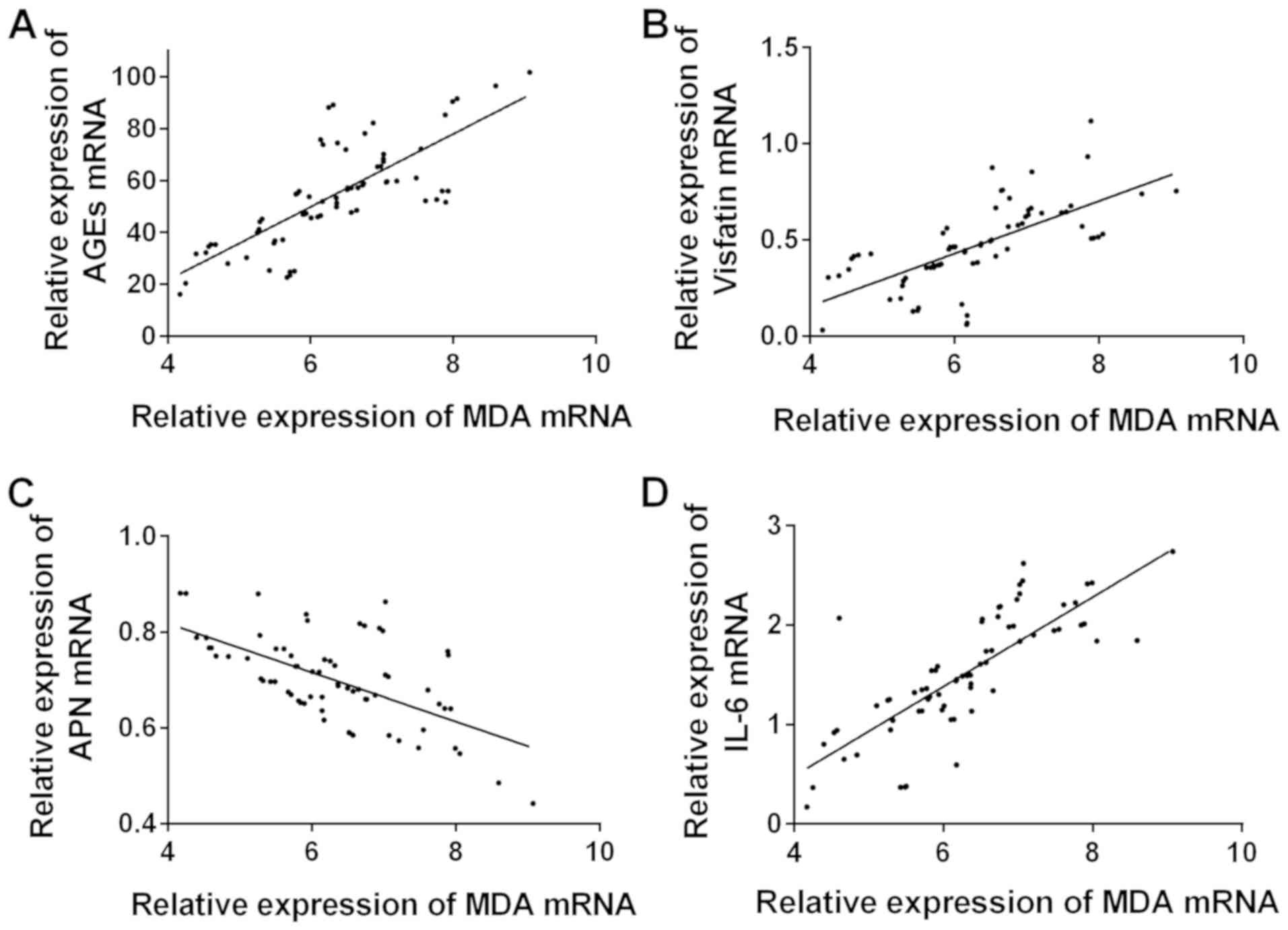
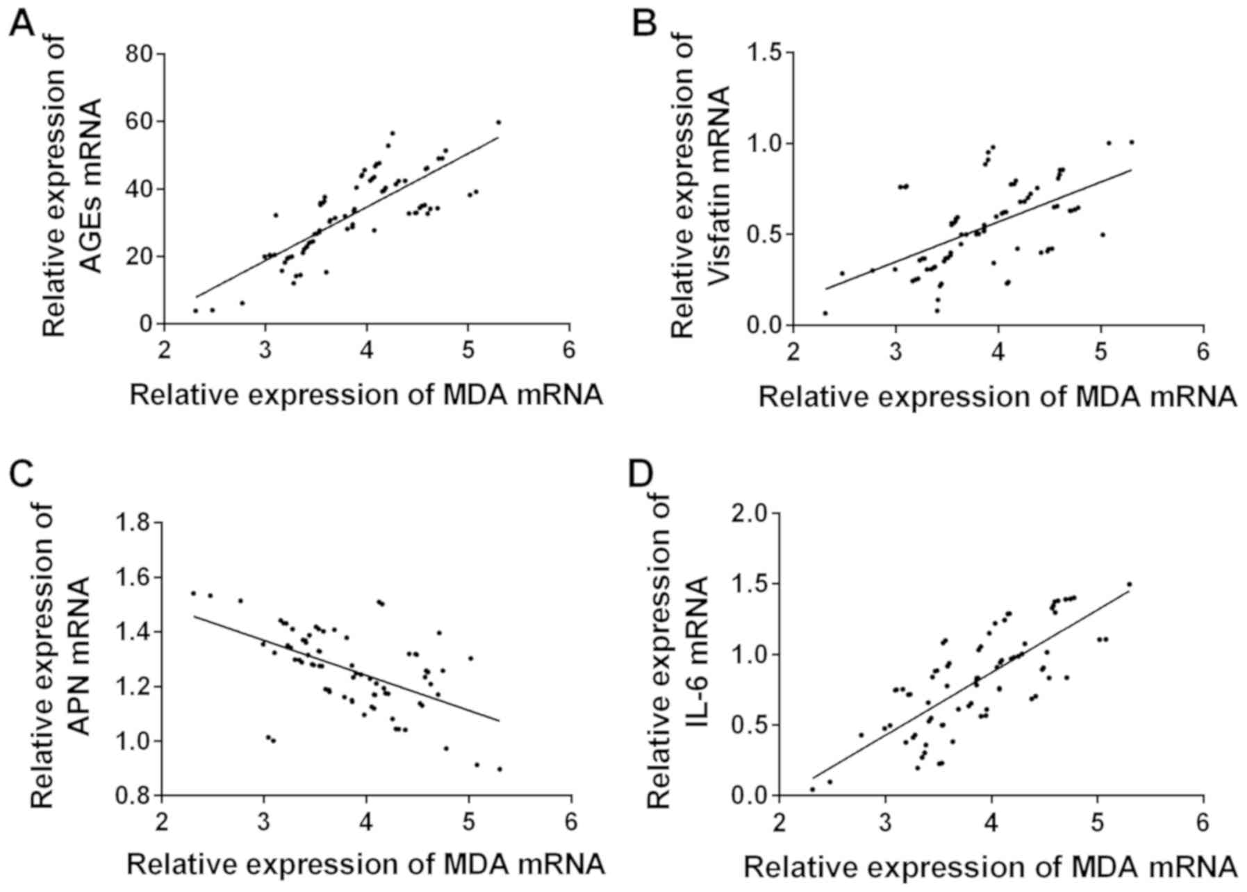
Correlation analysis between AGEs,
adipocytokines and expression of MDA
As shown in Figs. 1
and 2, the correlation between AGEs,
adipocytokines and expression of MDA in placental tissues of the
two groups was analyzed. The expression levels of AGEs, visfatin
and IL-6 in the two groups were positively correlated with MDA.
There was a significant negative correlation between APN and MDA in
the two groups (Table IV).
| Table IV.Correlation analysis between AGEs,
visfatin, APN, IL-6 and expression of MDA. |
Table IV.
Correlation analysis between AGEs,
visfatin, APN, IL-6 and expression of MDA.
|
| AGEs | Visfatin | APN | IL-6 |
|---|
|
|
|
|
|
|
|---|
| Groups | r | P-value | r | P-value | r | P-value | r | P-value |
|---|
| Observation
(n=72) | 0.757 | <0.001 | 0.454 | <0.001 | −0.602 | <0.001 | 0.792 | <0.001 |
| Control (n=80) | 0.794 | <0.001 | 0.573 | <0.001 | −0.544 | <0.001 | 0.763 | <0.001 |
Comparison of pregnancy outcomes
between the two groups
There was no neonatal malformation or perinatal
death in the two groups. The incidence of cesarean section,
neonatal hypoglycemia, fetal distress and macrosomia in the
observation group was significantly higher than that in the control
group (P<0.05). There was no significant difference in the
incidence of membrane rupture and preterm birth between the two
groups (P>0.05) (Table V).
| Table V.Comparison of pregnancy outcomes
between the two groups [n (%)]. |
Table V.
Comparison of pregnancy outcomes
between the two groups [n (%)].
| Group | Cesarean
section | Premature rupture
of membranes | Premature
birth | Neonatal
hypoglycemia | Fetal distress | Macrosomia |
|---|
| Observation group
(n=72) | 34 (47.2) | 8 (11.1) | 18 (25.0) | 9 (12.5) | 6 (8.3) | 14 (19.4) |
| Control group
(n=80) | 13 (16.3) | 6 (7.5) | 15 (18.6) | 2 (2.5) | 1 (1.3) | 5 (6.3) |
| χ2
value | 17.020 | 0.591 | 0.871 | 5.645 | 4.328 | 6.032 |
| P-value | <0.001 | 0.442 | 0.351 | 0.018 | 0.038 | 0.014 |
Discussion
GDM not only leads to metabolic disorders in
pregnant women, but may also lead to various early neonatal
diseases (19). When the balance
between the body's oxidative defense and free radical generation is
broken, the target tissue will be damaged, thus forming oxidative
stress (20). The function of tissue
cells in GDM patients will be affected by the peroxidation state,
and the function of islet β cells is most severely affected, so it
is extremely easy to cause cell damage or apoptosis (21).
AGEs is a stable covalent compound produced by
non-enzymatic glycation of proteins, lipids or nucleic acids in
long-term hyperglycemia (22). AGEs
can promote intracellular signal transduction and mediate diabetic
renal dysfunction (23). Visfatin
promotes the formation of fat, which is highly expressed in
visceral adipose tissues (24), and
can also upregulate the production of pro-inflammatory cytokines by
monocytes (25). APN is a plasma
protein (26), which can promote the
proliferation of mesangial cells in high-glucose environment, delay
their apoptosis, reduce cell damage, and have a protective effect
on cells (27). IL-6 is
overexpressed in obesity and inflammation (28). It has been reported that the
increased level of IL-6 in the placenta of GDM patients is caused
by oxidative stress and inflammatory changes caused by
hyperglycemia (24). MDA is one of
the most reliable indicators of oxidative stress, and the increase
of MDA level in diabetic patients is caused by the peroxidative
decomposition of phospholipids caused by free oxygen radicals
(29).
The expression levels of AGEs and adipocytokines in
placental tissues of parturients in the observation group and the
control group were detected. The results showed that the expression
levels of AGEs and IL-6 in placental tissues of the observation
group were significantly higher than those of the control group.
The expression level of APN in placental tissues of the observation
group was significantly lower than that of the control group. There
was no significant difference in expression of visfatin in
placental tissues between the two groups (P>0.05). It indicates
that AGEs and IL-6 are highly expressed in placental tissues of GDM
parturients, which is consistent with the studies of Voinov
(30), and Wang et al
(31). In this study, the expression
of APN in placental tissues of GDM patients was low, indicating
that there was abnormal regulation of adipokines in placenta.
Studies have shown that compared with pregnant women with normal
glucose tolerance, the expression of APN mRNA in GDM group was
significantly decreased, and the expression change of APN in
placental tissues was related to the expression of peroxisome
proliferator activated receptor (PPAR-γ) and IL-6 (32). Relevant literature shows that the
expression of visfatin in the female placental tissues of GDM was
significantly higher than that in the normal control group, and the
serum lipid concentration was positively correlated with the
expression of visfatin in the placenta (33), and there are reports that the
relative expression of visfatin mRNA in the placental tissues
showed no statistically significant difference between the GDM
group and the normal control group (34), and the results of this study are
similar. It has been reported that GDM in pregnant women presents
increased circulating oxidative stress hyperglycemia induction and
decreased antioxidant enzymes (35,36),
suggesting that increased oxidative stress may have adverse effects
on mother and fetus (37). In the
present study, the expression level of MDA in placental tissues of
the observation group and the control group was detected, and the
results showed that the expression level of MDA in placental
tissues of the observation group was significantly higher than that
of the control group. In addition, studies have shown that MDA is
highly expressed in the serum of pregnant women with GDM (16). This study showed that the expression
levels of AGEs, visfatin and IL-6 in placental tissues of the two
groups were positively correlated with MDA. There was a
significantly negative correlation between APN and MDA in the two
groups. The results suggested that AGEs, visfatin, IL-6 and APN
might be related to oxidative stress in pregnant women with GDM.
Bansal et al (38) indicated
that AGEs and MDA in serum in type 2 diabetes mellitus patients
with vascular complications were significantly positively
correlated. Studies have reported that adipokines leptin and
resistin are positively correlated with MDA in maternal blood of
GDM pregnant women, and APN and oxidative stress are antagonistic
to each other (39). However,
whether there is interaction in placental tissues or not remains to
be confirmed by further studies. There was no neonatal malformation
or perinatal death in the two groups. The incidence of cesarean
section, neonatal hypoglycemia, fetal distress and macrosomia in
the observation group was significantly higher than that in the
control group (P<0.05). There was no significant difference in
the incidence of membrane abruption and premature birth between the
two groups (P>0.05). The results of Dutta et al (40) showed that oxidative stress was
associated with cell cycle arrest and fetal membrane oxidative
injury, which jointly determined adverse pregnancy outcomes and
were important factors for premature rupture of membranes and
spontaneous premature birth. Studies on the relationship between
AGE level and perinatal outcome in pregnant women with GDM have
shown that high expression of AGEs is a risk factor for abnormal
perinatal outcome in GDM (30).
However, whether the AGEs, adipocytokines and oxidative stress
levels can be used as important indicators to predict the pregnancy
outcome with GDM requires further studies by expanding the number
of samples.
This investigation studied the correlation between
AGEs, adipocytokines and oxidative stress in pregnant women with
GDM, but the specific mechanism still needs to be further studied
to provide a clearer reference for the pathogenesis, clinical
diagnosis and treatment of GDM.
In conclusion, the expression levels of AGEs,
visfatin and IL-6 in placental tissues of GDM pregnant women were
positively correlated with MDA. APN and MDA showed a significant
negative correlation, which together played an important role in
the pathogenesis of GDM.
Acknowledgements
Not applicable.
Funding
No funding was received.
Availability of data and materials
The datasets used and/or analyzed during the present
study are available from the corresponding author on reasonable
request.
Authors' contributions
HL analyzed and interpreted the patients' data. AD
was responsible for ELISA and thiobarbituric acid method. XL helped
with statistical analysis. HL wrote the manuscript. All the authors
read and approved the final manuscript.
Ethics approval and consent to
participate
The study was approved by the Ethics Committee of
Binzhou City Center Hospital (Binzhou, China). Patients who
participated in this research had complete clinical data. The
signed informed consents were obtained from the patients or the
guardians.
Patient consent for publication
Not applicable.
Competing interests
The authors declare that they have no competing
interests.
References
|
1
|
Buchanan TA, Xiang A, Kjos SL and Watanabe
R: What is gestational diabetes? Diabetes Care. 30 (Suppl
2):S105–S111. 2007. View Article : Google Scholar : PubMed/NCBI
|
|
2
|
Ghodke B, Pusukuru R and Mehta V:
Association of lipid profile in pregnancy with preeclampsia,
gestational diabetes mellitus, and preterm delivery. Cureus.
9:e14202017.PubMed/NCBI
|
|
3
|
Kang MJ: The adiposity rebound in the 21st
century children: Meaning for what? Korean J Pediatr. 61:375–380.
2018. View Article : Google Scholar : PubMed/NCBI
|
|
4
|
Vargas-Terrones M, Nagpal TS and Barakat
R: Impact of exercise during pregnancy on gestational weight gain
and birth weight: An overview. Braz J Phys Ther. 23:164–169. 2019.
View Article : Google Scholar : PubMed/NCBI
|
|
5
|
Poprawski G, Pietryga M, Zawiejska A,
Iciek R, Wender-Ozegowska E and Brazert J: The impact of metabolic
control on uteroplacental circulation parameters in pregnancies
complicated by gestational hypertension and/or preeclampsia in
pregnant women with pregestational diabetes. Ginekol Pol.
86:811–820. 2015.(In Polish). View Article : Google Scholar : PubMed/NCBI
|
|
6
|
Schmidt AM: Highlighting diabetes
mellitus: The epidemic continues. Arterioscler Thromb Vasc Biol.
38:e1–e8. 2018. View Article : Google Scholar : PubMed/NCBI
|
|
7
|
Belfiore A, Malaguarnera R, Vella V,
Lawrence MC, Sciacca L, Frasca F, Morrione A and Vigneri R: Insulin
receptor isoforms in physiology and disease: An updated view.
Endocr Rev. 38:379–431. 2017. View Article : Google Scholar : PubMed/NCBI
|
|
8
|
van Rhijn-Brouwer FCC, Gremmels H,
Fledderus JO and Verhaar MC: Mesenchymal stromal cell
characteristics and regenerative potential in cardiovascular
disease: Implications for cellular therapy. Cell Transplant.
27:765–785. 2018. View Article : Google Scholar : PubMed/NCBI
|
|
9
|
Abell SK, De Courten B, Boyle JA and Teede
HJ: Inflammatory and other biomarkers: Role in pathophysiology and
prediction of gestational diabetes mellitus. Int J Mol Sci.
16:13442–13473. 2015. View Article : Google Scholar : PubMed/NCBI
|
|
10
|
Sudharshana Murthy KA, Bhandiwada A,
Chandan SL, Gowda SL and Sindhusree G: Evaluation of oxidative
stress and proinflammatory cytokines in gestational diabetes
mellitus and their correlation with pregnancy outcome. Indian J
Endocrinol Metab. 22:79–84. 2018.PubMed/NCBI
|
|
11
|
Badon SE, Zhu Y, Sridhar SB, Xu F, Lee C,
Ehrlich SF, Quesenberry CP and Hedderson MM: A pre-pregnancy
biomarker risk score improves prediction of future gestational
diabetes. J Endocr Soc. 2:1158–1169. 2018. View Article : Google Scholar : PubMed/NCBI
|
|
12
|
Low Birth Weight; Nephron Number Working
Group, : The Impact of kidney development on the life course: A
consensus document for action. Nephron. 136:3–49. 2017. View Article : Google Scholar : PubMed/NCBI
|
|
13
|
Lappas M, Permezel M and Rice GE: Release
of proinflammatory cytokines and 8-isoprostane from placenta,
adipose tissue, and skeletal muscle from normal pregnant women and
women with gestational diabetes mellitus. J Clin Endocrinol Metab.
89:5627–5633. 2004. View Article : Google Scholar : PubMed/NCBI
|
|
14
|
Ekun OA, Ogidi NO, Lawal RA, Ogunmuyiwa
OA, Umewune MC, Adefolaju FO, Oshundun MF and Oremosu AI:
Interrelationship between markers of oxidative stress, inflammation
and hematological parameters among preeclamptic Nigerian women. Med
Sci Monit Basic Res. 24:225–231. 2018. View Article : Google Scholar : PubMed/NCBI
|
|
15
|
Al-Gubory KH, Fowler PA and Garrel C: The
roles of cellular reactive oxygen species, oxidative stress and
antioxidants in pregnancy outcomes. Int J Biochem Cell Biol.
42:1634–1650. 2010. View Article : Google Scholar : PubMed/NCBI
|
|
16
|
Zhang C, Yang Y, Chen R, Wei Y, Feng Y,
Zheng W, Liao H and Zhang Z: Aberrant expression of oxidative
stress related proteins affects the pregnancy outcome of
gestational diabetes mellitus patients. Am J Transl Res.
11:269–279. 2019.PubMed/NCBI
|
|
17
|
Xin G, Du J, Wang YT and Liang TT: Effect
of oxidative stress on heme oxygenase-1 expression in patients with
gestational diabetes mellitus. Exp Ther Med. 7:478–482. 2014.
View Article : Google Scholar : PubMed/NCBI
|
|
18
|
Schäfer-Graf UM, Gembruch U, Kainer F,
Groten T, Hummel S, Hösli I, Grieshop M, Kaltheuner M, Bührer C,
Kautzky-Willer A, et al: Gestational diabetes mellitus (GDM) -
diagnosis, treatment and follow-up. Guideline of the DDG and DGGG
(S3 level, AWMF registry number 057/008, February 2018).
Geburtshilfe Frauenheilkd. 78:1219–1231. 2018.(In German).
View Article : Google Scholar : PubMed/NCBI
|
|
19
|
England L, Kotelchuck M, Wilson HG, Diop
H, Oppedisano P, Kim SY, Cui X and Shapiro-Mendoza CK: Estimating
the recurrence rate of gestational diabetes mellitus (GDM) in
Massachusetts 1998–2007: Methods and findings. Matern Child Health
J. 19:2303–2313. 2015. View Article : Google Scholar : PubMed/NCBI
|
|
20
|
Orr SK, Dachner N, Frank L and Tarasuk V:
Relation between household food insecurity and breast feeding in
Canada. CMAJ. 190:E312–E319. 2018. View Article : Google Scholar : PubMed/NCBI
|
|
21
|
Wells JCK, Figueiroa JN and Alves JG:
Maternal pelvic dimensions and neonatal size: Implications for
growth plasticity in early life as adaptation. Evol Med Public
Health. 2017:191–200. 2018. View Article : Google Scholar : PubMed/NCBI
|
|
22
|
Chen C, Gong W, Li C, Xiong F, Wang S,
Huang J, Wang Y, Chen Z, Chen Q, Liu P, et al: Sphingosine kinase 1
mediates AGEs-induced fibronectin upregulation in diabetic
nephropathy. Oncotarget. 8:78660–78676. 2017.PubMed/NCBI
|
|
23
|
Manigrasso MB, Juranek J, Ramasamy R and
Schmidt AM: Unlocking the biology of RAGE in diabetic microvascular
complications. Trends Endocrinol Metab. 25:15–22. 2014. View Article : Google Scholar : PubMed/NCBI
|
|
24
|
Briana DD and Malamitsi-Puchner A:
Reviews: Adipocytokines in normal and complicated pregnancies.
Reprod Sci. 16:921–937. 2009. View Article : Google Scholar : PubMed/NCBI
|
|
25
|
Moschen AR, Kaser A, Enrich B, Mosheimer
B, Theurl M, Niederegger H and Tilg H: Visfatin, an adipocytokine
with proinflammatory and immunomodulating properties. J Immunol.
178:1748–1758. 2007. View Article : Google Scholar : PubMed/NCBI
|
|
26
|
Galic S, Oakhill JS and Steinberg GR:
Adipose tissue as an endocrine organ. Mol Cell Endocrinol.
316:129–139. 2010. View Article : Google Scholar : PubMed/NCBI
|
|
27
|
Kukla M, Mazur W, Bułdak RJ and
Zwirska-Korczala K: Potential role of leptin, adiponectin and three
novel adipokines - visfatin, chemerin and vaspin - in chronic
hepatitis. Mol Med. 17:1397–1410. 2011. View Article : Google Scholar : PubMed/NCBI
|
|
28
|
Vozarova B, Weyer C, Hanson K, Tataranni
PA, Bogardus C and Pratley RE: Circulating interleukin-6 in
relation to adiposity, insulin action, and insulin secretion. Obes
Res. 9:414–417. 2001. View Article : Google Scholar : PubMed/NCBI
|
|
29
|
Decroli E, Manaf A, Syahbuddin S, Syafrita
Y and Dillasamola D: The correlation between malondialdehyde and
nerve growth factor serum level with diabetic peripheral neuropathy
score. Open Access Maced J Med Sci. 7:103–106. 2019. View Article : Google Scholar : PubMed/NCBI
|
|
30
|
Voinov VA: Therapeutic apheresis in
metabolic syndrome. Immunol Endocr Metab Agents Med Chem. 18:38–54.
2018. View Article : Google Scholar : PubMed/NCBI
|
|
31
|
Wang C, Guelfi KJ and Yang HX: Exercise
and its role in gestational diabetes mellitus. Chronic Dis Transl
Med. 2:208–214. 2016. View Article : Google Scholar : PubMed/NCBI
|
|
32
|
Pantham P, Aye IL and Powell TL:
Inflammation in maternal obesity and gestational diabetes mellitus.
Placenta. 36:709–715. 2015. View Article : Google Scholar : PubMed/NCBI
|
|
33
|
Ma Y, Cheng Y, Wang J, Cheng H, Zhou S and
Li X: The changes of visfatin in serum and its expression in fat
and placental tissue in pregnant women with gestational diabetes.
Diabetes Res Clin Pract. 90:60–65. 2010. View Article : Google Scholar : PubMed/NCBI
|
|
34
|
Luo JX, Liu XH, Zhang L and He GL: The
expression of visfatin in placenta in women with gestational
diabetes mellitus. Sichuan Da Xue Xue Bao Yi Xue Ban. 42:204–207.
2011.(In Chinese). PubMed/NCBI
|
|
35
|
Karacay O, Sepici-Dincel A, Karcaaltincaba
D, Sahin D, Yalvaç S, Akyol M, Kandemir O and Altan N: A
quantitative evaluation of total antioxidant status and oxidative
stress markers in preeclampsia and gestational diabetic patients in
24–36 weeks of gestation. Diabetes Res Clin Pract. 89:231–238.
2010. View Article : Google Scholar : PubMed/NCBI
|
|
36
|
Lappas M, Hiden U, Desoye G, Froehlich J,
Hauguel-de Mouzon S and Jawerbaum A: The role of oxidative stress
in the pathophysiology of gestational diabetes mellitus. Antioxid
Redox Signal. 15:3061–3100. 2011. View Article : Google Scholar : PubMed/NCBI
|
|
37
|
Sultan S, Alzahrani N and Al-Sakkaf K: The
postpartum effect of maternal diabetes on the circulating levels of
sirtuins and superoxide dismutase. FEBS Open Bio. 8:256–263. 2018.
View Article : Google Scholar : PubMed/NCBI
|
|
38
|
Bansal S, Chawla D, Siddarth M, Banerjee
BD, Madhu SV and Tripathi AK: A study on serum advanced glycation
end products and its association with oxidative stress and
paraoxonase activity in type 2 diabetic patients with vascular
complications. Clin Biochem. 46:109–114. 2013. View Article : Google Scholar : PubMed/NCBI
|
|
39
|
Brink HS, van der Lely AJ and van der
Linden J: The potential role of biomarkers in predicting
gestational diabetes. Endocr Connect. 5:R26–R34. 2016. View Article : Google Scholar : PubMed/NCBI
|
|
40
|
Dutta EH, Behnia F, Boldogh I, Saade GR,
Taylor BD, Kacerovský M and Menon R: Oxidative stress
damage-associated molecular signaling pathways differentiate
spontaneous preterm birth and preterm premature rupture of the
membranes. Mol Hum Reprod. 22:143–157. 2016. View Article : Google Scholar : PubMed/NCBI
|