Introduction
Ischemia stroke is the third leading cause of
mortality and the major cause of disability worldwide (1,2). The
cause of ischemia stroke is the obstruction of cerebral blood flow
with ischemia-reperfusion injury serving as its principal cause
(3,4). Following the onset of ischemia stroke,
local brain tissue becomes necrotic and apoptotic, which leads to
the corresponding neurological degeneration (5). Despite decades of research focusing on
ischemia stroke, the therapeutic strategies for the treatment of
this disease remain limited. Currently, the main treatment methods
for ischemia stroke are thrombolytic therapy and management of
symptoms (6). A previous study
indicated that the immune response and inflammation serve important
roles in brain tissue damage and the pathogenesis of ischemia
stroke (7). Therefore, targeting
ischemia stroke-induced neuroinflammation can be explored as a
potential therapeutic option for this disease.
MicroRNAs (miRNAs) are small non-coding RNAs that
are the key mediators for post-transcriptional gene silencing;
which is achieved by binding to the 3′untranslated regions of their
target mRNAs (8). A total of 30% of
all mammalian protein-encoding genes are regulated by miRNAs
(9,10), and have been demonstrated to serve
important functions in biological processes, including cell
proliferation, differentiation, apoptosis, cell cycle and cellular
stress response (11,12). Accumulating evidence shows that
miRNAs play critical roles in ischemia (13–15). The
expression of various miRNAs has been found to be altered in the
blood and brain of mammalian cells after the onset of stroke
(16–19). MiRNA expression can be modulated by
the application of external agents to alleviate stroke-induced loss
of biological function (16,20,21). In
addition, prior mechanistic studies have suggested that miRNAs
regulate the intracellular signaling of different inflammatory
mediators, thus performing a neuroprotective role in ischemia
stroke (22,23). However, the underlying mechanisms of
miRNA-mediated regulation of gene expression in relation to
inflammation and the signaling pathways involved in this process
remain poorly defined.
miR-183 was found to be expressed in the retina, and
its expression decreases after maturation (24). In zebrafish, miR-183 regulates
circadian rhythm by directly targeting E4 binding protein 4–6 and
arylalkylamine N-acetyltransferase 2 (25). Aberrant expression of miR-183 has
been illustrated to contribute to the symptoms of neurodegenerative
disorder in a mouse model (26).
Indeed, downregulation of miR-183 has been reported in ectopic and
eutopic tissues, and aberrant miR-183 expression may be associated
with the development of endometriosis (27). Previously, miR-183 has been reported
to be downregulated following stroke onset (28). In addition, depletion of miR-183 has
been suggested to lead to microglia activation (29). However, the biological function and
the mechanism of miR-183 expression associated with cerebral
ischemia-reperfusion injury remains largely unknown. Therefore, the
present study focuses on exploring the role and function of miR-183
in a rat model of cerebral ischemia-reperfusion injury by using
miR-183 agomir, and investigating the underlying molecular
mechanism.
Materials and methods
Experimental animals
A total of 36 specific pathogen-free (SPF) male
Sprague-Dawley rats (8–10 weeks old; 250±30 g) were purchased from
Jinan Peng Yue Experimental Animal Breeding Co., Ltd. [license
number, SCXK (Lu) 2014–0007; Shandong, China]. The animals received
food and water ad libitum, and were housed at a temperature
of 23±2°C, a humidity of 55±5% and a 12/12 h of light/dark cycle.
Animal experiments were performed under NIH guidelines (No, 85–23;
revised 1996) and approved by the Qingdao Central Hospital Animal
Protection and Use Committee.
Establishment of cerebral
ischemia-reperfusion model and animal groups
Rats were anesthetized by intraperitoneal injection
of 3% sodium pentobarbital (50 mg/kg). Establishment of middle
cerebral artery occlusion (MCAO) model was performed using the
modified nylon suture method (1).
Following 2 h of ischemia onset, the suture was gently pulled to
the distal/proximal end of the external carotid artery before the
rats were subsequently reperfused for 24 h. The sham-operated group
was destined for the separation and ligation of the blood vessels,
and therefore the insertion of suture was not performed in this
group. In accordance with the protocol described by Longa et
al (30), the animals were
scored 24 h after regaining consciousness and before the
neurological deficits were recorded. The scores were specified as
follows: 0 point (no symptoms of nerve damage were identified), 1
point (the contralateral forelimbs could not be fully extended when
the tail of the animals was lifted), 2 points (turned to the
temporal side when walking), 3 points (rats fell to the opposite
side of the lesion when walking) and 4 points (rats could not walk
spontaneously and lost their consciousness). An investigator who
was blind to the experimental setup performed the analysis. A total
of 36 rats were randomly divided into three separate groups: Sham
operation (Control), MCAO and MCAO + miR-183 agomir groups.
Intracerebroventrivular injection of miR-183 agomir (Guangzhou
Ruibo Biotechnology Co., Ltd.) for 2 h after ischemia onset was
performed in the MCAO + miR-183 agomir group, with the
concentration at 20 µmol/l and total volume of 10 µl. The sham and
MCAO groups were injected with corresponding amount of normal
saline.
Analysis of miR-183 mRNA expression in
brain tissue by reverse transcription (RT)-quantitative PCR
Following neurological function scoring, the rats
were anesthetized by intraperitoneal injection of 3% sodium
pentobarbital (50 mg/kg) and sacrificed by cervical dislocation.
The cerebellum and lower brainstem were rapidly removed from the
brain on ice, and the cerebral cortex of the ischemic tissues of 6
rats in each group were placed in liquid nitrogen for subsequent
experiments. Tissues (50 mg) were ground into powder with liquid
nitrogen, in a mortor, and centrifuged at 1,204 × g for 15 min at
4°C. According to the manufacturer's protocol, total RNA was
extracted using TRIzol (Thermo Fisher Scientific, Inc.). Absorption
ratios at 260 /280 nm of 1.8 and 2.0 were used to evaluate RNA
purity. SuperScript™ IV Reverse Transcriptase (cat. no. 18090010;
Thermo Fisher Scientific, Inc.) was used for the reverse
transcription of RNA into cDNA at 65°C, according to the
manufactures instruction. RT-qPCR was performed using SYBR Green I
(Thermo Fisher Scientific, Inc.) and a Mastercycler®
nexus X2 (Eppendorf). The thermocycling conditions were as follows:
95°C for 10 min, 95°C for 10 sec (40 cycles), 60°C for 1 min.
Results were quantified using the 2−ΔΔCq method
(31). The relative expression
levels of miR-183 were calculated using let-7a as internal control.
The following primers were used for RT-qPCR: miR-183, forward,
5′-AGGAGCAGAGGAGGTCTTT-3′ and reverse,
5′-TATGGCACTGGTAGAATTCACT-3′.
Tetrazolium chloride (TTC)
staining
The rats were anesthetized by intraperitoneal
injection of 3% sodium pentobarbital (50 mg/kg) and subsequently
sacrificed by cervical dislocation after neurological functions
were scored. Brain tissue was placed on ice, before the brain
tissues of 6 rats in each group were frozen at −20°C for 30 min.
The brain tissue was then cut into coronal sections, which were
quickly placed in 2% TTC (cat. no. G3005; Beijing Solarbio Science
& Technology Co., Ltd.) solution for incubation at 37°C for 15
min, before placement in 4% paraformaldehyde for fixation for 24 h,
at 25°C. The tissue was washed with PBS for 3–5 min, and images
were captured using a ×100 optical microscope (Olympus
Corporation). The image analysis software Image J 1.43 (National
Institutes of Health) was applied to measure the volume of cerebral
infarction.
Analysis of ionized calcium-binding
adaptor molecule 1 (IBA-1)-positive cells by
immunohistochemistry
Following TTC staining, the brain tissues were
dehydrated and embedded in paraffin, before being serially sliced
to a thickness of 5 µm. After dewaxed in xylene, the brain tissue
sections were rehydrated in a descending ethanol gradient, at room
temperature. At room temperature, the sections were subsequently
inactivated with a 3% H2O2 solution in
methanol for 20 min, citrate buffer (pH 6.0) at 65°C for 10 min,
before incubation with 5% bovine serum albumin (cat. no. A8020;
Beijing Solarbio Science & Technology Co., Ltd.) at 25°C for 20
min. The sections were then incubated using primary rabbit anti-rat
IBA-1 polyclonal antibody (1:200; orb336635; Biorbyt Ltd.) at 4°C
overnight. After PBS washing, the sections were incubated with
horseradish peroxidase-conjugated goat anti-rabbit IgG secondary
antibody (1:1,000; ABIN101988; antibodies-online GmbH, Aachen) at
37°C for 30 min. Following secondary antibody incubation,
3,3′-diaminobenzidine (DAB) staining was developed on the sections
for 5–10 min at room temperature, before counterstaining with
hematoxylin for 10 min at 37°C. Finally, the sections were
dehydrated by gradient alcohol for 5 min, treated twice with xylene
for 10 min, before being sealed with mounting medium.
The results were observed under a ×400 optical
microscope (Olympus Corporation) and counted using Aperio
Imagescope 11.1 software (Leica Microsystems Inc.). The percentage
(%) of positive cells was evaluated.
Evaluation of IL-1β, IL-6 and TNF-α
expression in brain tissue by ELISA
Brain tissue was thoroughly homogenized in ice cold
PBS, before centrifugation at 500 × g at 4°C for 15 min. The
supernatant was then assayed for IL-1β (orb79117; Biorbyt Ltd.),
IL-6 (orb79123; Biorbyt Ltd.) and TNF-α (orb79138-480; Biorbyt
Ltd.) according to manufacturer's protocols. The results were
obtained by measuring absorption at 450 nm on a microplate reader
(Model 680, Bio-Rad Laboratories, Inc.).
Western blot analysis of
NF-κB-associated protein expression in brain tissue
The cerebral cortex of the ischemic area was
grounded and homogenized before centrifugation (800 × g; 10 min;
4°C), and the supernatant was extracted. The bicinchoninic assay
(BCA) kit (Beijing Solarbio Science & Technology Co., Ltd.) was
used to quantify protein concentration. A total of 40 µg of each
protein sample was mixed with 10% of the SDS gel buffer at a 1:1
ratio. This mixture was then boiled at 95°C for 5 min. Following
denaturation, the protein samples were separated by SDS-PAGE (10%
gels) and transferred onto a polyvinylidene membrane (Merck KGaA,
Darmstadt) at 80 V for 30 min. The membranes were subsequently
blocked with 5% skim milk powder dissolved in TBS with Tween-20
(TBS-T) for 1 h at 4°C, before incubation with their respective
rabbit anti-rat polyclonal antibodies dissolved in TBS-T solution
containing 3% bovine serum albumin at 4°C overnight. The antibodies
used in this study were as follows: NF-κB p65 (1:500; orb11118;
Biorbyt Ltd.), IκBα (1:500; orb223182; Biorbyt Ltd.) and β-actin
(1:2,000; orb178392; Biorbyt Ltd.). The membranes were then washed
with TBS-T four times prior to incubation with horseradish
peroxidase-conjugated goat anti-rabbit IgG (1:1,000; ABIN101988;
antibodies-online GmbH) at room temperature for 1 h. The membranes
were subsequently washed with TBS-T four times and incubated with
ECL luminescent substrate (Thermo Fisher Scientific, Inc.) for 3–5
min for visualization. Levels of protein expression were normalized
to β-actin, and quantification was performed using Image J software
(v 1.51; National Institutes of Health).
Statistical analysis
All data was processed using the SPSS 19.0
statistical analysis software (IBM Corp.). Data analysis was
expressed as mean ± standard deviation (mean ± SD), and comparison
between groups was performed using one-way analysis of variance
(ANOVA) followed by least significance difference (LSD) test.
P<0.05 was considered to indicate a statistically significant
difference.
Results
miR-183 agomir treatment improves
neurological function in rat brain tissue after
ischemia-reperfusion injury
Neurological function was scored in
rats following cerebral ischemia onset
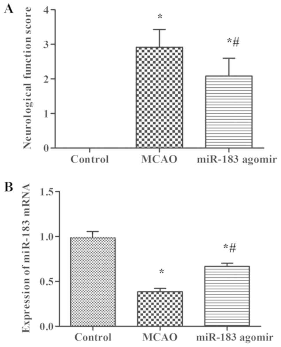
The control rats exhibited a neurological score of
zero when comparing with the MCAO group, which was used as a
positive control. The neurological scores of the MCAO + miR-183
agomir group were significantly reduced compared with those in the
MCAO group (Fig. 1A). The effect of
miR-183 agomir on miR-183 mRNA levels of brain tissue was measured
using RT-qPCR. Compared with the control group, the expression of
miR-183 mRNA in the brain tissue of rats in MCAO and MCAO + miR-183
agomir groups was significantly reduced (Fig. 1B). Following miR-183 agomir
treatment, the expression of miR-183 mRNA was significantly
increased in the brain tissue of rats compared with those in the
MCAO group (Fig. 1B).
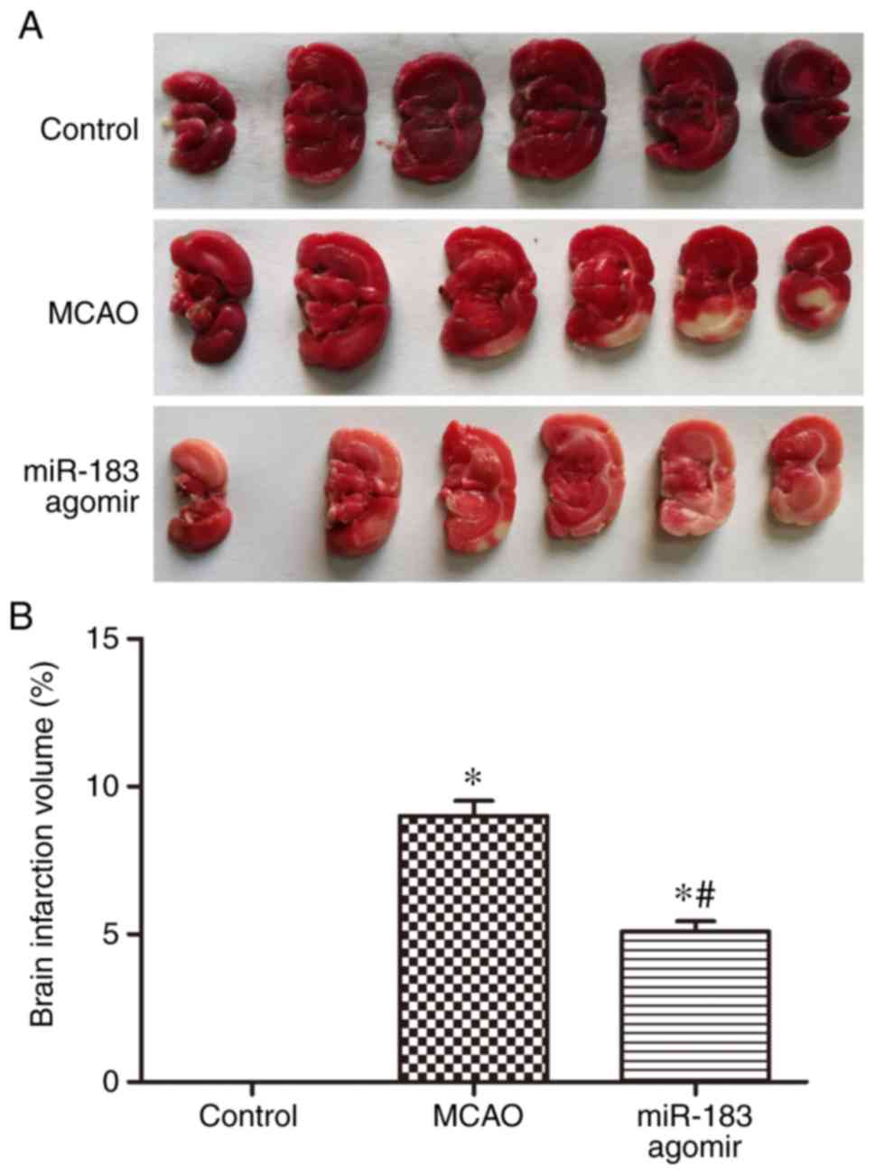
The effect of miR-183 on brain tissue damage was
determined by TTC staining following cerebral ischemia induction in
rats, with the non-infarcted areas staining red. The percentage of
cerebral infarction volume in the MCAO + miR-183 agomir group
appeared to be significantly lower compared with that in the MCAO
group (Fig. 2). This observation
suggests that miR-183 agomir treatment reduced brain tissue damage
in rats with cerebral ischemia reperfusion. These results
collectively indicate that cerebral miR-183 expression is reduced
by ischemia onset, an effect that can be partially rescued by
miR-183 agomir treatment. Subsequent miR-183 agomir treatment
increased miR-183 expression and improved neurological function,
suggesting that miR-183 serves a neuroprotective role in rats with
cerebral ischemia reperfusion.
miR-183 agomir treatment decreases the
expression of IBA-1 in rats with cerebral ischemia-reperfusion
injury
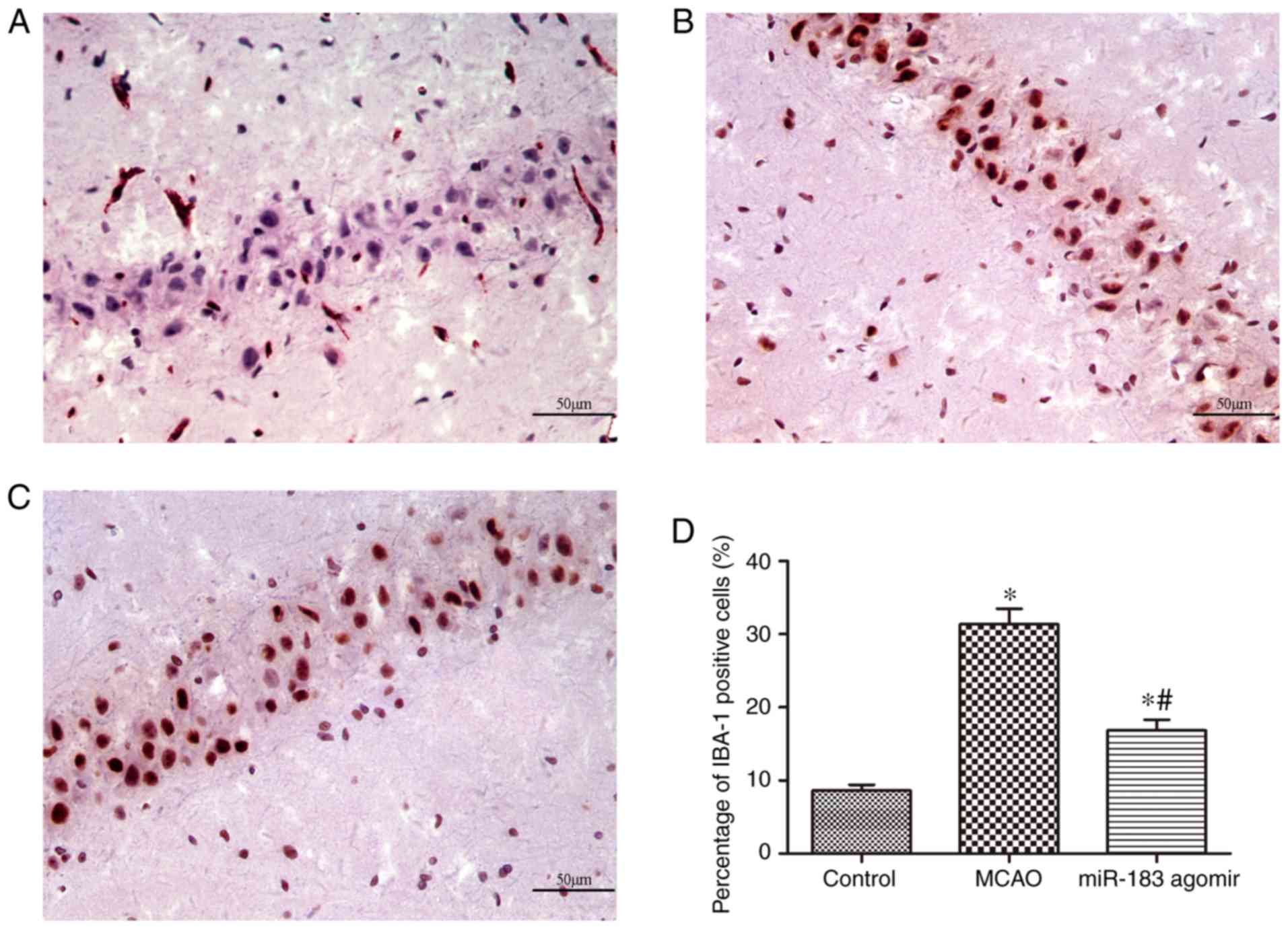
It has been reported that IBA-1 expression may be
increased in rat brain following the onset of cerebral ischemia
(32). As a result, IBA-1 expression
levels and the distribution of IBA-1-positive cells in the three
treatment groups were assessed using immunohistochemistry. Whilst
only a small number of IBA-1-positive cells (arrows) were found in
the hippocampal CA1 of the control group (Fig. 3A and D), a large number of
IBA-1-positive cells appeared in the CA1 region in the MCAO group
(Fig. 3B and D). The number of
IBA-1-positive cells observed in the hippocampal CA1 area was
significantly reduced following miR-183 agomir treatment (Fig. 3C and D), when compared with the MCAO
group. This finding suggests that miR-183 agomir treatment
decreases the expression of IBA-1 in rats following cerebral
ischemia-reperfusion injury.
miR-183 agomir treatment decreases the
expression of inflammatory cytokines IL-1β, IL-6 and TNF-α in brain
tissue
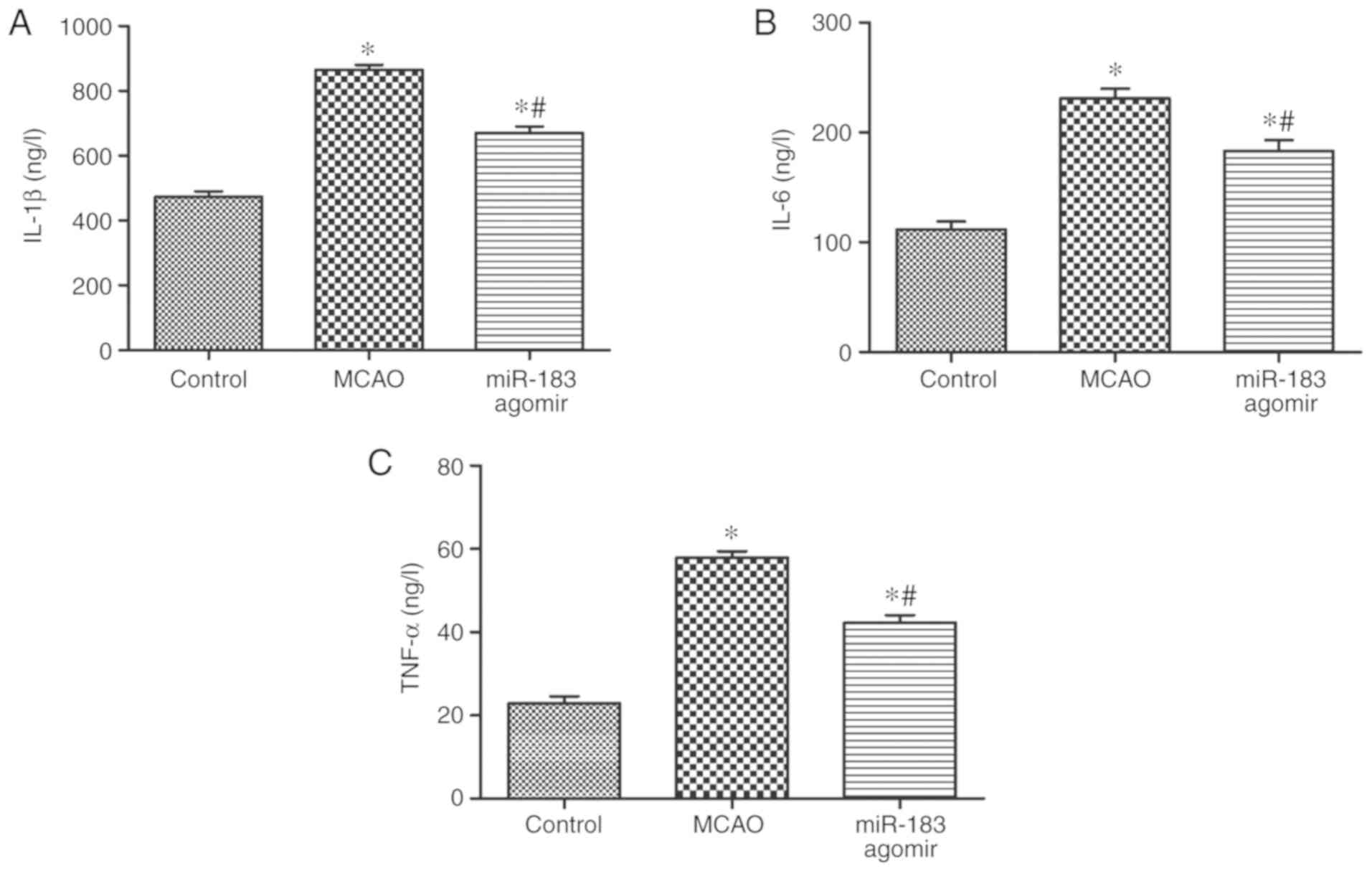
Results from the ELISA assay demonstrated that the
expression of inflammatory factors IL-1β, IL-6 and TNF-α in the
brain tissues of MCAO and MCAO + miR-183 agomir groups were
significantly increased following the cerebral ischemia onset
compared with the control group (Fig.
4A-C). After treatment with miR-183 agomir, the expressions of
IL-1β, IL-6 and TNF-α were significantly decreased in the brain
tissue of rats when compared with MCAO group (Fig. 4A-C). These results implicate miR-183
to be a suppressor of inflammation during cerebral ischemia.
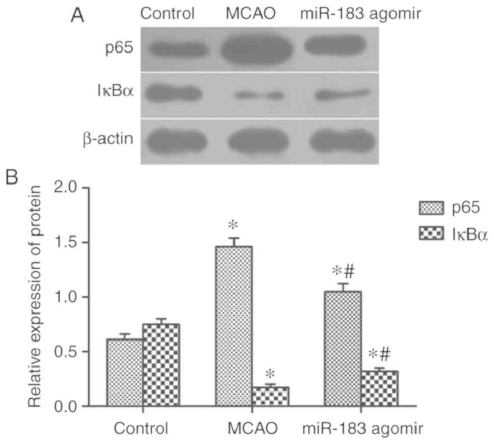
miR-183 agomir treatment inhibits
NF-κB signaling in brain tissue
Lastly, the effect of miR-183 on the regulation of
NF-κB signaling was investigated by measuring the protein levels of
the NF-κB subunit p65 and IκBα, a negative regulator of NF-κB
(33), in the brain tissues of the
three treatment groups. The expression of p65 in the brain tissue
of MCAO and MCAO + miR-183 agomir groups was significantly
increased compared with the control group, while the expression of
IκBα was significantly decreased (Fig.
5). In contrast, the expression of p65 in brain tissue of MCAO
+ miR-183 agomir group was significantly decreased, and the
expression of IκBα was significantly increased when compared with
MCAO group (Fig. 5). This finding
suggests that miR-183 agomir treatment inhibits NF-κB signaling
activation.
Discussion
miRNAs serve important roles in a number of
pathophysiological conditions by the virtue of binding to and
regulating target gene expression. Previously, a number of studies
reported that miRNAs can mediate ischemia injury, the suppression
of which by treatment with their respective mimic significantly
alleviated brain tissue damage in ischemia stroke models (34,35).
Indeed, Wang et al (35)
showed that miR-3473b expression was significantly increased, and
treatment with miR-3473b antagomir significantly reduced infarction
after MCAO in mice. In another study, Xiang et al (29) found that following ischemic
preconditioning for 15 min, the expression of miR-183 was
demonstrated to be upregulated after 3 and 24 h reperfusion.
However, the role and biological function of miR-183 remains
unknown. In the present study, the role of miR-183 in ischemia
reperfusion-induced brain damage was investigated by employing a
model of cerebral ischemia-reperfusion injury. Upon treatment with
miR-183 agomir, the neurological score was markedly decreased in
rats with cerebral ischemia-reperfusion injury. On the molecular
level, results from the present study indicated that miR-183
expression was significantly decreased in rats with cerebral
ischemia-reperfusion injury, an effect that was partially rescued
by miR-183 agomir treatment. Therefore, these findings suggest a
neuroprotective role of miR-183 agomir in the cerebral ischemia
model, which correlate well with previous studies (29).
Cerebral infarction volume is a long established
measure of outcome in clinical trials of acute ischemia stroke
therapies (36,37). Following miR-183 agomir treatment,
the cerebral infarction volume profoundly decreased compared with
the MCAO group, which indicated that tissue damage was alleviated
by miR-183 agomir treatment. IBA-1, a calcium binding protein,
which is specifically expressed in microglia, has been reported to
be a critical factor in microglia membrane ruffling, as well as
being involved in the regulation of Rho-GTPase, Rac and calcium
signaling pathways, and is required for cell mobility and
phagocytosis (38,39). In addition, high levels of IBA-1
expression in activated microglia suggest that IBA may be a
potential marker for microglia activation (40). Thus, it serves as one of the most
useful proteins for distinguishing microglia from other types of
brain tissues in studies of cerebral ischemia (38,41). In
this study, immunohistochemistry staining results showed that the
number of IBA-1-positive cells was significantly decreased in rats
treated with miR-183 agomir after cerebral ischemia-reperfusion
injury.
Next, the mechanism involved in the neuroprotection
of miR-183 agomir in cerebral ischemia-reperfusion rat models was
investigated. Inflammation and immune response serve a pivotal role
in stroke induced by tissue damage and repair (42). Numerous studies have reported that
the expression level of miRNAs was altered following ischemia
stroke onset (13,43,44).
Around 50 miRNAs have been found to be upregulated in the rat model
of ischemic stroke, a list which includes but not limited to
miR-17, miR-134 and miR-206 (16–20). On
the other hand, several miRNAs including miR-25, miR-138 and
miR-361 were found to be downregulated in a rat model of ischemia
(43). These miRNAs regulate the
intracellular pathways of a variety of inflammatory mediators
(43). In the present study, the
effect of miR-183 agomir on the expression of IL-6, IL-1β and
TNF-α, three prominent pro-inflammatory cytokines (35), was assessed. ELISA results reported
that the expression of all three were increased following the
induction of cerebral ischemia-reperfusion injury, an observation
that was partially reversed by miR-183 agomir treatment. Thus,
these results suggest that miR-183 agomir-mediated enhancement of
neurological function and reduction of tissue damage is at least in
part due to the downregulation of pro-inflammatory cytokine
expression.
The toll-like receptor 4 (TLR4)/NF-κB signaling
pathway was reported to be activated in cerebral
ischemia-reperfusion injury in rats (33). In this aforementioned study, the
expression of TLR4 and NF-κB was found to be increased along with
the activation of corresponding signaling pathway following
cerebral ischemia-reperfusion injury. Data from the present study
supports these results, the expression of p65 and IκBα in rats with
cerebral ischemia-reperfusion was evaluated. Furthermore, miR-183
agomir treatment partially reversed this effect. Altogether, this
study revealed that miR-183 regulates the activation of microglia
in cerebral ischemia-reperfusion injury in rats by inhibiting NF-κB
signaling pathway. Therefore, the present study uncovers a role for
miR-183 in the development of cerebral ischemia reperfusion-injury
in rats, along with further mechanistic insights. This information
may be useful for further clinical studies and treatment strategies
of patients with ischemia stroke. Thus, miR-183 agomir can be
explored as a potential therapeutic reagent for treatment of
ischemia stroke in the future clinically. A potential limitation of
this study is that only one concentration of miR-183 agomir was
tested. In future investigations, different concentrations will be
tested in vitro and in vivo to improve our
understanding of miR-183 as a therapeutic target.
The results of the current study indicated that
miR-183 agomir treatment significantly reduced neurological
function scores, and serves a neuroprotection role by reducing the
percentage of cerebral infarction volume, and decreasing the
IBA-1-positive cells in the CA1 area of the hippocampus in response
to rat cerebral ischemia-reperfusion injury. Furthermore, miR-183
agomir treatment decreased the expression of pro-inflammatory
proteins and regulated the activation of microglia in cerebral
ischemia-reperfusion injury in rats by inhibiting NF-κB signaling
pathway. Therefore, these results uncover the role of miR-183 in
cerebral ischemia-reperfusion. miR-183 may be used as a therapeutic
target for the treatment of cerebral ischemia-reperfusion.
Acknowledgements
Not applicable.
Funding
The present study was supported by National Major
Special Projects for the Major New Drug Creation (grant no.
2012ZX09103- 101-015).
Availability of data and materials
All data generated or analyzed during this study are
included in this published article.
Authors' contributions
BX, PZ and HY conceived and designed the study. LF,
XW, and YS performed the experiments. BX wrote the paper. PZ
reviewed and edited the manuscript. All authors read and approved
the manuscript.
Ethics approval and consent to
participate
Animal experiments were performed under NIH
guidelines (publication no. 85-123; revised 1996) and approved by
the Qingdao Central Hospital Animal Protection and Use
Committee.
Patient consent for publication
Not applicable.
Competing interest
The authors declare that they have no competing
interests.
References
|
1
|
Mendis S, Davis S and Norrving B:
Organizational update: The world health organization global status
report on noncommunicable diseases 2014; One more landmark step in
the combat against stroke and vascular disease. Stroke.
46:e121–122. 2015. View Article : Google Scholar : PubMed/NCBI
|
|
2
|
Zhang R, Zhang Z and Chopp M: Function of
neural stem cells in ischemic brain repair processes. J Cereb Blood
Flow Metab. 36:2034–2043. 2016. View Article : Google Scholar : PubMed/NCBI
|
|
3
|
Schaller B and Graf R: Cerebral ischemia
and reperfusion: The pathophysiologic concept as a basis for
clinical therapy. J Cereb Blood Flow Metab. 24:351–371. 2004.
View Article : Google Scholar : PubMed/NCBI
|
|
4
|
Baird AE, Donnan GA, Austin MC, Fitt GJ,
Davis SM and McKay WJ: Reperfusion after thrombolytic therapy in
ischemic stroke measured by single-photon emission
computed-tomography. Stroke. 25:79–85. 1994. View Article : Google Scholar : PubMed/NCBI
|
|
5
|
Lee JM, Grabb MC, Zipfel GJ and Choi DW:
Brain tissue responses to ischemia. J Clin Invest. 106:723–731.
2000. View
Article : Google Scholar : PubMed/NCBI
|
|
6
|
Blakeley JO and Llinas RH: Thrombolytic
therapy for acute ischemic stroke. J Neurol Sci. 261:55–62. 2007.
View Article : Google Scholar : PubMed/NCBI
|
|
7
|
Khoshnam SE, Winlow W, Farzaneh M, Farbood
Y and Moghaddam HF: Pathogenic mechanisms following ischemic
stroke. Neurol Sci. 38:1167–1186. 2017. View Article : Google Scholar : PubMed/NCBI
|
|
8
|
Zhang CX: MicroRNomics: A newly emerging
approach for disease biology. Physiol Genomics. 33:139–147. 2008.
View Article : Google Scholar : PubMed/NCBI
|
|
9
|
Friedman RC, Farh KK, Burge CB and Bartel
DP: Most mammalian mRNAs are conserved targets of microRNAs. Genome
Res. 19:92–105. 2009. View Article : Google Scholar : PubMed/NCBI
|
|
10
|
Griffiths-Jones S, Saini HK, van Dongen S
and Enright AJ: miRBase: Tools for microRNA genomics. Nucleic Acids
Res 36 (Database Issue). D154–D158. 2008.
|
|
11
|
Kloosterman WP and Plasterk RH: The
diverse functions of MicroRNAs in animal development and disease.
Dev Cell. 11:441–450. 2006. View Article : Google Scholar : PubMed/NCBI
|
|
12
|
Felekkis K, Touvana E, Stefanou CH and
Deltas C: microRNAs: A newly described class of encoded molecules
that play a role in health and disease. Hippokratia. 14:236–240.
2010.PubMed/NCBI
|
|
13
|
Ouyang YB, Stary CM, Yang GY and Giffard
R: microRNAs: Innovative targets for cerebral ischemia and stroke.
Curr Drug Targets. 14:90–101. 2013. View Article : Google Scholar : PubMed/NCBI
|
|
14
|
Saugstad JA: MicroRNAs as effectors of
brain function with roles in ischemia and injury, neuroprotection,
and neurodegeneration. J Cereb Blood Flow Metab. 30:1564–1576.
2010. View Article : Google Scholar : PubMed/NCBI
|
|
15
|
Li GW, Morris-Blanco KC, Lopez MS, Yang T,
Zhao H, Vemuganti R and Luo Y: Impact of microRNAs on ischemic
stroke: From pre- to post-disease. Prog Neurobiol. 163-164:59–78.
2018. View Article : Google Scholar : PubMed/NCBI
|
|
16
|
Jeyaseelan K, Lim KY and Armugam A:
MicroRNA expression in the blood and brain of rats subjected to
transient focal ischemia by middle cerebral artery occlusion.
Stroke. 39:959–966. 2008. View Article : Google Scholar : PubMed/NCBI
|
|
17
|
Dharap A, Bowen K, Place R, Li LC and
Vemuganti R: Transient focal ischemia induces extensive temporal
changes in rat cerebral MicroRNAome. J Cereb Blood Flow Metab.
29:675–687. 2009. View Article : Google Scholar : PubMed/NCBI
|
|
18
|
Sørensen SS, Nygaard AB, Nielsen MY,
Jensen K and Christensen T: miRNA expression profiles in
cerebrospinal fluid and blood of patients with acute ischemic
stroke. Transl Stroke Res. 5:711–718. 2014. View Article : Google Scholar : PubMed/NCBI
|
|
19
|
Li SH, Su SY and Liu JL: Differential
regulation of microRNAs in patients with ischemic stroke. Curr
Neurovasc Res. 12:214–221. 2015. View Article : Google Scholar : PubMed/NCBI
|
|
20
|
Saugstad JA: Non-coding RNAs in stroke and
neuroprotection. Front Neurol. 6:502015. View Article : Google Scholar : PubMed/NCBI
|
|
21
|
Liu da Z, Jickling GC, Ander BP, Hull H,
Zhan X, Cox C, Shroff N, Dykstra-Aiello C, Stamova B and Sharp FR:
Elevating microRNA-122 in blood improves outcomes after temporary
middle cerebral artery occlusion in rats. J Cereb Blood Flow Metab.
36:1374–1383. 2016. View Article : Google Scholar : PubMed/NCBI
|
|
22
|
Khoshnam SE, Winlow W and Farzaneh M: The
Interplay of MicroRNAs in the inflammatory mechanisms following
ischemic stroke. J Neuropathol Exp Neurol. 76:548–561. 2017.
View Article : Google Scholar : PubMed/NCBI
|
|
23
|
Liu P, Zhao H, Wang R, Wang P, Tao Z, Gao
L, Yan F, Liu X, Yu S, Ji X and Luo Y: MicroRNA-424 protects
against focal cerebral ischemia and reperfusion injury in mice by
suppressing oxidative stress. Stroke. 46:513–519. 2015. View Article : Google Scholar : PubMed/NCBI
|
|
24
|
Dambal S, Shah M, Mihelich B and Nonn L:
The microRNA-183 cluster: The family that plays together stays
together. Nucleic Acids Res. 43:7173–7188. 2015. View Article : Google Scholar : PubMed/NCBI
|
|
25
|
Wienholds E, Kloosterman WP, Miska E,
Alvarez-Saavedra E, Berezikov E, de Bruijn E, Horvitz HR, Kauppinen
S and Plasterk RH: MicroRNA expression in zebrafish embryonic
development. Mechanisms Dev. 122 (Suppl):S149–S150. 2005.
|
|
26
|
Kye MJ, Niederst ED, Wertz MH, Gonçalves
Ido C, Akten B, Dover KZ, Peters M, Riessland M, Neveu P, Wirth B,
et al: SMN regulates axonal local translation via miR-183/mTOR
pathway. Hum Mol Genet. 23:6318–6331. 2014. View Article : Google Scholar : PubMed/NCBI
|
|
27
|
Shi XY, Gu L, Chen J, Guo XR and Shi YL:
Downregulation of miR-183 inhibits apoptosis and enhances the
invasive potential of endometrial stromal cells in endometriosis.
Int J Mol Med. 33:59–67. 2014. View Article : Google Scholar : PubMed/NCBI
|
|
28
|
Sepramaniam S, Jun-Rong T, Kay-Sin T,
Deidre Ann De S, Subramaniam T, Fung-Peng W, Chee-Woon W, Fung-Lin
Y, Dwi-Setyowati K, Prameet K, et al: Circulating MicroRNAs as
Biomarkers of Acute Stroke. Int J Mol Sci. 15:1418–1432. 2014.
View Article : Google Scholar : PubMed/NCBI
|
|
29
|
Xiang L, Chen XJ, Wu KC, Zhang CJ, Zhou
GH, Lv JN, Sun LF, Cheng FF, Cai XB and Jin ZB: miR-183/96 plays a
pivotal regulatory role in mouse photoreceptor maturation and
maintenance. Proc Natl Acad Sci USA. 114:6376–6381. View Article : Google Scholar : PubMed/NCBI
|
|
30
|
Longa EZ, Weinstein PR, Carlson S and
Cummins R: Reversible middle cerebral artery occlusion without
craniectomy in rats. Stroke. 20:84–91. 1989. View Article : Google Scholar : PubMed/NCBI
|
|
31
|
Livak KJ and Schmittgen TD: Analysis of
relative gene expression data using real-time quantitative PCR and
the 2(-Delta Delta C(T)) method. Methods. 25:402–408. 2001.
View Article : Google Scholar : PubMed/NCBI
|
|
32
|
Ito D, Tanaka K, Suzuki S, Dembo T and
Fukuuchi Y: Enhanced expression of Iba1, ionized calcium-binding
adapter molecule 1, after transient focal cerebral ischemia in rat
brain. Stroke. 32:1208–1215. 2001. View Article : Google Scholar : PubMed/NCBI
|
|
33
|
Chen J, Yang C, Xu X, Yang Y and Xu B: The
effect of focal cerebral ischemia-reperfusion injury on TLR4 and
NF-κB signaling pathway. Exp Ther Med. 15:897–903. 2018.PubMed/NCBI
|
|
34
|
Ni J, Wang X, Chen S, Liu H, Wang Y, Xu X,
Cheng J, Jia J and Zhen X: MicroRNA let-7c-5p protects against
cerebral ischemia injury via mechanisms involving the inhibition of
microglia activation. Brain Behav Immun. 49:75–85. 2015. View Article : Google Scholar : PubMed/NCBI
|
|
35
|
Wang X, Chen S, Ni J, Cheng J, Jia J and
Zhen X: miRNA-3473b contributes to neuroinflammation following
cerebral ischemia. Cell Death Dis. 9:112018. View Article : Google Scholar : PubMed/NCBI
|
|
36
|
van der Worp HB, Claus SP, Bär PR, Ramos
LM, Algra A, van Gijn J and Kappelle LJ: Reproducibility of
measurements of cerebral infarct volume on CT scans. Stroke.
32:424–430. 2001. View Article : Google Scholar : PubMed/NCBI
|
|
37
|
Han M, Choi JW, Rim NJ, Kim SY, Suh HI,
Lee KS, Hong JM and Lee JS: Cerebral infarct volume measurements to
improve patient selection for endovascular treatment. Medicine
(Baltimore). 95:e47022016. View Article : Google Scholar : PubMed/NCBI
|
|
38
|
Imai Y, Ibata I, Ito D, Ohsawa K and
Kohsaka S: A novel gene iba1 in the major histocompatibility
complex class III region encoding an EF hand protein expressed in a
monocytic lineage. Biochem Biophys Res Commun. 224:855–862. 1996.
View Article : Google Scholar : PubMed/NCBI
|
|
39
|
Ohsawa K, Imai Y, Kanazawa H, Sasaki Y and
Kohsaka S: Involvement of Iba1 in membrane ruffling and
phagocytosis of macrophages/microglia. J Cell Sci. 113:3073–3084.
2000.PubMed/NCBI
|
|
40
|
Ito D, Imai Y, Ohsawa K, Nakajima K,
Fukuuchi Y and Kohsaka S: Microglia-specific localisation of a
novel calcium binding protein, Iba1. Brain Res Mol Brain Res.
57:1–9. 1998. View Article : Google Scholar : PubMed/NCBI
|
|
41
|
Ito D, Tanaka K, Suzuki S, Dembo T and
Fukuuchi Y: Enhanced expression of Iba1, ionized calcium-binding
adapter molecule 1, after transient focal cerebral ischemia in rat
brain. Stroke. 32:1208–1215. 2001. View Article : Google Scholar : PubMed/NCBI
|
|
42
|
Ma Y, Wang J, Wang Y and Yang GY: The
biphasic function of microglia in ischemic stroke. Prog Neurobiol.
157:247–272. 2017. View Article : Google Scholar : PubMed/NCBI
|
|
43
|
Rink C and Khanna S: MicroRNA in ischemic
stroke etiology and pathology. Physiol Genomics. 43:521–528. 2011.
View Article : Google Scholar : PubMed/NCBI
|
|
44
|
Hamzei Taj S, Kho W, Riou A, Wiedermann D
and Hoehn M: MiRNA-124 induces neuroprotection and functional
improvement after focal cerebral ischemia. Biomaterials.
91:151–165. 2016. View Article : Google Scholar : PubMed/NCBI
|