Introduction
Laparoscopic sleeve gastrectomy (LSG) is a popular
bariatric procedure in obese patients for weight loss and improves
obesity-related comorbidities such as arterial hypertension,
diabetes mellitus, obstructive sleep apnea and dyslipidemia
(1,2). The major complications of LSG are leak
and staple-line bleeding despite technical developments and
surgical improvements in bariatric surgery (3,4). The
incidence of leak after LSG and staple-line hemorrhage has been
reported to be 0–8 and 0–8.7% respectively (5,6). In
order to prevent these major complications, several synthetic or
biological reinforcement materials, such as fibrin sealant and/ or
gelatin matrix agents, Seamguard, clips and sutures has been used
following LSG (4,7,8).
However, there are controversial studies regarding the best method
for staple-line reinforcement or its necessity in the current
literature (4,8,9).
Fibrin sealant (FS; Tisseel) is an agent that
provides hemostasis, sealing and adhesion and it has a wide area of
use in various surgical procedures (10,11). In
their study, Coşkun H and Yardımcı (4) suggested that FS is a reliable and
useful agent to reinforce the staple-line and may prevent twists in
the sleeved stomach.
Ankaferd blood stopper® (ABS) is a
previously developed topical hemostatic agent containing a
standardized mixture of the plants Thymus vulgaris, Glycyrrhiza
glabra, Vitis vinifera, Alpinia officinarum and Urtica
dioica (12). Through its
effects on the endothelium, blood cells, angiogenesis, cellular
proliferation, vascular dynamics and cell mediators; ABS is shown
to have hemostatic and regenerative proliferation effects in in
vivo and in vitro studies (13,14). The
topical use of ABS has been approved by the Turkish Ministry of
Health for the management of postsurgical bleeding (12).
In this experimental study, the aim was to compare
the effects of ABS and FS in terms of bursting pressure
measurement, hydroxyproline and histological examination in
resected sleeve gastrectomy specimens of rats. To the best of our
knowledge, this is the first study investigating the potential
effects of ABS as well as FS on staple line healing in an
experimental study.
Materials and methods
This animal study was performed after the approval
of Akdeniz University Experimental Animal Studies Local Ethical
Committee (Ethical committee no: B.30.2.AKD.0.05.07.00/120). A
total of 30 adult female Wistar Albino rats weighing between 200
and 250 g were obtained from the Akdeniz University Department of
Experimental Animals Care and Production Unit (Konyaaltı/Antalya,
Turkey). The rats were kept in standard colony cages (15×25×40 cm)
under controlled conditions including temperature (18°C), light
(12-h light-dark cycle) and humidity (50–55%). The rats were fed
with the standard rat chow and tap water ad libitum during
the experimental procedure except 12 h fasting period before the
surgery.
After a 12-h fast, subjects were anesthetized using
an intraperitoneal injection of 10 mg/kg xylazine (Bayer AG,
Leverkusen, Germany) and 50 mg/kg of ketamine hydrochloride
(Eczacibasi Holding A.S., Istanbul, Turkey). All surgeries were
performed by a single surgeon blind to the subjects' grouping.
After cleaning the skin with 10% povidone iodine (Central
Laboratory, Istanbul, Turkey), a 3.5–4 cm midline laparotomy was
made to access the abdominal cavity. The stomach was identified and
the gastrosplenic ligament was ligated with 6/0 polyglycolic
sutures and divided. The great omentum was ligated with 6/0
polyglycolic sutures and divided down to the level of pylorus. A
part of the pylorus was left intact by starting the excision of the
stomach 3–5 mm above the level of the pylorus, in order to maintain
the free passage of the food to the duodenum. Stitches were placed
defining the incision line, which contained the gastric fundus. The
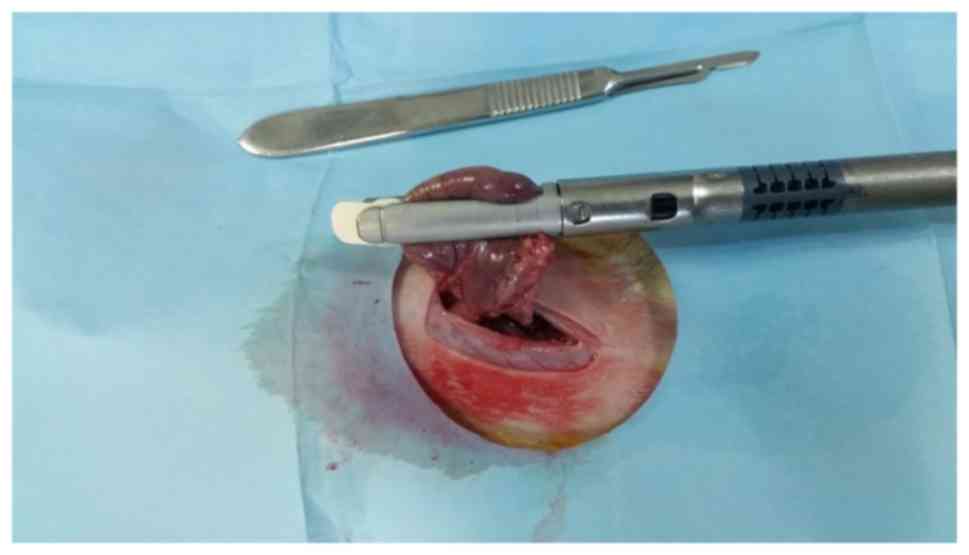
stapler (two rows, 3.5 mm height-1.5 mm when closed; Echelon Flex
powered vasculer stapler; Ethicon, Endo-Surgery, Inc., Cincinnati,
OH, USA) was positioned on that line and the great curvature along
with the gastric fundus was removed, leading to a drastic reduction
of the gastric volume of ~70–80% (Sleeve gastrectomy model;
Fig. 1). An orogastric tube was not
used during the excision of the stomach.
There were 3 experimental groups studied: Group A
(control group)-only sleeve gastrectomy was performed (n=10); Group
B-was administered a 4 ml box FS on staple-line after sleeve
gastrectomy by spray method (n=10); and Group C-was administered
0.2 cc of ABS topically using an insulin injector on staple-line
after sleeve gastrectomy (n=10). The abdominal wall was closed with
running 3/0 polyglycolic suture and the skin was sewed with a 4/0
running intracutaneus suture.
Measurement of staple-line bursting
pressure
On postoperative day 5, the abdomen was reopened and
the sleeved stomach was located. Thereafter, total gastrectomy was
made in all subjects. A 8-French catheter was fixed with 2/0 silk
suture through in pylorus which was connected to a metilen
perfusion pump (Samtronic ST670 infusion pump; Samtronic, São
Paolo, Brazil). An intracet was also fixed with 2/0 silk suture
through the esophagus that is connected to a monitor (Datex-Ohmeda,
Compact Anesthesia Monitor; GE Healthcare, Chicago, IL, USA) by a
pressure transducer (Transpac IV; Abbbott Laboratories, IL, USA).
In order to measure bursting pressure, 6 ml/min of methylene blue
was infused into the gastric lumen through the catheter, which is
inserted into the pylorus using the perfusion pump and was
monitored. The pressure level at the time of fluid leakage or a
sudden pressure drop occurred was recorded. After the bursting
pressure had been measured, the staple line segment was resected
and divided into two equal parts passing through the anastomosis.
One part was put in 10% formaldehyde solution for 12 h and
maintained at 4°C for histopathological examination and the other
was frozen at −80°C for measurement of hydroxyproline levels.
Hydroxyproline analysis
Tissue samples were weighed, cut into small pieces
and homogenized in a PBS (pH 7.4) with a sonicator for 10 min at
20°C and 35 kHz (Bandelin Electronic, GmbH & Co. KG., Berlin,
Germany) for hydroxyproline levels determination. The homogenates
were immediately centrifuged at 1,008 × g for 20 min, at 4°C and
supernatants were collected. The supernatants were kept on ice.
Supernatants were used for the measurement of hydroxyproline levels
on the same day. Hydroxyproline levels were measured
spectrophotometrically (Multiscan Spectrum; Thermo Labsystems,
Santa Rosa, CA, USA) using a SunRed Rat Hydroxyproline ELISA kit
(Shanghai SunRed Biological Technology Co., Ltd., Shanghai, China;
cat. no. 201-11-0512). The hydroxyproline values were expressed as
microgram per gram of tissue (µg/g).
Histopathological examination
Formalin-fixed tissue samples were embedded in
paraffin and 4.5 µm sections were cut. Replicate sections were
either stained with hematoxylin and eosin for the evaluation of
morphological features under a light microscope (BX51; Olympus
Corporation, Tokyo, Japan) by a single pathologist blind to the
sections' grouping. Tissues taken from the perianastomotic section
were histopathologically examined for scoring of mucosal ischemia,
anastomotic wound healing, granulation tissue formation and
histological changes in local inflammatory response according to
the criteria described by Biert et al (15) and modified by Verhofstad et al
(16).
Statistical analysis
Descriptive statistics (mean ± standard deviation)
were determined and the results were used to produce charts in
which the 95% confidence interval error bars indicate the means.
The results of the analysis of the bursting pressure and the tissue
hydroxyproline level were analyzed using the Shapiro-Wilks.
Nonparametric measurements and comparisons of the three groups in
terms of pathology were performed using the Kruskal-Wallis test.
Comparisons of the two groups in terms of tissue hydroxyproline
levels, bursting pressure and pathology were made using the
Mann-Whitney U test with Bonferroni correction. IBM SPSS Statistics
ver. 22.0 PASW 22 (IBM Corp., Chicago, IL, USA), was used to
determine the statistical significance of the results. P<0.05
was considered to indicate a statistically significant
difference.
Results
There was no mortality because of anesthesia and
surgical operations in any group during the study period. Bursts
had occurred from staple-line anastomosis in all subjects. The
fluid leakage site was the gastric fundus in all rats. Although the
mean bursting pressure value was increased in group C compared with
in groups A and B, no statistically significant differences was
found among the group values (P=0.754; Table I). Additionally there was no
significant statistically difference of binary comparison of groups
(Table II).
 | Table I.Staple-line bursting pressure
measurements. |
Table I.
Staple-line bursting pressure
measurements.
| Group | Mean ± SD (mmHg) | P-valuea |
|---|
| Group A
(Control) | 7±5.98 | 0.754 |
| Group B (FS) | 10±13.35 |
|
| Group C (ABS) | 23.9±33.32 |
|
| Table II.Binary comparison of Staple-line
bursting pressure levels among all groups. |
Table II.
Binary comparison of Staple-line
bursting pressure levels among all groups.
| Measurements | Pa | Pb | Pc |
|---|
| Tissue bursting
pressure (mmHg) | 0.909 | 0.061 | 0.074 |
The results of the hydroxyproline levels among all
groups are summarized in Tables
III and IV. Analysis of the
mean tissue hydroxyproline levels revealed statistically
significant differences between groups A and B and between groups A
and C (P<0.001 and P<0.001, respectively). Group C had the
highest mean level however there was no statistically significant
differences between group B and C (P>0.05). Based on these data,
it was concluded that administration of ABS and FS has significant
effect on the sleeved stapled-line in terms of tissue
hydroxyproline levels.
| Table III.Tissue hydroxyproline levels (µg/g wet
tissue). |
Table III.
Tissue hydroxyproline levels (µg/g wet
tissue).
| Group | Mean ± SD | P-valuea |
|---|
| Group A
(Control) | 1.919±0.366 | P<0.001 |
| Group B (FS) | 5.639±1.031 |
|
| Group C (ABS) | 5.89±0.87 |
|
| Table IV.Binary comparison of the tissue
hydroxyproline levels among the study groups. |
Table IV.
Binary comparison of the tissue
hydroxyproline levels among the study groups.
| Measurements | Pa | Pb | Pc |
|---|
| Hydroxyproline/wet
tissue weight (µg/g) | <0.001 | <0.001 | >0.05 |
The results of histopathological examination scores
of the groups are given in Table V.
Analysis of the groups in terms of necrosis, polymorphonuclear
leukocyte, lymphocyte intensity, edema, mucosal epithelial damage
and submucosal-mucosal bridging scores revealed the absence of any
statistically significant differences among the groups. Group A had
the highest macrophage score among all groups and there was
statistically significant differences between groups (P=0.002). In
the binary comparison of the macrophage score between groups, there
was statistically significant differences between group A and C
(P=0.002; Table VI).
| Table V.Results of histopathologic examination
scores. |
Table V.
Results of histopathologic examination
scores.
| Variable | Group A (mean ±
SD) | Group B (mean ±
SD) | Group C (mean ±
SD) | Pa |
|---|
| Necrosis | 1.1±0.88 | 0.5±0.53 | 0.4±0.52 | 0.109 |
| PMNL | 1.6±0.7 | 1.3±0.48 | 1.6±0.84 | 0.350 |
| Lymphocyte | 1.6±0.70 | 1.9±0.57 | 1.6±0.84 | 0.413 |
| Macrophage | 1.5±0.71 | 1.1±0.57 | 0.3±0.48 | 0.002 |
| Edema | 1.2±0.63 | 0.9±0.57 | 0.8±0.42 | 0.254 |
| Mucosal
epithelium | 1.3±0.95 | 1.2±0.79 | 1.2±0.79 | 0.894 |
|
Submucosal-mucosal | 1.3±0.82 | 1.0±0.67 | 1.1±0.74 | 0.602 |
| Table VI.Binary comparison of the
histopathologic examination scores of the groups. |
Table VI.
Binary comparison of the
histopathologic examination scores of the groups.
| Variable | Pa | Pb | Pc |
|---|
| Necrosis | 0.104 | 0.061 | 0.661 |
| PMNL | 0.159 | 0.531 | 0.276 |
| Lymphocyte | 0.251 | 0.866 | 0.249 |
| Macrophage | 0.131 | 0.002 | 0.006 |
| Edema | 0.264 | 0.111 | 0.689 |
| Mucosal
epitelium | 0.681 | 0.681 | 0.999 |
|
Submucosal-mucosal | 0.328 | 0.516 | 0.737 |
Discussion
In this experimental study, the effects of FS and
ABS on staple-line bursting pressure measurement, hydroxyproline
level and histopathological examination in a sleeve gastrectomy
model have been evaluated. It has been demonstrated that ABS and FS
had better results on bursting pressure levels and tissue
hydroxyproline levels in sleeved staple-line of rats. However, both
ABS and FS had no effect at histopathological examinations of all
groups. To the best of our knowledge, there is no study
investigating the effects of FS and ABS in an experimental model of
sleeve gastrectomy.
Staple-line leak and bleeding are the most common
complications of LSG associated with significant morbidity and
mortality (17). The hematoma
formation and/or postoperative bleeding at the sleeved staple-line
of the stomach can interfere with wound healing and could lead to a
leak (18). In order to reduce these
major complications of LSG, different surgical techniques and
stapler line reinforcement materials have been defined and used.
Although a number of studies have been carried on this topic, there
is no enough evidence to support the necessity of reinforcement
and/or hemostatic agents in LSG (9,19).
ABS has been shown to have hemostatic and
regenerative proliferation effects in in vivo and in
vitro studies (13,14). In addition, ABS has also been
reported to have antimicrobial, antifungal, wound healing-enhancing
effects and antiseptic properties in a small number of animal
studies and case reports (13,14). It
has been known that collagen production is the main indicator of
the healing process. Hydroxyproline is a measure of collagen
content that has a positive correlation with the anastomose
strength (20). In their study,
Gulden et al (20)
investigated the effects of ABS on colon anastomosis in an
experimental study. The authors found that the levels of tissue
hydroxyproline, an important indicator of anastomosis healing, were
statistically significantly higher in ABS administered group
(20). In the present study it was
seen that ABS increased the bursting pressure on the stapler-line.
Similar to Gulden et al study, it was demonstrated that
topical use of ABS had improved tissue hydroxyproline levels on
sleeved staple-line. In contrast to the Gulden et al study,
although higher bursting pressure levels were observed in ABS
administered group, there was no significant difference between the
groups. The present study found that histopathological parameters
were similar between the groups, except macrophage scores. The
lowest macrophage score was found in group C. Previous studies
reported that the higher macrophage score is correlated with poor
wound healing (15,16). As a result of this the present study
concluded that ABS may improve wound healing. Since the rats were
scarified on postoperative day 5, further analysis with other
markers of histopathological examination could provide different
results in the same setting.
In the present study, apart from ABS and the control
group, the FS group was formed as a third group. FS is one of the
most commonly used reinforcement agent for the staple line
reinforcement in bariatric surgery, although it is efficacy is
under debate in studies (4,9). For this reason, the present
experimental study was designed to investigate the compatible
efficiencies of ABS and FS. It was demonstrated that staple-line
burst pressure and tissue hydroxyproline levels were increased in
the ABS group compared with the FS group. The histopatological
scores were similar also in the FS group compared with other
groups. Based on these data, it was concluded that ABS may be an
alternative to FS as a reinforcement agent in bariatric
surgery.
The present study has several limitations. First of
all, the present study was an animal study carrying all bias
associated with the differences in animal and human metabolisms.
Secondly, in this experimental study, a formal sleeve gastrectomy
was not performed in rats as in humans. Third, only hydroxyproline
levels were evaluated as a separate biochemical parameter and no
immuno-histochemical study was conducted. In addition a light
microscope was used for the evaluation of histopathological
features. Further histopathological analyses under electron
microscopy could give different results. As a result of these
limitations, results similar to the present study may not be
obtained in humans.
In conclusion, it was first demonstrated that ABS as
well as FS improved staple line healing in terms of burst pressure
and at tissue hydroxyproline level in an experimental study.
Although there was no statistically significant difference between
effects of ABS and FS on staple-line healing, it was suggested that
ABS can be used with similar efficacy instead of FS as a novel
reinforcement agent in bariatric surgery. The results of the
present study should be supported by further experimental and
clinical studies.
Acknowledgements
Not applicable.
Funding
No funding was received.
Availability of data and materials
The datasets used and/or analyzed during the present
study are available from the corresponding author on reasonable
request.
Authors' contributions
AS conceived and designed the present study; TB
conceived/designed the current study, collected data and wrote the
article. BM acquired, analyzed and interpreted the data. TO made
critical revisions to the manuscript and conceived/designed the
current study. All authors read and approved the final version of
the manuscript.
Ethics approval and consent to
participate
The present animal study was performed after the
approval of Akdeniz University Experimental Animal Studies Local
Ethical Committee (Ethical committee no:
B.30.2.AKD.0.05.07.00/120).
Patient consent for publication
Not applicable.
Conflict of interest
The authors declare that they have no competing
interests.
References
|
1
|
Choi YY, Bae J, Hur KY, Choi D and Kim YJ:
Reinforcing the staple line during laparoscopic sleeve gastrectomy:
Does it have advantages? A Meta-analysis. Obes Surg. 22:1206–1213.
2012. View Article : Google Scholar : PubMed/NCBI
|
|
2
|
D'Hondt M, Vanneste S, Pottel H, Devriendt
D, Van Rooy F and Vansteenkiste F: Laparoscopic sleeve gastrectomy
as a single-stage procedure for the treatment of morbid obesity and
the resulting quality of life, resolution of comorbidities, food
tolerance, and 6-year weight loss. Surg Endosc. 25:2498–2504. 2011.
View Article : Google Scholar : PubMed/NCBI
|
|
3
|
Aurora AR, Khaitan L and Saber AA: Sleeve
gastrectomy and the risk of leak: A systematic analysis of 4888
patients. Surg Endosc. 26:1509–1515. 2012. View Article : Google Scholar : PubMed/NCBI
|
|
4
|
Coşkun H and Yardımcı E: Effects and
results of fibrin sealant use in 1000 laparoscopic sleeve
gastrectomy cases. Surg Endosc. 31:2174–2179. 2017. View Article : Google Scholar : PubMed/NCBI
|
|
5
|
Moon Han S, Kim WW and Oh JH: Results of
laparoscopic sleeve gastrectomy (LSG) at 1 year in morbidly obese
Korean patients. Obes Surg. 15:1469–1475. 2005. View Article : Google Scholar : PubMed/NCBI
|
|
6
|
Gagner M, Deitel M, Kalberer TL, Erickson
AL and Crosby RD: The second international consensus summit for
sleeve gastrectomy, March 19–21, 2009. Surg Obes. Relat Dis.
5:476–485. 2009.
|
|
7
|
Consten EC, Gagner M, Pomp A and Inabnet
WB: Decreased bleeding after laparoscopic sleeve gastrectomy with
or without duodenal switch for morbid obesity using a stapled
buttresses absorbable polymer membrane. Obes Surg. 14:1360–1366.
2004. View Article : Google Scholar : PubMed/NCBI
|
|
8
|
Gentileschi P, Camperchioli I, D'Ugo S,
Benavoli D and Gaspari AL: Staple-line reinforcement during
laparoscopic sleeve gastrectomy using three different techniques: A
randomized trial. Surg Endosc. 26:2623–2629. 2012. View Article : Google Scholar : PubMed/NCBI
|
|
9
|
Chen B, Kiriakopoulos A, Tsakayannis D,
Wachtel MS, Linos D and Frezza EE: Reinforcement does not
necessarily reduce the rate of staple line leaks after sleeve
gastrectomy. A review of the literature and clinical experiences.
Obes Surg. 19:166–172. 2009. View Article : Google Scholar : PubMed/NCBI
|
|
10
|
Spotnitz WD: Fibrin sealant: The only
approved hemostat, sealant, and adhesive-a laboratory and clinical
perspective. ISRN Surg 2014. 2039432014.
|
|
11
|
Silecchia G, Boru CE, Mouiel J, Rossi M,
Anselmino M, Morino M, Toppino M, Gaspari A, Gentileschi P,
Tacchino R and Basso N: The use of fibrin sealent to prevent major
complications following laparoscopic gastric bypass: Results of a
multicenter, randomized trial. Surg Endosc. 22:2492–2497. 2008.
View Article : Google Scholar : PubMed/NCBI
|
|
12
|
Gungor G, Goktepe MH, Biyik M, Polat I,
Tuna T, Ataseven H and Demir A: Efficacy of Ankaferd blood stopper
application on non-variceal upper gastrointestinal bleeding. World
J Gastrointest Endosc. 4:556–560. 2012. View Article : Google Scholar : PubMed/NCBI
|
|
13
|
Deveci A, Coban AY, Tanrıverdi Çaycı Y,
Acicbe O, Taşdelen Fışgın N, Akgüneş A, Ozatlı D, Uzun M and
Durupınar B: In Vitro Effect of AnkaferdBloodStopper a Plant
Extract Against Mycobacterium tuberculosis Isolates Mikrobiyol Bul.
47:71–78. 2013.PubMed/NCBI
|
|
14
|
Isler SC, Demircan S, Cakarer S, Cebi Z,
Keskin C, Soluk M and Yüzbaşioğlu E: Effects of folk medicinalplant
extract Ankaferd blood stopper on early bone healing. J Appl Oral
Sci. 18:409–414. 2010. View Article : Google Scholar : PubMed/NCBI
|
|
15
|
Biert J, Seifert WF, Verhofstad AA, Wobbes
T, de Man BM, Hoogenhout J and Hendriks T: A semiquantitative
histological analysis of repair of anastomoses in the rat colon
after combined preoperative irradiation and local hyperthermia.
Radiat Res. 149:372–371, 998. View
Article : Google Scholar : PubMed/NCBI
|
|
16
|
Verhofstad MH, Lange WP, van der Laak JA,
Verhofstad AA and Hendriks T: Microscopic analysis of anastomotic
healing in the intestine of normal and diabetic rats. Dis Colon
Rectum. 44:423–431. 2001. View Article : Google Scholar : PubMed/NCBI
|
|
17
|
Buchwald H, Avidor Y, Braunwald E, Jensen
MD, Pories W, Fahrbach K and Schoelles K: Bariatric surgery: A
systematic review and meta-analysis. JAMA. 292:1724–1737. 2004.
View Article : Google Scholar : PubMed/NCBI
|
|
18
|
De Angelis F, Abdelgawad M, Rizzello M,
Mattia C and Silecchia G: Perioperative hemorrhagic complications
after laparoscopic sleeve gastrectomy: Four-year experience of a
bariatric center of excellence. Surg Endosc. 31:3547–3551. 2017.
View Article : Google Scholar : PubMed/NCBI
|
|
19
|
Wang Z, Dai X, Xie H, Feng J Li Z and Lu
Q: The efficacy of staple line reinforcement during laparoscopic
sleeve gastrectomy: A meta-analysis of randomized controlled
trials. Int J Surg. 25:145–152. 2016. View Article : Google Scholar : PubMed/NCBI
|
|
20
|
Gulden C, Teksoz S, Aytac E, Arikan AE,
Erman H, Uzun H, Ozden F, Aydin O and Ozcan M: Effects of Ankaferd
on Anastomotic Healing of Colon. J Invest Surg. 27:1–6. 2014.
View Article : Google Scholar : PubMed/NCBI
|