Introduction
Esophageal cancer is one of the most aggressive
cancers in the world. According to the latest epidemiological data,
in 2018, the number of incidences of esophageal cancer and the
number of death from esophageal cancer were reported to be 572,034
and 508,585 worldwide, respectively, which makes it the ninth and
sixth highest rates for incidence and mortality among all the
malignant tumors (1). Therefore,
there is an immediate need to find more effective methods to
diagnose, treat, and predict the prognosis of esophageal
cancer.
Yes-associated protein (YAP), a candidate oncogene
located on chromosome 11q22, is a negative regulator of the Hippo
pathway, which has been previously reported as a tumor suppressor
pathway in Drosophila and mammals (2,3).
Previous studies suggest that the Hippo-Yap pathway plays an
important role in the genesis and progression of tumors (4,5).
Increased YAP expression has been associated with the progression
of several human cancers, including cervical cancer (6), pancreatic ductal adenocarcinoma
(7) and human urothelial carcinoma
of the bladder (8). In addition, YAP
overexpression is associated with poor prognosis in human
urothelial carcinoma of the bladder (8), ovarian cancer (9), and colorectal cancer (10). Our previous studies have shown that
YAP overexpression plays vital roles in the progression and
metastasis of prostate and pancreatic cancer (11,12).
However, a previous study has reported that the overexpression of
YAP was associated with poor overall survival (OS) in esophageal
cancer in Japan (13). Further
research is required to understand the role of the expression level
of YAP in esophageal cancer and its importance in prognosis
prediction.
Epithelial-mesenchymal transition (EMT) is a process
described as the transition of cells from epithelial phenotype to
mesenchymal phenotype and during this process cells gain more
migratory and invasive properties (14). Although YAP has previously been
reported to be associated with tumor metastasis via EMT in several
tumors, including non-small cell lung cancer (15), breast cancer (16), pancreatic ductal adenocarcinoma
(17), and hepatocellular carcinoma
(18), its involvement in ESCC still
remains unclear. There are no reports, so far, with regard to YAP
regulation of tumor migration and invasion through EMT in ESCC.
Since one previous study reported that
downregulation of YAP inhibits proliferation and induces apoptosis
in ESCC cells (19), the exact role
of YAP in ESCC remains largely unclear. The present study aimed to
investigate the role of YAP expression in tissue samples from
patients with ESCC by performing a stratified analysis based on
pathological TNM stage to provide further insights into the
influence of YAP expression for prognosis of patients with ESCC,
with respect to both OS and disease-free survival (DFS).
Furthermore, small interfering RNA (siRNA) was transfected into
Eca109 and Kyse150 cells and the effects of YAP inhibition on these
ESCC cells was investigated to explore the underlying molecular
mechanism.
Materials and methods
Patients and samples
A total of 107 paraffin-embedded tumor tissue
samples from patients who underwent esophagectomy at Qilu Hospital
of Shandong University (Jinan, China) were collected between
January 2008 and October 2008. All cases were confirmed as ESCC by
three people as stated in the immunohistochemical (IHC) analysis
section. Samples were excluded from the study as follows: i) If the
patient received radiation therapy or chemotherapy before surgery;
ii) if the patient died or was untraceable during follow-up and
iii) if the patient was diagnosed with more than one primary tumor.
The clinicopathological data, including age, sex, history of
smoking and alcohol consumption, differentiation degree, invasion
depth (T stage), lymph node metastasis (N stage), pathological TNM
(pTNM) (20), and adjuvant treatment
after surgery were obtained from the clinical or pathological
records. The present study was approved by The Ethics Committee of
Qilu Hospital. All patients in the study were anonymous and
provided written informed consent.
Follow-up
In the first 2 years after surgery, patients were
contacted by telephone every 3 months to enquire about their
recovery and assess the level of recurrence, if any. More detailed
instructions were provided according to tumor progression if
recurrence occurred. After 2 years, information regarding the
patients was collected every 6 months until November 2013, unless
they were either untraceable or had died.
IHC analysis
Tumor tissues of patients with ESCC were fixed in
10% formalin for 12 h at 4°C and embedded in paraffin. The
paraffin-embedded tissues were cut into 5 µm thick sections, and
IHC staining was performed using the Streptavidin-BiotinComplex kit
according to the manufacturer's protocol (cat. no. SA1022; Wuhan
Boster Biological Technology Co., Ltd.). Tissue sections were
mounted on to the microslides and incubated for 1 h at 60°C before
de-waxing in xylene and hydrated in graded concentrations of
alcohol (95, 90, 85, 80 and 75%). Then the sections were placed in
a solution of sodium citrate (pH 6.0) and were heated to 93°C, and
maintained at 90°C for 15 min to retrieve the antigen. The solution
was cooled to room temperature and 3% H2O2
was subsequently added at room temperature for 10 min, to block the
non-specific protein binding sites and inactivate the endogenous
peroxidase. After blocking with 5% BSA (Wuhan Boster Biological
Technology Co., Ltd.) for 20 min at room temperature, the sections
were incubated overnight at 4°C with mouse polyclonal primary
antibody against YAP (dilution 1:300; cat. no. 4912; Cell Signaling
Technology, Inc.). Then, the sections were incubated with
biotinylated goat anti-rabbit antibody from the kit for 20 min at
37°C. Finally, the slides were counterstained with hematoxylin at
25°C for 2 min dehydrated in graded alcohol solution (75, 80, 85,
90, 95 and 100%) and xylene, and covered with coverslips using
neutral balsam.
The IHC images were scored for positive staining
intensity in five high-power fields using a light microscope
(magnification, ×400) independently by three people, including a
pathologist and two authors of the current study who did not
participate in the IHC staining (LZ and YJ). The intensity score
was graded as follows: i) None, 0; ii) mild, 1; iii) moderate, 2;
and iv) intense, 3. The percentage of positive tumor cells was
assessed according to the following patterns: i) No staining, 0;
ii) ≤10%, 1; iii) 10–50%, 2; iv) and >50% 3. The staining
intensity was assessed as follows: i) Negative, 0; ii) weak, 1;
iii) moderate, 2; and iv) strong, 3. The final score was the
combination of the intensity score and the positive percentage
score for each section. A score of 1–5 was designated as low
expression and an overall score of 6–9 was designated as high
expression of YAP in ESCC tissues (21).
Cell culture and transfection
Human esophageal squamous carcinoma cell lines
Eca109, TE-10 and TE-11 were obtained from Procell Life Science
& Technology Co., Ltd. The Kyse150 cell line was purchased from
Cell Bank, Shanghai Institutes for Biological Sciences, Chinese
Academy of Sciences, and Kyse140 was kindly provided by Professor
Xinyuan Guan (Department of Clinical Oncology, Li Ka Shing Faculty
of Medicine, University of Hong Kong, Hong Kong, China). The cells
were grown in RPMI-1640 medium (Gibco; Thermo Fisher Scientific,
Inc.), and supplemented with 10% FBS (Gibco; Thermo Fisher
Scientific, Inc.) and 1% penicillin-streptomycin antibiotic
solution. Human esophageal squamous carcinoma Eca109 cells were
cultured in RPMI-1640 supplemented with only 10% FBS. The cells
were incubated at 37°C in a humidified incubator containing 5%
CO2.
Eca109 and Kyse150 cell lines were transfected with
YAP-specific siRNA oligonucleotides synthesized by GenePharma using
EndoFectin™ MAX (GeneCopoeia Inc.). YAP-specific siRNA
oligonucleotides were synthesized according to the following target
sequences: YAP#1, 5′-CAGGTGATACTATCAACCAAA-3′ and YAP#2,
5′-GACCAATAGCTCAGATCCTTT-3′. Non-targeting siRNA (silencer negative
control siRNA, siNC, forward: 5′-CCCAUUCAUUGUUGUCACUTT-3′, reverse:
5′-AGUGACAACAAUGAAUGGGTT-3′) was also transfected into Eca109 and
Kyse150 cell lines as the negative control. The final concentration
of the siRNA used was 50 nM. After transfection for 48 h, the cells
were used in the subsequent experiment.
Western blot analysis
After transfection for 48 h, cells were lysed, and
proteins were extracted using RIPA (cat. no. P0013C; Beyotime
Institute of Biotechnology) and determined by a BCA protein assay.
The proteins (25 µg/lane) were separated by SDS-PAGE (5% gel for
concentration and 10% gel for separation). Then, the separated
proteins were transferred onto polyvinylidene difluoride membranes.
The membrane was blocked with 5% skimmed milk in TBST (pH 7.4) at
room temperature for 1 h, and incubated with primary antibodies
against YAP (dilution 1:1,000), vimentin (dilution 1:1,000; cat.
no. 5741; Cell Signaling Technology, Inc.), E-cadherin (dilution
1:1,000; cat. no. ab15148; Abcam), N-cadherin (dilution 1:1,000;
cat. no. 22018-1-AP; ProteinTech Group, Inc.) and GAPDH (dilution
1:1,000; cat. no. sc-47724; Santa Cruz Biotechnology, Inc.)
overnight at 4°C, then incubated with horseradish
peroxidase-conjugated goat anti-rabbit (cat. no. s0001) and goat
anti-mouse (cat. no. s0002) immunoglobulin G secondary antibodies
(diluted 1:5,000; Affinity Biosciences) for 1 h at 25°C.
Immunoreactivity was detected using an enhanced chemiluminescence
reaction kit (Pierce, Thermo Fisher Scientific, Inc.) and the bands
were quantified by densitometry using ImageJ software (version
1.8.0; National Institutes of Health). GAPDH was used as the
loading control.
Transwell migration and invasion
assay
ESCC cells were resuspended in serum-free RPMI-1640
medium and added to the upper chamber of Transwell inserts (8-µm
pore size, 6.5-mm diameter; Costar; Corning, Inc.) at a density of
3.0×10⁵ cells/ml. The lower chamber contained RPMI-1640
medium with 15% FBS. After incubation for 24 h, a cotton-tipped
swab was used to swab the cells on the upper chamber. The migrated
cells, which were attached to the lower surface of the membrane,
were fixed with pure methanol for 30 min at 25°C and stained with
0.1% crystal violet (Sigma-Alrich; Merck KGaA) for 20 min at 25°C.
The numbers of migrated cells were counted (5 fields per filter)
using an inverted light microscope at a magnification of ×100 and
the mean number was subsequently calculated. The experiments were
performed in triplicate and repeated three times. The procedure for
the Transwell invasion assay was similar to the migration assay
except that the membrane was precoated with Matrigel (Corning,
Inc.) and the time of incubation was increased to 36 h.
Statistical analysis
χ2 test was performed to evaluate the
association between YAP expression and clinicopathological factors
in ESCC. Kaplan-Meier analysis and log-rank test was used to
calculate the survival rate and assess the difference between the
two subgroups (the YAP overexpression and YAP low expression
groups), respectively. Cox regression model was used in univariate
and multivariate analyses, to identify significant independent
prognostic factors associated with ESCC. For cell experiments, the
data are presented as the mean ± standard deviation and three
individual experiments were performed in triplicate. One-way
analysis of variance followed by Tukey's post hoc test was used to
compare the data from different groups. All statistical analyses
were performed using SPSS v17.0 software (SPSS, Inc.). P<0.05
was considered to indicate a statistically significant
difference.
Results
Clinicopathological characteristics of
patients with ESCC
Clinicopathological characteristics of the 107
patients with ESCC are shown in Table
I. Out of the 107 patients, a total of 20 (18.7%) were females
and 87 (81.3%) were males, with a median age of 61 years, ranging
from 32 to 84 years. A total of 54 (50.5%) patients had a former or
current cigarette smoking history and 49 (45.8%) had a history of
alcohol consumption. The median follow-up time was 49.50 months
(range, 4.40–0.40 months).
 | Table I.Baseline characteristics of the 107
esophageal squamous cell carcinoma patients. |
Table I.
Baseline characteristics of the 107
esophageal squamous cell carcinoma patients.
| Characteristics | Value, n (%) |
|---|
| Sex |
|
|
Female | 20 (18.7) |
| Male | 87 (81.3) |
| Age |
|
| Mean ±
SD | 61.20±9.266 |
| Median, n
(range) | 61 (32–84) |
| Smoking |
|
| Yes | 54 (50.5) |
| No | 53 (49.5) |
| Drinking |
|
| Yes | 49 (45.8) |
| No | 58 (54.2) |
| Differentiation
degree |
|
| Well | 27 (25.2) |
|
Middle | 55 (51.4) |
| Poor | 25 (23.4) |
| T stage |
|
| T1 | 4 (3.7) |
| T2 | 42 (39.3) |
| T3 | 56 (52.3) |
| T4 | 5 (4.7) |
| N stage |
|
| N0 | 56 (52.3) |
|
N1-3 | 51 (47.7) |
| pTNM stage |
|
| I | 5 (4.7) |
|
III | 56 (52.3) |
|
III | 46 (43.0) |
| Follow-up time |
|
| Mean ±
SD | 43.20±22.83 |
| Median,
n (range) | 49.50 |
|
| (4.40–70.40) |
| YAP expression |
|
|
Low | 65 (60.7) |
|
Over | 42 (39.3) |
| Adjuvant
treatment |
|
| None | 63 (58.9) |
|
Radiotherapy | 17 (15.9) |
|
Chemotherapy | 9 (8.4) |
|
CRT | 18 (16.8) |
YAP expression level in ESCC
tissues
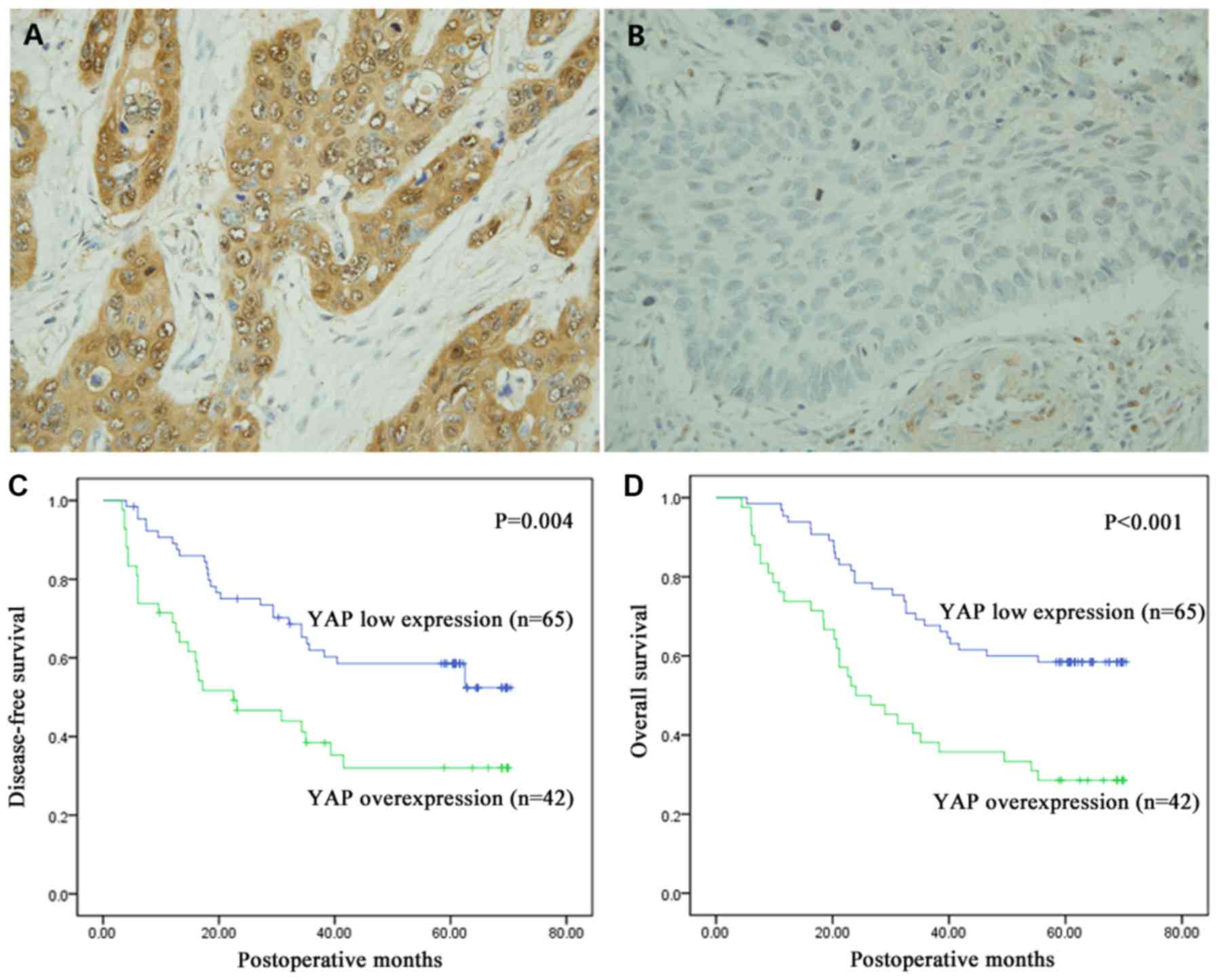
YAP expression level in the tumor tissues was
investigated using IHC staining and shown in brown. As shown in
Fig. 1A and B, YAP was mainly
positively expressed in the cytoplasm, but it was also observed in
the nucleus. IHC staining showed that YAP was overexpressed in
39.3% (42/107) of the samples and its expression was low in 60.7%
(65/107) of the samples.
The association of YAP expression with
clinicopathological features
To better understand the role of YAP expression
level in the progression of ESCC, the data was analyzed using
χ2 test. Overexpression of YAP was significantly
associated with increased N stage of ESCC (P=0.029). However, there
were no significant associations with the other clinicopathological
features (P>0.05; Table II).
| Table II.Association between
clinicopathological features of esophageal squamous cell carcinoma
and YAP expression in tumor tissues. |
Table II.
Association between
clinicopathological features of esophageal squamous cell carcinoma
and YAP expression in tumor tissues.
|
| YAP expression |
|
|---|
|
|
|
|
|---|
| Clinicopathological
features | Low, n=65 | Over, n=42 |
P-valuea |
|---|
| Age |
|
|
|
|
<60 | 28 | 22 | 0.428 |
|
≥60 | 37 | 20 |
|
| Sex |
|
|
|
|
Male | 51 | 36 | 0.449 |
|
Female | 14 | 6 |
|
| Smoking |
|
|
|
| No | 34 | 19 | 0.554 |
|
Yes | 31 | 23 |
|
| Drinking |
|
|
|
| No | 35 | 23 | 0.926 |
|
Yes | 30 | 19 |
|
|
Differentiation |
|
|
|
|
Well | 18 | 9 | 0.146 |
|
Moderate | 36 | 19 |
|
|
Poor | 11 | 14 |
|
| T stage |
|
|
|
|
T1-2 | 31 | 15 | 0.238 |
|
T3-4 | 34 | 27 |
|
| N stage |
|
|
|
| N0 | 40 | 16 | 0.029b |
|
N1-3 | 25 | 26 |
|
| pTNM |
|
|
|
|
I–II | 42 | 19 | 0.071 |
|
III | 23 | 23 |
|
| Adjuvant
treatment |
|
|
|
|
None | 35 | 28 | 0.417 |
|
Radiotherapy | 13 | 4 |
|
|
Chemotherapy | 5 | 4 |
|
|
CRT | 12 | 6 |
|
The prognostic value of YAP expression
in ESCC
The mean survival time of all 107 patients was
43.20±22.83 months (range 4.40–70.40 months). Kaplan-Meier analysis
was performed along with log-rank test to investigate the
relationship between YAP expression and prognosis. Compared to low
expression of YAP, its overexpression was significantly associated
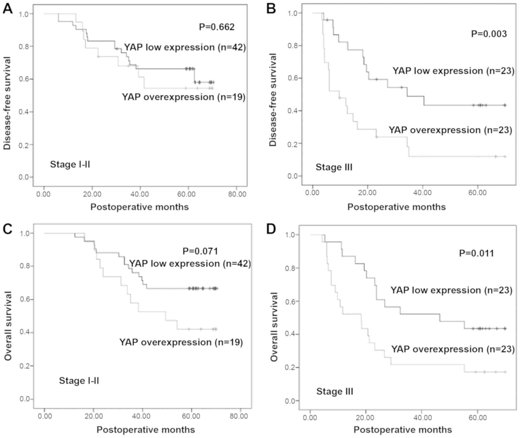
with decreased DFS (P=0.004) and OS (P<0.001; Fig. 1C and D). Furthermore, the prognostic
significance of YAP expression in patient subgroups stratified by
pTNM stage (I and II vs. III) was also investigated, and the
results revealed significant associations between YAP
overexpression and poor survival in patients with late stage ESCC,
but not in early stage ESCC (I and II; Fig. 2).
Univariate and multivariate survival
analyses
To further identify the role of YAP expression,
univariate and multivariate analyses were performed. Univariate
analysis showed that age, N stage, pTNM and YAP expression were
statistically significantly associated with OS and DFS (P<0.05).
Since previous studies reported that sex, smoking status, alcohol
consumption, T stage, differentiation degree, and adjuvant
treatment might play critical roles in prognosis of ESCC patients
(22,23), all factors were included in the
multivariate analysis, and the result indicated YAP overexpression
is an independent prognostic factor for OS (HR, 2.727; 95% CI
1.556–4.780; P<0.001) and DFS (HR, 2.161; 95% CI, 1.223–3.818;
P=0.008; Table III).
| Table III.Univariate and multivariate analysis
of prognostic variables for esophageal squamous cell carcinoma. |
Table III.
Univariate and multivariate analysis
of prognostic variables for esophageal squamous cell carcinoma.
|
| Overall
survival | Disease-free
survival |
|---|
|
|
|
|
|---|
|
| Univariate
analysis | Multivariate
analysis | Univariate
analysis | Multivariate
analysis |
|---|
| Variable | P-value | HR (95% CI) | P-value | P-value | HR (95% CI) | P-value |
|---|
| Sex |
| 1.528 |
|
| 0.699 |
|
| Male
vs. female | 0.598 | (0.670–3.484) | 0.313 | 0.999 | (0.303–1.613) | 0.402 |
| Age |
| 0.559 |
|
| 0.941 |
| <60
vs. ≥60 | 0.030a | (0.318–0.981) | 0.043a | 0.585 | (0.533–1.662) | 0.835 |
| Smoking |
| 0.931 |
|
| 0.639 |
| Yes vs.
no | 0.921 | (0.469–1.847) | 0.838 | 0.212 | (0.335–1.218) | 0.174 |
| Drinking |
| 1.910 |
|
| 0.951 |
| Yes vs.
no | 0.282 | (0.957–3.812) | 0.067 | 0.762 | (0.488–1.851) | 0.882 |
| T stage |
| 0.872 |
|
| 0.785 |
| T1-2
vs. T3 −4 | 0.445 | (0.466–1.631) | 0.668 | 0.452 | (0.422–1.458) | 0.443 |
| N stage |
| 1.530 |
|
| 1.035 |
| N0 vs.
N1-3 |
<0.001a | (0.533–4.395) | 0.430 | 0.001a | (0.365–2,937) | 0.948 |
|
Differentiation |
| 0.783 |
|
| 0.885 |
| Well
vs. moderate vs. poor | 0.658 | (0.526–1.165) | 0.227 | 0.915 | (0.595–1.316) | 0.545 |
| pTNM |
| 1.805 |
|
| 3.108 |
| I–II
vs. III |
<0.001a | (0.608–5.357) | 0.287 |
<0.001a | (1.038–9.310) | 0.043a |
| YAP expression |
| 2.727 |
|
| 2.161 |
| Low vs.
over | 0.001a | (1.556–4.780) |
<0.001a | 0.005a | (1.223–3.818) | 0.008a |
| CRT |
| 0.974 |
|
| 0.907 |
| None
vs. RT vs. | 0.785 | (0.768–1.237) | 0.831 | 0.620 | (0.708–1.163) | 0.443 |
| CT vs. RT+CT |
Expression analysis of YAP protein in
ESCC cell lines
Five common ESCC cell lines (TE-10, TE-11, Kyse150,
Kyse140 and Eca109) were used to screen for cell lines with high
YAP expression. Since Kyse150 and Eca109 expressed higher levels of
YAP than other three cell lines, they were selected for further
experiments. The results of the screening by western blot are shown
in Fig. S1.
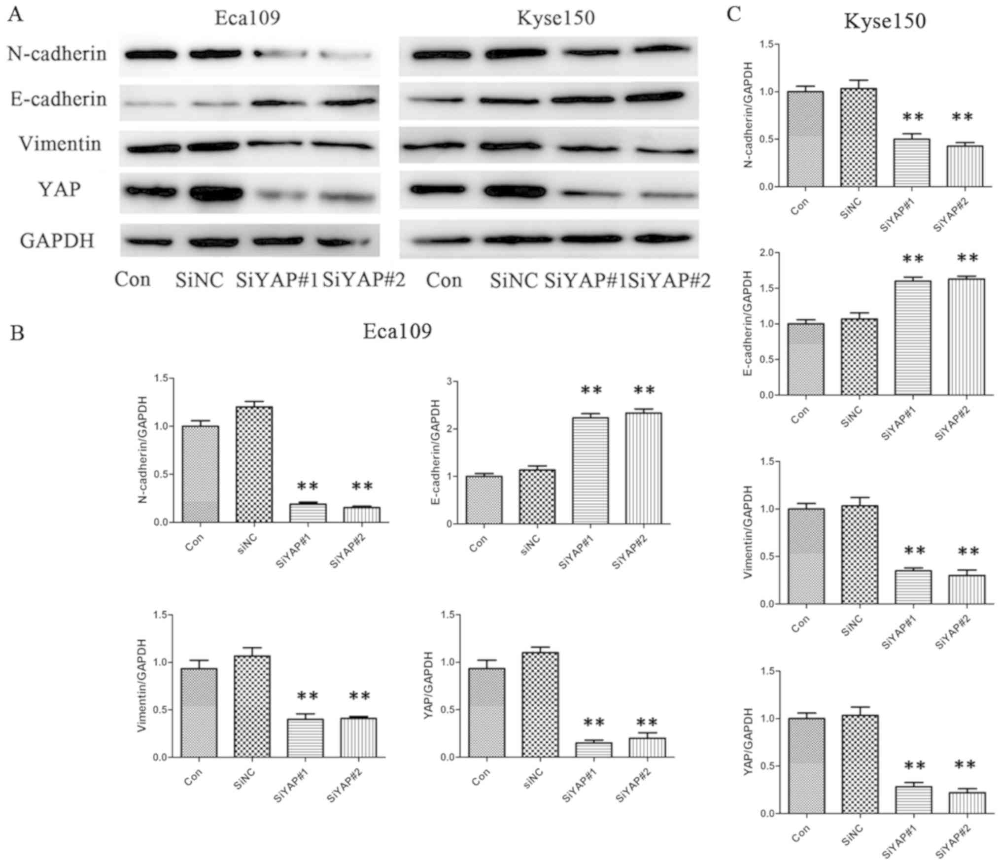
Downregulation of YAP protein
expression inhibits the expression of EMT markers in ESCC
To investigate the function of YAP in ESCC, siRNA
was used to specifically knockdown YAP expression in ESCC cells.
YAP siRNA and siNC were successfully transfected into the cells,
and western blot analysis was performed. The analysis showed that
YAP expression was successfully downregulated by siYAP but not by
siNC, at the protein level (Fig. 3).
Western blot analysis was also performed to detect levels of
EMT-related proteins. Compared with the control group, levels of
vimentin and N-cadherin were significantly reduced when YAP was
downregulated, while that of E-cadherin was significantly
increased, in both cell lines (Eca109 and Kyse150). Western
blotting results showed that YAP could regulate levels of
EMT-related proteins in ESCC cells.
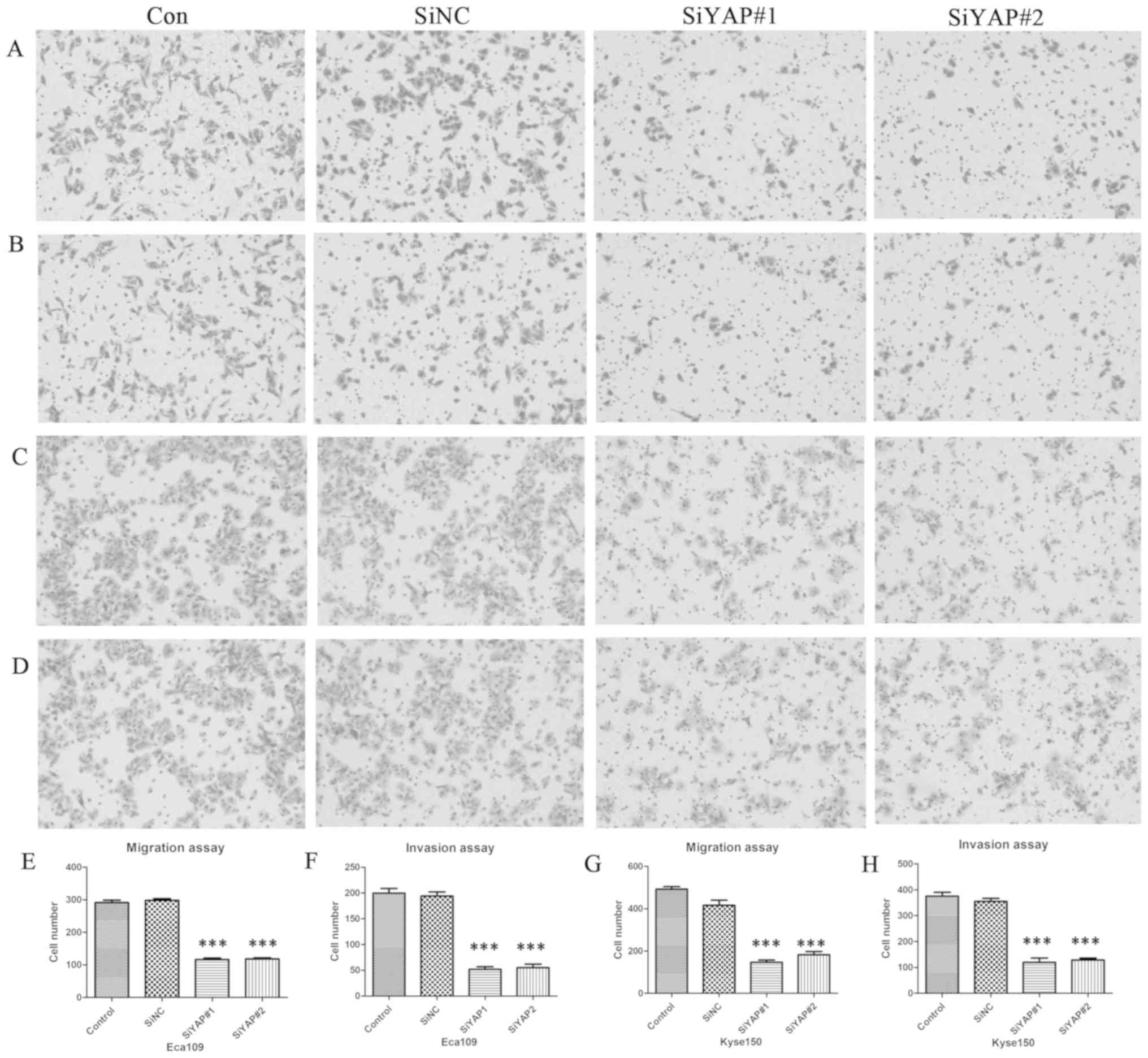
Downregulation of YAP inhibits cell
migration and invasion of ESCC cells
To identify whether YAP affects the ability of cell
migration and invasion in Eca109 and Kyse150 cells, transwell
migration and invasion assays were performed. Compared with the
control group, cell migration and invasion were both significantly
decreased by the downregulation of YAP after siYAP#1 and siYAP#2
transfection (P<0.001), in both cell lines. However, no
significant difference was found between the control and siNC
groups (Fig. 4).
Discussion
The oncogenic role of Hippo-YAP signaling pathway
has been reported in various human malignancies (6–9,24). Our previous research revealed that
YAP overexpression in prostate and pancreatic cancers promoted
tumor progression and metastasis (11,12).
However, there are few reports regarding YAP expression in ESCC. In
the present study, the level of YAP expression was detected in ESCC
tissues and its association with clinicopathological features was
analyzed. The analysis showed that YAP expression was only
associated with N stage of tumor, however this could be due to the
small sample size. Consistent with observations by previous
studies, the results also indicate that compared with low
expression of YAP, overexpression of YAP predicted poorer OS and
DFS (6–9,24).
Different statistical methods suggested that YAP expression level
could serve as an independent factor for predicting poor prognosis,
especially in patients with stage III ESCC, when stratified based
on pTNM stage.
The mechanisms underlying YAP-regulated progression
and prognosis of ESCC have not been verified. YAP was found to aid
esophageal cancer cells develop cancer stem cell-like properties
through driving SRY-box 9 expression (21). There is a previous report suggesting
that downregulation of YAP inhibits proliferation and induces
apoptosis in Eca109 cells (19).
Other possible mechanisms of YAP regulation have been reported in
other cell lines and tumors. It has been proven that YAP
oncoprotein can overcome inhibition caused by high cell-contact and
promote cell proliferation in NIH-3T3 cells and MCF10A human breast
epithelial cell line (25). YAP
overexpression was further confirmed to trigger EMT in MCF10A cells
(3). In papillary thyroid cancer
B-CPAP and KI cell lines, knockdown of YAP was found to inhibit the
proliferation, migration, and invasion, and cause cell cycle arrest
and induce autophagy of tumor cells (24). In addition, in prostate cancer, YAP
acted as a regulator of cell motility, invasion,
castration-resistant growth and hence might be a potential
therapeutic target (11). In gastric
cancer, it was found that Netrin-1 promotes metastasis of gastric
cancer by upregulating YAP expression via its transmembrane
receptor neogenin (26). In
addition, YAP could regulate the initiation and progression of
cervical (6) and ovarian cancer
(9) through the ErbB signaling
pathway. Previous studies have reported that activated YAP could
cause the upregulation of TGF-α, amphiregulin, and epidermal growth
factor receptor, facilitating the formation of a positive signaling
loop to promote cervical and ovarian cancer cell proliferation
(6,9). There are reports regarding the tumor
suppressive effect of YAP, either by inhibiting WNT signaling
(27) or by triggering DNA
damage-induced apoptosis (28).
Studies have shown that the YAP signaling pathway was associated
with EMT (15–18). Nevertheless, the mechanism of YAP
overexpression in ESCC tissues and further influence on the
prognosis remain to be elucidated. The present study presents the
potential mechanism of YAP regulation in ESCC, possibly through
EMT.
There are several limitations in the present study.
First, as a retrospective cohort study, the sample number was
small, which might have influenced the reliability of the results.
Second, there could have been more suitable cutoff values for
determining the overexpression and the reduced expression of YAP,
which could improve predicting the prognosis. Third, more detailed
elucidation of the mechanism of YAP expression affecting the
prognosis remains to be discovered. Further studies are required to
propose suitable methods to target the elevated expression of YAP
and thereby improve the prognosis.
Supplementary Material
Supporting Data
Acknowledgements
The authors would like to thank Dr. Xinyuan Guan
(Department of Clinical Oncology Cancer Research Center, University
of Hong Kong, Hong Kong, China) for offering us the ESCC cell
line.
Funding
The present study was supported by National Natural
Science Foundation of China (grant nos. 81773228, 81572958 and
81602007), Natural Science Foundation of Shandong Province (grant
no. ZR2016HB68), Nanshan Project of Yantai for Shandong University
(grant no. 2014QLKY31) and Key Research and Development Program of
Shandong Province (grant no. 2017GSF18153).
Availability of data and materials
The datasets used and/or analyzed during the current
study are available from the corresponding author upon reasonable
request.
Authors' contributions
YQ, LZ, JW and YJ performed the experiments,
analysis and wrote the manuscript. PC, CW and WY generated the data
and performed the analyses. ZW, QS, BT and YC interpreted the data,
drafted the manuscript and made critical revisions. All authors
discussed the results and reviewed the manuscript.
Ethics approval and consent to
participate
The present study was approved by The Ethics
Committee of Qilu Hospital. All the patients in the study were
anonymous and provided written informed consent.
Patient consent for publication
Not applicable.
Competing interests
The authors declare that they have no competing
interests.
References
|
1
|
Bray F, Ferlay J, Soerjomataram I, Siegel
RL, Torre LA and Jemal A: Global cancer statistics 2018: GLOBOCAN
estimates of incidence and mortality worldwide for 36 cancers in
185 countries. CA Cancer J Clin. 68:394–424. 2018. View Article : Google Scholar : PubMed/NCBI
|
|
2
|
Zender L, Spector MS, Xue W, Flemming P,
Cordon-Cardo C, Silke J, Fan ST, Luk JM, Wigler M, Hannon GJ, et
al: Identification and validation of oncogenes in liver cancer
using an integrative oncogenomic approach. Cell. 125:1253–1267.
2006. View Article : Google Scholar : PubMed/NCBI
|
|
3
|
Overholtzer M, Zhang J, Smolen GA, Muir B,
Li W, Sgroi DC, Deng CX, Brugge JS and Haber DA: Transforming
properties of YAP, a candidate oncogene on the chromosome 11q22
amplicon. Proc Natl Acad Sci USA. 103:12405–12410. 2006. View Article : Google Scholar : PubMed/NCBI
|
|
4
|
Pan D: The hippo signaling pathway in
development and cancer. Dev Cell. 19:491–505. 2010. View Article : Google Scholar : PubMed/NCBI
|
|
5
|
Moroishi T, Hansen CG and Guan KL: The
emerging roles of YAP and TAZ in cancer. Nat Rev Cancer. 15:73–79.
2015. View
Article : Google Scholar : PubMed/NCBI
|
|
6
|
He C, Mao D, Hua G, Lv X, Chen X,
Angeletti PC, Dong J, Remmenga SW, Rodabaugh KJ, Zhou J, et al: The
Hippo/YAP pathway interacts with EGFR signaling and HPV
oncoproteins to regulate cervical cancer progression. EMBO Mol Med.
7:1426–1449. 2015. View Article : Google Scholar : PubMed/NCBI
|
|
7
|
Zhang W, Nandakumar N, Shi Y, Manzano M,
Smith A, Graham G, Gupta S, Vietsch EE, Laughlin SZ, Wadhwa M, et
al: Downstream of mutant KRAS, the transcription regulator YAP is
essential for neoplastic progression to pancreatic ductal
adenocarcinoma. Sci Signal. 7:ra422014. View Article : Google Scholar : PubMed/NCBI
|
|
8
|
Liu JY, Li YH, Lin HX, Liao YJ, Mai SJ,
Liu ZW, Zhang ZL, Jiang LJ, Zhang JX, Kung HF, et al:
Overexpression of YAP 1 contributes to progressive features and
poor prognosis of human urothelial carcinoma of the bladder. BMC
Cancer. 13:3492013. View Article : Google Scholar : PubMed/NCBI
|
|
9
|
He C, Lv X, Hua G, Lele SM, Remmenga S,
Dong J, Davis JS and Wang C: YAP forms autocrine loops with the
ERBB pathway to regulate ovarian cancer initiation and progression.
Oncogene. 34:6040–6054. 2015. View Article : Google Scholar : PubMed/NCBI
|
|
10
|
Wang L, Shi S, Guo Z, Zhang X, Han S, Yang
A, Wen W and Zhu Q: Overexpression of YAP and TAZ Is an independent
predictor of prognosis in colorectal cancer and related to the
proliferation and metastasis of colon cancer cells. PLoS One.
8:e655392013. View Article : Google Scholar : PubMed/NCBI
|
|
11
|
Zhang L, Yang S, Chen X, Stauffer S, Yu F,
Lele SM, Fu K, Datta K, Palermo N, Chen Y and Dong J: The hippo
pathway effector YAP regulates motility, invasion, and
castration-resistant growth of prostate cancer cells. Mol Cell
Biol. 35:1350–1362. 2015. View Article : Google Scholar : PubMed/NCBI
|
|
12
|
Yang SP, Zhang L, Purohit V, Shukla SK,
Chen X, Yu F, Fu K, Chen Y, Solheim J, Singh PK, et al: Active YAP
promotes pancreatic cancer cell motility, invasion and
tumorigenesis in a mitotic phosphorylation-dependent manner through
LPAR3. Oncotarget. 6:36019–36031. 2015. View Article : Google Scholar : PubMed/NCBI
|
|
13
|
Muramatsu T, Imoto I, Matsui T, Kozaki K,
Haruki S, Sudol M, Shimada Y, Tsuda H, Kawano T and Inazawa J: YAP
is a candidate oncogene for esophageal squamous cell carcinoma.
Carcinogenesis. 32:389–398. 2011. View Article : Google Scholar : PubMed/NCBI
|
|
14
|
Chen T, You Y, Jiang H and Wang ZZ:
Epithelial-mesenchymal transition (EMT): A biological process in
the development, stem cell differentiation, and tumorigenesis. J
Cell Physiol. 232:3261–3272. 2017. View Article : Google Scholar : PubMed/NCBI
|
|
15
|
Jin D, Wu Y, Shao C, Gao Y, Wang D and Guo
J: Norcantharidin reverses cisplatin resistance and inhibits the
epithelial mesenchymal transition of human nonsmall lung cancer
cells by regulating the YAP pathway. Oncol Rep. 40:609–620.
2018.PubMed/NCBI
|
|
16
|
Kulkarni M, Tan TZ, Syed Sulaiman NB,
Lamar JM, Bansal P, Cui J, Qiao Y and Ito Y: RUNX1 and RUNX3
protect against YAP-mediated EMT, stem-ness and shorter survival
outcomes in breast cancer. Oncotarget. 9:14175–14192. 2018.
View Article : Google Scholar : PubMed/NCBI
|
|
17
|
Thongon N, Castiglioni I, Zucal C, Latorre
E, D'Agostino V, Bauer I, Pancher M, Ballestrero A, Feldmann G,
Nencioni A and Provenzani A: The GSK3β inhibitor BIS I reverts
YAP-dependent EMT signature in PDAC cell lines by decreasing SMADs
expression level. Oncotarget. 7:26551–26566. 2016. View Article : Google Scholar : PubMed/NCBI
|
|
18
|
Yu S, Jing L, Yin XR, Wang MC, Chen YM,
Guo Y, Nan KJ and Han LL: MiR-195 suppresses the metastasis and
epithelial-mesenchymal transition of hepatocellular carcinoma by
inhibiting YAP. Oncotarget. 8:99757–99771. 2017.PubMed/NCBI
|
|
19
|
Cui M and Li Z: Downregulation of YAP
inhibits proliferation and induces apoptosis in Eca-109 cells. Exp
Ther Med. 15:1048–1052. 2018.PubMed/NCBI
|
|
20
|
Rice TW, Ishwaran H, Ferguson MK,
Blackstone EH and Goldstraw P: Cancer of the Esophagus and
Esophagogastric Junction: An Eighth Edition Staging Primer. J
Thorac Oncol. 12:36–42. 2017. View Article : Google Scholar : PubMed/NCBI
|
|
21
|
Song S, Ajani JA, Honjo S, Maru DM, Chen
Q, Scott AW, Heallen TR, Xiao L, Hofstetter WL, Weston B, et al:
Hippo coactivator YAP1 upregulates SOX9 and endows esophageal
cancer cells with stem-like properties. Cancer Res. 74:4170–4182.
2014. View Article : Google Scholar : PubMed/NCBI
|
|
22
|
Lin WC, Ding YF, Hsu HL, Chang JH, Yuan
KS, Wu ATH, Chow JM, Chang CL, Chen SU and Wu SY: Value and
application of trimodality therapy or definitive concurrent
chemoradiotherapy in thoracic esophageal squamous cell carcinoma.
Cancer. 123:3904–3915. 2017. View Article : Google Scholar : PubMed/NCBI
|
|
23
|
Liu B, Cheng B, Wang C, Chen P and Cheng
Y: The prognostic significance of metabolic syndrome and weight
loss in esophageal squamous cell carcinoma. Sci Rep. 8:101012018.
View Article : Google Scholar : PubMed/NCBI
|
|
24
|
Liu Z, Zeng W, Wang S, Zhao X, Guo Y, Yu
P, Yin X, Liu C and Huang T: A potential role for the Hippo pathway
protein, YAP, in controlling proliferation, cell cycle progression,
and autophagy in BCPAP and KI thyroid papillary carcinoma cells. Am
J Transl Res. 9:3212–3223. 2017.PubMed/NCBI
|
|
25
|
Zhao B, Wei X, Li W, Udan RS, Yang Q, Kim
J, Xie J, Ikenoue T, Yu J, Li L, et al: Inactivation of YAP
oncoprotein by the Hippo pathway is involved in cell contact
inhibition and tissue growth control. Genes Dev. 21:2747–2761.
2007. View Article : Google Scholar : PubMed/NCBI
|
|
26
|
Yin K, Dang S, Cui L, Fan X, Xie R, Qu J,
Shang M and Chen J: Corrigendum to ‘Netrin-1 promotes metastasis of
gastric cancer by regulating YAP activity’ [Biochem. Biophys. Res.
Commun.496 (1) (2018 Jan 29) 76–82]. Biochem Biophys Res Commun.
498:2622018. View Article : Google Scholar : PubMed/NCBI
|
|
27
|
Attisano L and Wrana JL: Signal
integration in TGF-β, WNT, and Hippo pathways. F1000Prime Rep.
5:172013. View
Article : Google Scholar : PubMed/NCBI
|
|
28
|
Cottini F, Hideshima T, Xu C, Sattler M,
Dori M, Agnelli L, ten Hacken E, Bertilaccio MT, Antonini E, Neri
A, et al: Rescue of Hippo coactivator YAP1 triggers DNA
damage-induced apoptosis in hematological cancers. Nat Med.
20:599–606. 2014. View
Article : Google Scholar : PubMed/NCBI
|