Introduction
Amblyopia is a vision development disorder caused by
abnormal visual experience during the sensitive period, affecting
~3% of the world's population. The neural mechanism underlying
amblyopia remains to be determined; however, a number of studies
have aimed to identify the localization of the neurological deficit
(1–17).
Previous studies reported reduced visual cortex gray
matter volume (1–3), as well as poor white-matter development
among patients with amblyopia (3,4). These
morphological alterations of the visual cortex may be associated
with the visual deficits in amblyopia.
Functional magnetic resonance imaging (fMRI) is a
non-invasive method frequently used to determine the functional
alterations of cerebral areas. The majority of previous studies on
amblyopia used task-based fMRI. Certain studies indicated that this
disease may originate from the striate cortex (5,6);
however, neurological deficits were also identified in the lateral
geniculate nucleus (7,8) and extrastriate cortex (9,10).
Task-based fMRI is a modified fMRI analysis based on
stimulation or task performance. Lack of patient cooperation and
challenges associated with task standardization limit the clinical
applicability of task-based fMRI. Therefore, this type of fMRI is
not extensively used in the diagnosis of human diseases and
associated research. The human brain constitutes ~2% of the human
body weight; however, it consumes ~20% of the total energy
expenditure. The greatest amount of energy is required for
information transfer between neurons. Energy consumption during
cognitive task performance increases by 5% compared with
resting-state energy expenditure (11). Therefore, task-based fMRI
investigates part of the brain activity, whereas a comprehensive
brain analysis may be performed using resting-state fMRI.
A number of studies investigated the underlying
neurological mechanisms of amblyopia using resting-state fMRI. Lin
et al (12) investigated
alterations in resting-state local spontaneous brain activity among
patients with anisometropic amblyopia using regional homogeneity
(ReHo) values. The results of this study indicated decreased
spontaneous brain activity represented by low ReHo values of
certain brain regions, including the precuneus, medial prefrontal
cortex and cerebellum (12).
However, compensatory plasticity was detected in other areas of the
brain, including the bilateral conjunction area of the post-central
and pre-central gyri, paracentral lobule and superior temporal
gyrus (12). Ding et al
(13) further investigated the
functional connectivity of the primary visual area of patients with
amblyopia using resting-state fMRI. The results demonstrated
altered functional connectivity of the cerebellum, inferior
parietal lobule and primary visual cortex among individuals with
amblyopia (13). Wang et al
(14) used functional-connectivity
density mapping and an ultrafast data-driven method based on
resting-state fMRI to investigate alterations in cortical
functional connectivity among patients with anisometropic
amblyopia. This study identified impaired short-range functional
connections in the visual pathways of the brain and in the frontal
cortex, and altered long-range functional connections among the
visual regions, posterior parietal and frontal cortices of patients
with amblyopia (14). A previous
study by our group used resting-state fMRI combined with amplitude
of low-frequency fluctuation (ALFF) analysis to investigate the
intrinsic brain activity patterns of patients with amblyopia and
the results indicated an impaired function of certain brain regions
and increased ALFF of other brain areas (15). Similar data were reported in Liang
et al (16). Recently, a
novel voxel-wise image analysis method, voxel-mirrored homotopic
connectivity (VMHC), was used to assess the alterations in
resting-state functional connectivity in patients with amblyopia
(17). The results revealed
similarities and differences in interhemispheric functional
connectivity between patients with anisometropic and strabismic
amblyopia (17).
Based on the hypothesis that brain activity occurs
in clusters, as opposed to a single voxel, Zang et al
(18) proposed that the ReHo index
may be used for the analysis of brain activity patterns during the
resting state. The ReHo index evaluates the temporal homogeneity
between the time series of each voxel and its nearest neighbors, to
reflect the regional activity of the whole brain. The ReHo index
indirectly reflects the homogeneity of blood oxygen level-dependent
signals, and alterations of ReHo values may suggest abnormal
activity of neurons.
The ReHo index has been used to evaluate diverse
neurological and psychiatric diseases, including schizophrenia
(19), attention deficit
hyperactivity disorder (20),
Parkinson's disease (21) and
Alzheimer's disease (22).
Furthermore, this index has been used in the field of ophthalmology
to investigate blindness (23),
glaucoma (24), strabismus (25) and anisometropic amblyopia (12). The above studies used the ReHo index
to obtain novel neuropathological data.
The ReHo values of spontaneous brain activity of
patients with strabismic amblyopia remain elusive. Furthermore,
strabismic and anisometropic amblyopia are characterized by
different early abnormal visual experiences. A previous study
revealed differences between these conditions using resting-state
fMRI combined with VMHC analysis (17). Therefore, the spontaneous activity
pattern of patients with strabismic amblyopia may differ from that
of patients with anisometropic amblyopia. In the present study, the
ReHo index was used to analyze the brain activity of patients with
strabismic amblyopia.
Patients and methods
Patients
Patients with strabismic amblyopia were recruited
between June 2015 and November 2017 from the Department of
Ophthalmology of West China Hospital of Sichuan University
(Chengdu, China) and diagnosed using the Expert Consensus on
Amblyopia Diagnosis guidelines (26). The normal control group included
healthy student volunteers recruited from Sichuan University
between January 2016 and December 2017.
All participants were subjected to eye examination,
including a visual acuity test, ocular tonometry, slit-lamp
examination, dilated fundus examination, cover test and ocular
movement examination. Furthermore, the near point of convergence,
stereoacuity and refraction were determined for each patient. A
total of 12 patients with strabismic amblyopia and 34 normal
controls were enrolled in the present study. A total of 1 patient
with strabismic amblyopia and 7 healthy volunteers were excluded
due to excessive head motion. The patients included in the present
study were right-handed and aged 18–35 years. Patients with
strabismic amblyopia exhibited an ocular deviation of ≥15Δ and
normal controls exhibited naked eye or corrected visual acuity of
≥1.0 in the bilateral eyes. The exclusion criteria were as follows:
i) A history of other ocular diseases; ii) strabismus identified
among control patients; iii) a history of neurological diseases;
iv) claustrophobia; and v) inability to cooperate.
The protocol of the present study was approved by
the Ethics Committee of West China Hospital of Sichuan University
(Chengdu, China) and complied with the principles of the
Declaration of Helsinki. Written informed consent was voluntarily
provided by all participants after the purpose and methods of the
present study were explained verbally and in writing.
MRI data acquisition
All MRI data analyses were performed at the MR
Research Center of West China Hospital using the Magnetom Trio 3.0T
MRI system (Siemens Healthineers) equipped with a standard
eight-channel quadrature head coil. The imaging area covered the
entire brain. All participants were informed of the
contraindications of the MRI examination and other associated
matters of importance. The MRI examination followed the
standardized scanning procedures (27).
MRI scout images and three-dimensional structural
T1-weighted images were initially obtained, followed by
resting-state fMRI scanning. Structural T1-weighted images were
obtained using a magnetization-prepared rapid gradient-echo
sequence with the following parameters: Echo time (TE), 2.62 msec;
repetition time (TR), 2,250 msec; field of view (FOV), 25.6×25.6
cm; flip angle (FA), 9°; matrix, 256×256. Resting-state functional
images were obtained using an echo-planar imaging sequence with the
following parameters: TE, 35 msec; TR, 2,000 msec; FOV, 20.8×20.8
cm; FA, 68°; matrix, 64×64.
Data pre-processing
Prior to data pre-processing, data in DICOM format
were converted to the NIFTI format using MRIcron software v.
12/2012 (www.cabiatl.com/mricro/mricron/dcm2nii.html).
Pre-processing was performed using the Data Processing Assistant
for Resting State fMRI software 2.2 (rfmri.org/DPARSF) based on Statistical Parametric
Mapping 8 (www.fil.ion.ucl.ac.uk/spm).
Due to initial transient signal fluctuations and
adaptation to the scanning process, the data obtained from the
first 10 volumes of each functional time series were discarded and
the remaining 210 volumes were processed with slice timing and
realignment for head motion correction. Participants with head
motion ≤2.0 mm or ≤2.0° rotation in any axis were included. One
patient with strabismic amblyopia and seven healthy volunteers were
excluded due to excessive head motion.
All realigned data were spatially normalized
according to the standard Montreal Neurological Institute (MNI)
coordinates and resampled to 3×3×3 mm3 voxels. To reduce
the effects of linear tendency due to the long duration of the
scan, linear drift was removed from the data. Furthermore, the
regressed images were smoothed with an 8-mm full width at half
maximum to remove spatial noise. Finally, a temporal bandpass
filter (0.01–0.08 Hz) was used to minimize the effects of
low-frequency drift and high-frequency noise.
ReHo index measurement
The ReHo indices of 12 patients with strabismic
amblyopia and 34 normal controls were calculated. When calculating
the ReHo index, it was assumed that each voxel has a high temporal
correlation with its neighboring voxels in the same cerebral
region. Kendall's coefficient of concordance (KCC) was used to
describe the similarity of time series in a voxel with its 26
neighboring voxels. KCC values range from 0 to 1, and an increase
in the KCC value indicates an increase in regional homogeneity
(18). The whole-brain map of
regional brain activity was obtained following calculation of the
ReHo index of each voxel. To minimize the influence of individual
variability, the ReHo value for each voxel was divided by the mean
ReHo of the whole brain for normalization. Normalized ReHo values
indicated regional brain activity across the entire brain.
Statistical analysis
Demographic data were compared between patients with
strabismic amblyopia and normal controls using SPSS software
(version 18.0; SPSS, Inc.). An independent-samples t-test was used
for the analysis of numerical variables and a χ2 test
was used for categorical variables.
Independent-samples t-tests were performed using
Resting State fMRI Data Analysis Toolkit software (version 1.8;
restfmri.net/forum/REST_V1.8) to
determine the differences in the normalized whole-brain ReHo values
between patients with strabismic amblyopia and healthy controls.
Initially, P<0.01 was considered to indicate a statistically
significant difference. Subsequently, AlphaSim multiple-comparison
correction was used and the P-value cut-off was adjusted to 0.05
with a cluster size of at least 77 voxels. Subsequently, the size,
cerebral region and MNI coordinates of the clusters with
significantly different ReHo values were obtained.
The present study aimed to determine whether the
ReHo values varied with disease progression among patients with
strabismic amblyopia. Pearson's correlation analysis was used to
determine the association between the ReHo indices of the
identified regions and the best-corrected visual acuity.
Furthermore, to evaluate the association between ReHo values and
age, the same correlation analysis was performed. P<0.05 was
used to indicate a statistically significant difference. Finally,
receiver operating characteristic (ROC) curve analysis was
performed to evaluate the sensitivity and specificity of ReHo
values from altered brain regions to distinguish subjects with
strabismic amblyopia from normal subjects.
Results
Comparison of demographic parameters
between normal controls and patients with amblyopia
As presented in Table
I, the patients with amblyopia included four males and eight
females aged 23.92±5.31 years. A total of 5 patients were affected
in the left eye and 7 patients were affected in the right eye. A
total of 7 patients exhibited esotropic amblyopia and 5 patients
exhibited exotropic amblyopia. The normal control group included 12
males and 22 females aged 24.09±2.78 years. The gender distribution
(χ2=0.015; P=1.00) and age (t=0.107; P=0.917) were not
significantly different between the two groups.
 | Table I.Basic characteristics of strabismic
amblyopia group and normal control group. |
Table I.
Basic characteristics of strabismic
amblyopia group and normal control group.
| Characteristic | SA (n=12) | NC (n=34) |
|---|
| Gender (M/F) | 4/8 | 12/22 |
| Age (years) | 23.92±5.31 | 24.09±2.78 |
| AE or NDE
(L/R) | 5/7 | 24/10 |
| BCVA (logMAR) of AE
or NDE | 1.19±0.44 | 0.00±0.00 |
Differences in the ReHo values between
the two groups
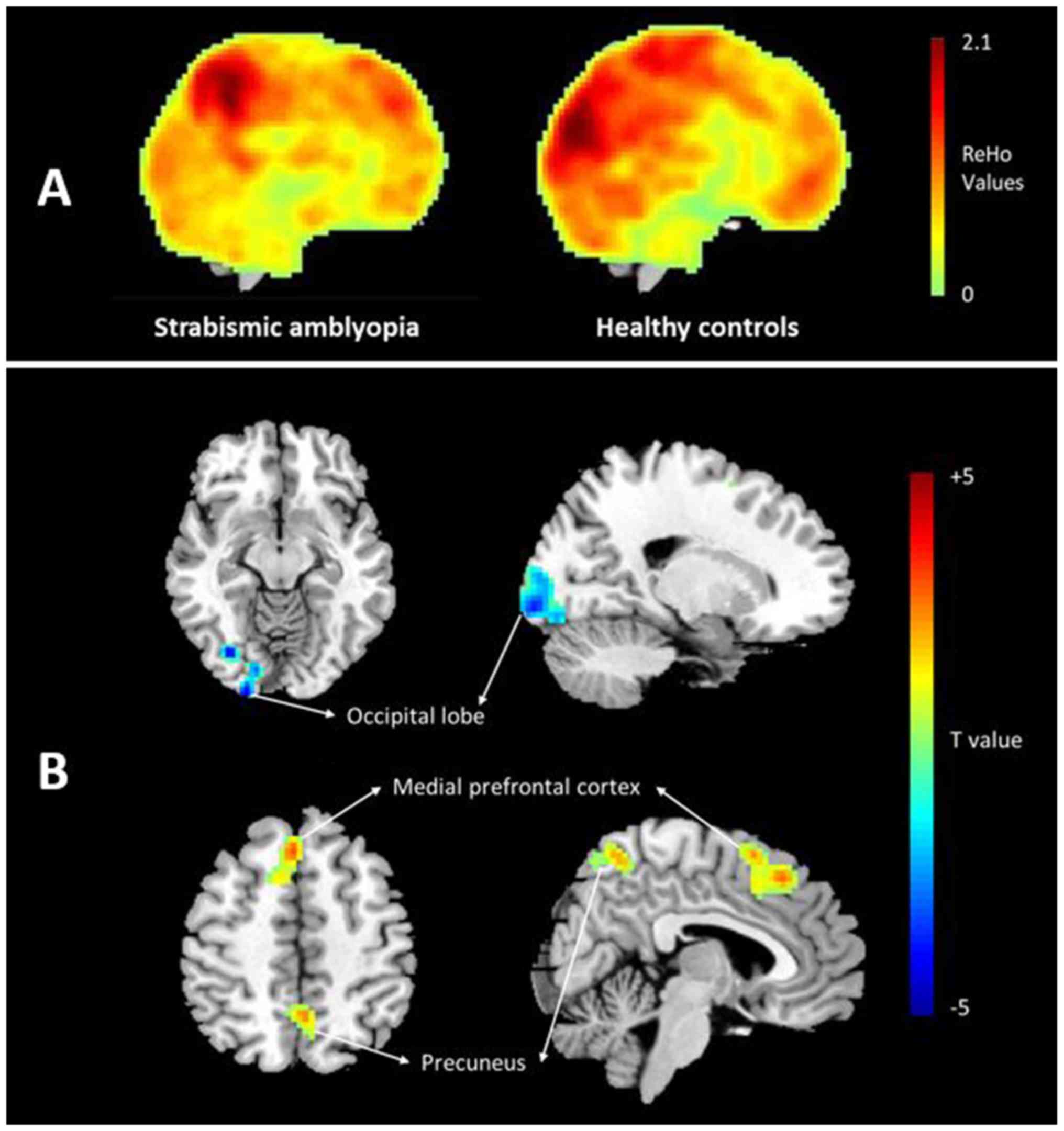
Significant differences in the ReHo values of the
visual cortices were identified between patients and controls
(Table II). Compared with those in
the control group, patients with strabismic amblyopia exhibited
significantly decreased ReHo values of certain parts of the
occipital lobe, including the cuneus, lingual gyrus and superior
occipital gyrus (Fig. 1). However,
certain brain areas exhibited significantly increased ReHo values,
including the precuneus and medial prefrontal cortex (Fig. 1).
| Table II.Differences of ReHo in brain
functional activities between the strabismic amblyopia group and
normal control group. |
Table II.
Differences of ReHo in brain
functional activities between the strabismic amblyopia group and
normal control group.
| Brain region | Brodmann
region | T-value | Cluster size
(voxels) | Coordinates in MNI
(x, y, z) |
|---|
| Decreased ReHo in
strabismic amblyopia |
| Certain
parts of the occipital lobe | 17/18 | −3.6055 | 128 | (21,-99,-9) |
| (cuneus, lingual
gyrus and superior occipital gyrus) |
| Increased ReHo in
strabismic amblyopia |
|
Precuneus | 7 | 4.3857 | 104 | (0,-57,54) |
| Medial
prefrontal cortex | 10 | 4.8984 | 136 | (3,27,48) |
| (superior frontal
gyrus and middle frontal gyrus) |
Association between the ReHo values
and clinical manifestations of patients with strabismic
amblyopia
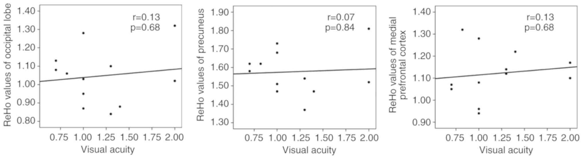
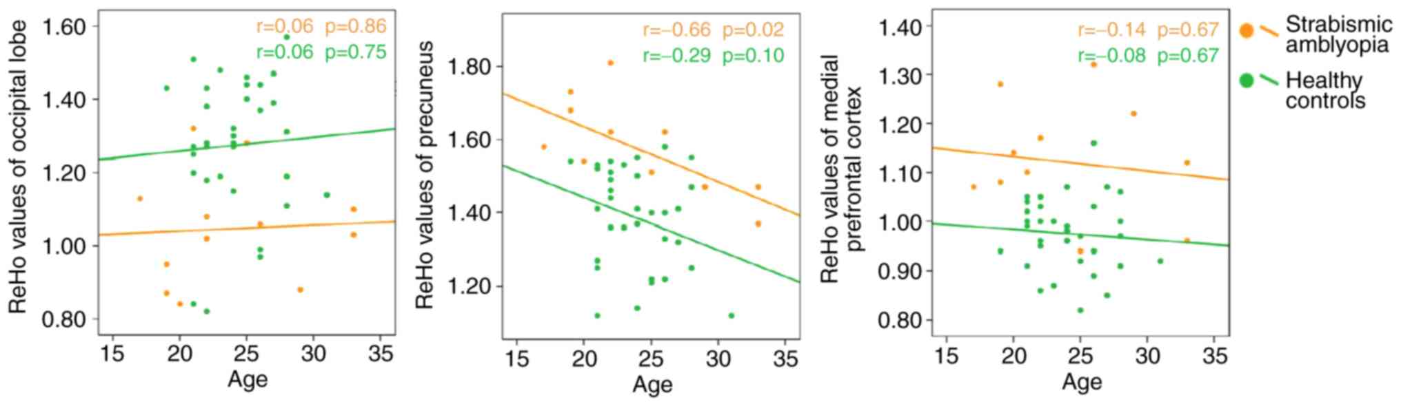
There was no correlation between the ReHo index and
visual acuity of patients with strabismic amblyopia (Fig. 2). The ReHo index of the precuneus was
negatively correlated with age (r=−0.664, P=0.019) in the amblyopic
group, but no such correlation was present in the occipital lobe
and medial prefrontal cortex (Fig.
3). Furthermore, there was no correlation between the ReHo
index and age in any of the above-mentioned regions in the healthy
controls (Fig. 3).
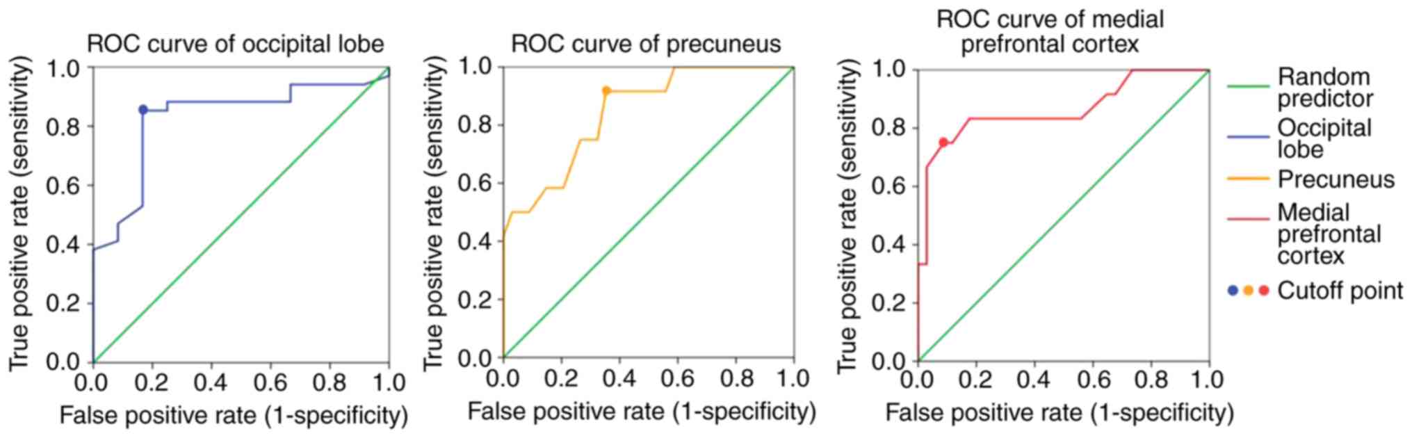
ROC curve analysis revealing ReHo in
the medial prefrontal cortex is able to distinguish strabismic
amblyopia from normal controls
As presented in Fig.
4, ROC curve analysis demonstrated that the greatest area under
curve (AUC) value was in the medial prefrontal cortex (AUC=0.864),
and the ReHo value was able to discriminate patients with
strabismic amblyopia from normal controls with a sensitivity of 75%
and specificity of 91.2%. The AUC of precuneus and occipital lobe
was 0.846 and 0.828, respectively.
Discussion
To the best of our knowledge, the ReHo index was not
previously used to investigate resting-state spontaneous brain
activity in patients with strabismic amblyopia. Decreased
spontaneous brain activity represented by reduced ReHo values was
detected in certain parts of the occipital lobe, including the
cuneus, lingual gyrus and superior occipital gyrus. However,
increased ReHo values were determined in the precuneus and certain
parts of the prefrontal cortex, including the superior frontal
gyrus and middle frontal gyrus of patients with strabismic
amblyopia compared with normal controls.
Mendola et al (2) proposed that amblyopia may be associated
with structural brain abnormalities. This study used voxel-based
analysis of MRI and detected decreased visual cortex gray-matter
volume among children and adult patients with amblyopia (2). A previous study by our group used
magnetic resonance diffusion tensor imaging and diffusion tensor
tractography to identify decreased compactability, integrity and
directivity of optic radiations among patients with anisometropic
amblyopia (28). Furthermore,
previous studies identified decreased white-matter volume among
patients with amblyopia (3,4). These structural abnormalities may
contribute to alterations of spontaneous brain activity detected in
the present study using resting-state fMRI.
In the present study, patients with strabismic
amblyopia exhibited significantly decreased ReHo values in certain
parts of the occipital lobe, including the cuneus, lingual gyrus
and superior occipital gyrus, which belong to the Brodmann area
(BA) 17 and 18. BA 17, also known as the striate cortex, V1 area or
primary visual cortex, is used for visual information processing.
BA 18, also known as the V2 area, is a part of the extrastriate
cortex used for further processing of visual information
transformed from the striate cortex. The present results are
consistent with those of numerous previous studies. Wiesel and
Hubel (29) proposed that form
deprivation including occlusion, congenital cataract and ptosis may
affect the visual cortex and identified amblyopia-associated
alterations in the V1 area. Subsequently, numerous studies
demonstrated that abnormal visual experiences may lead to
morphological and functional alterations of the V1 area (30–35). A
previous study by our group used task-based fMRI to further
demonstrate that the activation of the V1 and V2 areas was reduced
among patients with amblyopia (10).
These results suggested that the visual information processing of
the brain is deteriorated in strabismic amblyopia.
However, using the same ReHo analysis method based
on resting-state fMRI as that described in the present study, Lin
et al (12) did not identify
any alterations in the ReHo index of the V1 area among patients
with anisometropic amblyopia. Furthermore, increased ReHo values
were identified in the V2 and V3 areas (12). The above results may reflect the
differences in the neural deficit between patients with
anisometropic and strabismic amblyopia.
In the present study, patients with strabismic
amblyopia exhibited increased ReHo values in the precuneus and
medial prefrontal cortex, including the superior frontal gyrus and
middle frontal gyrus, compared with those in normal controls. The
precuneus is located in the medial wall of BA 7, a component of the
dorsal visual pathway receiving visual information from the middle
temporal area, also known as area V5. The major functions of the
dorsal stream, also known as the ‘where pathway’ or the ‘how
pathway’, include motion detection, object localization and arm-eye
coordination (13). The precuneus
participates in a number of highly integrated tasks, including
visuomotor coordination (36,37),
visuospatial imagery, episodic memory retrieval and self-processing
operations, including taking of the first-person perspective and
the experience of agency (38).
Niechwiej-Szwedo et al (39,40)
identified abnormal visuomotor processes in patients with
strabismic amblyopia using behavioral experiments. Contrary to the
results of those behavioral experiments, the increased ReHo values
detected in the present study in the precuneus of patients with
strabismic amblyopia may reflect the plasticity of the human brain
compensating for visuomotor coordination and visuospatial imagery
deficits.
The medial prefrontal cortex is located in BA 10, an
essential component of the default-mode network associated with
consciousness (41). Of note,
strabismic amblyopes frequently suffer from high anxiety (42). First, their appearance is changed due
to deviation. Furthermore, strabismic amblyopes have an increased
risk of blindness when the fellow eye is injured or diseased, due
to the low visual acuity of the amblyopic eye, leading to patients
becoming more anxious. This may be associated with the increased
ReHo values in the medial prefrontal cortex, and may also reflect
compensatory plasticity.
The potential compensatory mechanism has also been
reported in patients with visual impairments, including blindness
(43,44), optic neuritis (45) and amblyopia, as indicated by the
results of task-based fMRI (46).
In the present study, no correlation between the
standardized ReHo values and the visual acuity of patients with
amblyopia was determined. Furthermore, a number of previous
task-based fMRI studies revealed no association between visual
acuity and the activation of cerebral regions under visual
stimulation among patients with amblyopia (47,48). In
addition, a resting-state fMRI study by Liang et al
(16) revealed no correlation
between the ALFF values and the visual acuity of patients with
amblyopia. These results suggested that the visual impairment of
patients with amblyopia may not be associated with functional
deficits in the cortex. Among adult patients with amblyopia, visual
acuity alone did not accurately reflect cortical defects. In the
present study, the ReHo index of the precuneus was negatively
correlated with age, which suggested that the plasticity of the
precuneus decreases with age.
ROC curve analysis revealed that the ReHo value of
the medial prefrontal cortex is the best value to distinguish
patients with strabismic amblyopia from normal subjects.
Of note, the present study had several limitations.
The number of patients included in the analysis was small due to
difficulties in the recruitment of individuals with adult
strabismic amblyopia. Thus, no multi-parameter analysis was
performed and the results should be considered preliminary. In
addition, the present study did not differentiate between right-
and left-eye defects due to the small sample size, and the results
should be interpreted with caution. Furthermore, patient age may
influence brain plasticity and the ReHo characteristics of
pediatric patients with amblyopia remain elusive; however, the
present study included age-matched patient and control groups.
Finally, the enrolled subjects were all relatively young and the
correlation between ReHo values and age was only assessed in a
relatively narrow age range. Future studies on amblyopia should
include spontaneous brain activity data from a large cohort of
pediatric and adult patients, and add further parameters, including
the duration of strabismic amblyopia in patients, into the
correlation analysis.
In conclusion, in the present study, resting-state
fMRI and ReHo analysis were used to detect visual information
processing impairment in the V1 and V2 areas in adult anisometropic
amblyopia. The ReHo index of the precuneus was negatively
correlated with age. However, no correlation was observed between
ReHo values and patient visual acuity. ROC curve analysis
demonstrated that the ReHo in the medial prefrontal cortex was able
to distinguish strabismic amblyopia from normal controls. The
results suggested that among adult patients with strabismic
amblyopia, brain plasticity compensated for visuomotor coordination
and visuospatial imagery deficits.
Acknowledgements
Not applicable.
Funding
The present study was supported by the Sichuan
Science and Technology Program (grant no. 2018SZ0146).
Availability of data and materials
The datasets used and/or analyzed during the current
study are available from the corresponding author on reasonable
request.
Authors' contributions
XY and LL designed the method. XY drafted the
manuscript. QL collected patient data. QL and LL analyzed the data.
LL, QG and XH conceptualized the topic of the current study and
interpreted the data. LL revised the manuscript. All authors read
and approved the final manuscript.
Ethics approval and consent to
participate
The experimental protocol was approved by the
institutional review board of West China Hospital of Sichuan
University (Chengdu, China; approval no. 201433). All subjects
provided written informed consent to participate in the study.
Patient consent for publication
The participants provided written informed consent
for the publication of their MRI images.
Competing interests
The authors declare that they have no competing
interests.
References
|
1
|
Xiao JX, Xie S, Ye JT, Liu HH, Gan XL,
Gong GL and Jiang XX: Detection of abnormal visual cortex in
children with amblyopia by voxel-based morphometry. Am J
Ophthalmol. 143:489–493. 2007. View Article : Google Scholar : PubMed/NCBI
|
|
2
|
Mendola JD, Conner IP, Roy A, Chan ST,
Schwartz TL, Odom JV and Kwong KK: Voxel-based analysis of MRI
detects abnormal visual cortex in children and adults with
amblyopia. Hum Brain Mapp. 25:222–236. 2005. View Article : Google Scholar : PubMed/NCBI
|
|
3
|
Li Q, Jiang Q, Guo M, Li Q, Cai C and Yin
X: Grey and white matter changes in children with monocular
amblyopia: Voxel-based morphometry and diffusion tensor imaging
study. Br J Ophthalmol. 97:524–529. 2013. View Article : Google Scholar : PubMed/NCBI
|
|
4
|
Yan X, Lin X, Wang Q, Zhang Y, Chen Y,
Song S and Jiang T: Dorsal visual pathway changes in patients with
comitant extropia. PLoS One. 5:e109312010. View Article : Google Scholar : PubMed/NCBI
|
|
5
|
Goodyear BG, Nicolle DA, Humphrey GK and
Menon RS: BOLD fMRI response of early visual areas to perceived
contrast in human amblyopia. J Neurophysiol. 84:1907–1913. 2000.
View Article : Google Scholar : PubMed/NCBI
|
|
6
|
Barnes GR, Hess RF, Dumoulin SO, Achtman
RL and Pike GB: The cortical deficit in humans with strabismic
amblyopia. J Physiol. 533:281–297. 2001. View Article : Google Scholar : PubMed/NCBI
|
|
7
|
Hess RF, Thompson B, Gole G and Mullen KT:
Deficient responses from the lateral geniculate nucleus in humans
with amblyopia. Eur J Neurosci. 29:1064–1070. 2009. View Article : Google Scholar : PubMed/NCBI
|
|
8
|
Miki A, Liu GT, Goldsmith ZG, Liu CS and
Haselgrove JC: Decreased activation of the lateral geniculate
nucleus in a patient with anisometropic amblyopia demonstrated by
functional magnetic resonance imaging. Ophthalmologica.
217:365–369. 2003. View Article : Google Scholar : PubMed/NCBI
|
|
9
|
Ho CS and Giaschi DE: Low- and high-level
motion perception deficits in anisometropic and strabismic
amblyopia: Evidence from fMRI. Vision Res. 49:2891–2901. 2009.
View Article : Google Scholar : PubMed/NCBI
|
|
10
|
Li H, Yang X, Gong Q, Chen H, Liao M and
Liu L: BOLD responses to different temporospatial frequency stimuli
in V1 and V2 visual cortex of anisometropic amblyopia. Eur J
Ophthalmol. 23:147–155. 2013. View Article : Google Scholar : PubMed/NCBI
|
|
11
|
Raichle ME and Mintun MA: Brain work and
brain imaging. Annu Rev Neurosci. 29:449–476. 2006. View Article : Google Scholar : PubMed/NCBI
|
|
12
|
Lin X, Ding K, Liu Y, Yan X, Song S and
Jiang T: Altered spontaneous activity in anisometropic amblyopia
subjects: Revealed by resting-state FMRI. PLoS One. 7:e433732012.
View Article : Google Scholar : PubMed/NCBI
|
|
13
|
Ding K, Liu Y, Yan X, Lin X and Jiang T:
Altered functional connectivity of the primary visual cortex in
subjects with amblyopia. Neural Plast. 2013:6120862013. View Article : Google Scholar : PubMed/NCBI
|
|
14
|
Wang T, Li Q, Guo M, Peng Y, Li Q, Qin W
and Yu C: Abnormal functional connectivity density in children with
anisometropic amblyopia at resting-state. Brain Res. 1563:41–51.
2014. View Article : Google Scholar : PubMed/NCBI
|
|
15
|
Tang A, Chen T, Zhang J, Gong Q and Liu L:
Abnormal spontaneous brain activity in patients with anisometropic
amblyopia using resting-state functional magnetic resonance
imaging. J Pediatr Ophthalmol Strabismus. 54:303–310. 2017.
View Article : Google Scholar : PubMed/NCBI
|
|
16
|
Liang M, Xie B, Yang H, Yu L, Yin X, Wei L
and Wang J: Distinct patterns of spontaneous brain activity between
children and adults with anisometropic amblyopia: A resting-state
fMRI study. Graefes Arch Clin Exp Ophthalmol. 254:569–576. 2016.
View Article : Google Scholar : PubMed/NCBI
|
|
17
|
Liang M, Xie B, Yang H, Yin X, Wang H, Yu
L, He S and Wang J: Altered interhemispheric functional
connectivity in patients with anisometropic and strabismic
amblyopia: A resting-state fMRI study. Neuroradiology. 59:517–524.
2017. View Article : Google Scholar : PubMed/NCBI
|
|
18
|
Zang Y, Jiang T, Lu Y, He Y and Tian L:
Regional homogeneity approach to fMRI data analysis. Neuroimage.
22:394–400. 2004. View Article : Google Scholar : PubMed/NCBI
|
|
19
|
Liao H, Wang L, Zhou B, Tang J, Tan L, Zhu
X, Yi J, Chen X and Tan C: A resting-state functional magnetic
resonance imaging study on the first-degree relatives of persons
with schizophrenia. Brain Imaging Behav. 6:397–403. 2012.
View Article : Google Scholar : PubMed/NCBI
|
|
20
|
Yoo JH, Oh Y, Jang B, Song J, Kim J, Kim
S, Lee J, Shin HY, Kwon JY, Kim YH, et al: The effects of
equine-assisted activities and therapy on resting-state brain
function in attention-deficit/hyperactivity disorder: A pilot
study. Clin Psychopharmacol Neurosci. 14:357–364. 2016. View Article : Google Scholar : PubMed/NCBI
|
|
21
|
Wu T, Long X, Zang Y, Wang L, Hallett M,
Li K and Chan P: Regional homogeneity changes in patients with
Parkinson's disease. Hum Brain Mapp. 30:1502–1510. 2009. View Article : Google Scholar : PubMed/NCBI
|
|
22
|
Sorg C, Riedl V, Perneczky R, Kurz A and
Wohlschläger AM: Impact of Alzheimer's disease on the functional
connectivity of spontaneous brain activity. Curr Alzheimer Res.
6:541–553. 2009. View Article : Google Scholar : PubMed/NCBI
|
|
23
|
Liu C, Liu Y, Li W, Wang D, Jiang T, Zhang
Y and Yu C: Increased regional homogeneity of blood oxygen
level-dependent signals in occipital cortex of early blind
individuals. Neuroreport. 22:190–194. 2011. View Article : Google Scholar : PubMed/NCBI
|
|
24
|
Song Y, Mu K, Wang J, Lin F, Chen Z, Yan
X, Hao Y, Zhu W and Zhang H: Altered spontaneous brain activity in
primary open angle glaucoma: A resting-state functional magnetic
resonance imaging study. Plos One. 9:e894932014. View Article : Google Scholar : PubMed/NCBI
|
|
25
|
Huang X, Li SH, Zhou FQ, Zhang Y, Zhong
YL, Cai FQ, Shao Y and Zeng XJ: Altered intrinsic regional brain
spontaneous activity in patients with comitant strabismus: A
resting-state functional MRI study. Neuropsychiatr Dis Treat.
12:1303–1308. 2016. View Article : Google Scholar : PubMed/NCBI
|
|
26
|
Society CO: Expert consensus on amblyopia
diagnosis. Chin J Ophthalmol. 47:7682011.
|
|
27
|
Hou Y, Yang J, Luo C, Ou R, Song W, Liu W,
Gong Q and Shang H: Patterns of striatal functional connectivity
differ in early and late onset Parkinson's disease. J Neurol.
263:1993–2003. 2016. View Article : Google Scholar : PubMed/NCBI
|
|
28
|
Song HY, Qi S, Tang HH, Yu FJ and Liu LQ:
MR DTI and DTT study on the development of optic radiation in
patients with anisometropia amblyopia. Sichuan Da Xue Xue Bao Yi
Xue Ban. 41:648–651. 2010.(In Chinese). PubMed/NCBI
|
|
29
|
Wiesel TN and Hubel DH: Single-cell
responses in striate cortex of kittens deprived of vision in one
Eye. J Neurophysiol. 26:1003–1017. 1963. View Article : Google Scholar : PubMed/NCBI
|
|
30
|
Tychsen L, Wong AM and Burkhalter A:
Paucity of horizontal connections for binocular vision in V1 of
naturally-strabismic macaques: Cytochrome- oxidase compartment
specificity. J Comp Neurol. 474:261–275. 2004. View Article : Google Scholar : PubMed/NCBI
|
|
31
|
Kiorpes L: Visual processing in amblyopia:
Animal studies. Strabismus. 14:3–10. 2006. View Article : Google Scholar : PubMed/NCBI
|
|
32
|
Mitchell DE, Sengpiel F, Hamilton DC,
Schwarzkopf DS and Kennie J: Protection against deprivation
amblyopia depends on relative not absolute daily binocular
exposure. J Vis. 11:132011. View
Article : Google Scholar : PubMed/NCBI
|
|
33
|
Mitchell DE, Kennie J, Schwarzkopf DS and
Sengpiel F: Daily mixed visual experience that prevents amblyopia
in cats does not always allow the development of good binocular
depth perception. J Vis. 9:22.1–7. 2009. View Article : Google Scholar
|
|
34
|
Sengpiel F: Experimental models of
amblyopia: Insights for prevention and treatment. Strabismus.
19:87–90. 2011. View Article : Google Scholar : PubMed/NCBI
|
|
35
|
Norcia AM, Hale J, Pettet MW, McKee SP and
Harrad RA: Disparity tuning of binocular facilitation and
suppression after normal versus abnormal visual development. Invest
Ophthalmol Vis Sci. 50:1168–1175. 2009. View Article : Google Scholar : PubMed/NCBI
|
|
36
|
Hadjidimitrakis K, Breveglieri R, Placenti
G, Bosco A, Sabatini SP and Fattori P: Fix your eyes in the space
you could reach: Neurons in the macaque medial parietal cortex
prefer gaze positions in peripersonal space. PLoS One.
6:e233352011. View Article : Google Scholar : PubMed/NCBI
|
|
37
|
Milner AD and Goodale MA: Two visual
systems re-viewed. Neuropsychologia. 46:774–785. 2008. View Article : Google Scholar : PubMed/NCBI
|
|
38
|
Cavanna AE and Trimble MR: The precuneus:
A review of its functional anatomy and behavioural correlates.
Brain. 129:564–583. 2006. View Article : Google Scholar : PubMed/NCBI
|
|
39
|
Niechwiej-Szwedo E, Goltz HC, Chandrakumar
M and Wong AM: Effects of strabismic amblyopia on visuomotor
behavior: Part II. Visually guided reaching. Invest Ophthalmol Vis
Sci. 55:3857–3865. 2014. View Article : Google Scholar : PubMed/NCBI
|
|
40
|
Niechwiej-Szwedo E, Goltz HC, Chandrakumar
M and Wong AM: Effects of strabismic amblyopia and strabismus
without amblyopia on visuomotor behavior: III. Temporal eye-hand
coordination during reaching. Invest Ophthalmol Vis Sci.
55:7831–7838. 2014. View Article : Google Scholar : PubMed/NCBI
|
|
41
|
Frith C: The role of the prefrontal cortex
in self-consciousness: The case of auditoryhallucinations. Philos
Trans R Soc Lond B Biol Sci. 351:1505–1512. 1996. View Article : Google Scholar : PubMed/NCBI
|
|
42
|
Sim B, Yap GH and Chia A: Functional and
psychosocial impact of strabismus on Singaporean children. J AAPOS.
18:178–182. 2014. View Article : Google Scholar : PubMed/NCBI
|
|
43
|
Yu C, Liu Y, Li J, Zhou Y, Wang K, Tian L,
Qin W, Jiang T and Li K: Altered functional connectivity of primary
visual cortex in early blindness. Hum Brain Mapp. 29:533–543. 2008.
View Article : Google Scholar : PubMed/NCBI
|
|
44
|
Yu C, Shu N, Li J, Qin W, Jiang T and Li
K: Plasticity of the corticospinal tract in early blindness
revealed by quantitative analysis of fractional anisotropy based on
diffusion tensor tractography. Neuroimage. 36:411–417. 2007.
View Article : Google Scholar : PubMed/NCBI
|
|
45
|
Liang P, Liu Y, Jia X, Duan Y, Yu C, Qin
W, Dong H, Ye J and Li K: Regional homogeneity changes in patients
with neuromyelitis optica revealed by resting-state functional MRI.
Clin Neurophysiol. 122:121–127. 2011. View Article : Google Scholar : PubMed/NCBI
|
|
46
|
Conner IP, Odom JV, Schwartz TL and
Mendola JD: Retinotopic maps and foveal suppression in the visual
cortex of amblyopic adults. J Physiol. 583:159–173. 2007.
View Article : Google Scholar : PubMed/NCBI
|
|
47
|
Li C, Cheng L, Yu Q, Xie B and Wang J:
Relationship of visual cortex function and visual acuity in
anisometropic amblyopic children. Int J Med Sci. 9:115–120. 2012.
View Article : Google Scholar : PubMed/NCBI
|
|
48
|
Leguire LE, Algaze A, Kashou NH, Lewis J,
Rogers GL and Roberts C: Relationship among fMRI, contrast
sensitivity and visual acuity. Brain Res. 1367:162–169. 2011.
View Article : Google Scholar : PubMed/NCBI
|