Introduction
The hepatitis B virus (HBV) infection rate remains
high throughout the world. Hundreds of millions of infected
individuals have chronic hepatitis B (CHB), which represents a
serious threat to public health (1,2). China
is a moderately endemic area of HBV infection, the treatment of
which is thus one of the serious challenges for China's medical
community (3,4). After initial acute infection, certain
individuals continue to be infected, the disease gradually turns
into CHB. Untimely treatment may lead to cirrhosis and even liver
cancer (5). At present, the major
therapeutic measures for patients with CHB include nucleotide drugs
and IFN-α, the latter of which is more advantageous due to exerting
anti-viral and immunomodulatory effects with low drug resistance
(6,7). At present, the goal of HBV treatment is
to inhibit viral replication for a long time, thereby reducing
hepatic inflammatory necrosis, reducing liver tissue fibrosis and
improving the prognosis in terms of CHB (8). However, the efficacy of IFN therapy
remains limited. The hepatitis B e antigen (HBeAg)-negative rate of
patients with HBeAg-positive CHB is low and the treatment cost is
high (9,10). Therefore, it is important to
effectively evaluate the efficacy of IFN-α in the treatment of
patients with HBeAg-positive CHB.
Dendritic cells (DCs) are potent antigen-presenting
cells that activate primary T cells and participate in immune
response processes, including anti-viral response (11). Plasmacytoid DCs (pDCs), which secrete
a large amount of type I IFN, are among the naturally occurring
type I IFN cells and therefore have strong anti-viral effects
(12,13). Toll-like receptors (TLRs) are pattern
recognition receptors that participate in the innate immune
response by recognizing pathogen-associated molecular patterns. TLR
family members include TLR1-TLR11 (14). TLR9 is a member of the TLR family and
has an important role in the anti-viral response. It has been
identified that pDCs express TLR9 on their surface, and a TLR9
expression deficit leads to inability of pDCs to secrete type I IFN
during viral infection (15,16). However, in the process of IFN-α
treatment of HBeAg-positive CHB patients, no previous study has
reported on the detection of pDCs and the expression of TLR9, to
the best of our knowledge. Therefore, the aim of the present study
was to analyze the correlation of pDCs and TLR9 expression with HBV
DNA in HBeAg-positive CHB patients receiving IFN therapy in order
to evaluate the predictive value regarding treatment response.
Materials and methods
General patient information
A total of 178 patients with HBeAg-positive CHB who
were not previously treated for HBV were enrolled at Xiamen
Hospital of Traditional Chinese Medicine (Xiamen, China) between
January 2014 and December 2015. The patients had a mean age of
34.83±12.3 years (range, 15–62 years). The enrolled patients were
treated with recombinant type I IFN-α; a variety of different forms
of IFN-α are under development and clinical investigation (17).
The inclusion criteria were as follows: Compliance
with diagnostic criteria based on the Guidelines for the Prevention
and Treatment of Chronic Hepatitis B published by Chinese Medical
Association Liver Diseases Branch and the Chinese Medical
Association Infectious Diseases Branch (18); IFN-α treatment (subcutaneous
injection at 5MU, 3 times a week) for 48 weeks, and in all of the
responder cases, alanine aminotransferase (ALT) levels were normal
after 6 months of IFN-α treatment (19). The exclusion criteria were as
follows: Combination with other type of hepatitis virus infection
or HIV infection; combination with autoimmune diseases, including
hyperthyroidism, thyroiditis or systemic lupus erythematosus;
combination with severe heart disease, tumor, or dysfunction or
failure of a vital organ; recent anti-viral or immunomodulatory
therapy; recent liver protection or enzyme-lowering therapy;
pregnancy; mental disorders that limit the ability of the patient
to cooperate with the research; and poor compliance. The patients
were divided into a response group and non-responders group based
on treatment outcomes (levels of HBV DNA). A total of 40 healthy
individuals with matched age and gender were selected and used as
the normal controls. The current study was approved by the Medical
Ethics Committee of Xiamen Hospital of Traditional Chinese Medicine
(Xiamen, China) and all subjects had provided written informed
consent.
Major reagents and instruments
The hepatitis B surface antigen (HBsAg) enhanced
chemiluminescence detection kit was purchased from Roche
Diagnostics. RNA extraction kit (cat. no. AP-MN-MS-RNA-250) was
from Axygen; Corning Inc., and reverse transcription (RT) kit was
purchased from Takara Biotechnology Co., Ltd. (cat. no. RR037A).
Mouse anti-human BDCA-2 (cat. no. 748001) and ILT7 monoclonal
antibodies (cat. no. 562500) were purchased from BD Pharmigen; BD
Biosciences. Goat anti-mouse biotin-labeled secondary antibody
(cat. no. 31800), diaminobenzidine (DAB) chromogenic reagent and
immunohistochemistry (IHC) streptavidin peroxidase conjugated
method (SP) kit were purchased from Zymed (Thermo Fisher
Scientific, Inc.). The Labsystem Version 1.3.1 microplate reader
was purchased from Bio-Rad Laboratories, Inc. The ABI 7700 Fast
Quantitative PCR Reactor was from ABI (Thermo Fisher Scientific,
Inc.). The Cobas c311 automatic biochemical analyzer was purchased
from Roche Diagnostics. The C6015-2 B-ultrasonic instrument was
purchased from SonoSite, Inc.
Sample collection
Peripheral blood was collected from patients with
CHB prior to and following 48 weeks of treatment. Peripheral blood
(8 ml) was drawn into tubes coated with EDTA. Within 30 min,
samples were centrifuged at 820 × g for 10 min at room temperature,
generating the one-step centrifugation plasma sample (~4 ml).
Aliquots of 1 ml of the plasma were transferred to 1.5-ml tubes and
centrifuged at 16,000 × g for 10 min at room temperature.
Subsequently, white particles (e.g. cellular debris) were removed
from the bottom of the tube and the supernatant, the two-step
centrifugation plasma sample, was obtained and transferred to a
fresh tube (~2 ml). Liver tissue was collected by liver biopsy for
IHC staining under the guidance of liver B-ultrasound for
localization. A 18G cook biopsy needle was used. The liver tissue
specimen was >1.5 cm in length, or with intact hepatic lobules
containing >3 portal areas.
Liver routine indicator test
The ALT content in plasma was detected using an
automated biochemical analyzer. HBsAg was quantified using
chemiluminescence. RT-quantitative (RT-q) PCR was used to detect
HBV DNA with a sensitivity of 500 copies/ml.
RT-qPCR
Total RNA was extracted from peripheral blood
mononuclear cells with TRIzol reagent according to the
manufacturer's protocol. Next, total RNA was reverse transcribed to
complementary DNA using the RT kit. Rox SYBR Master Mix
(Eurogentec) was used for qPCR. The primers used were designed
using PrimerPremier 6.0 software (Premier Biosoft International).
The PCR thermocycling conditions were as follows: 55°C for 1 min,
followed by 35 cycles of 92°C for 30 sec, 60°C for 30 sec and 72°C
for 30 sec. GAPDH was used as a loading control. The relative
expression was calculated using the 2−ΔΔCq method
(20). The sequences of the PCR
primers (5′-3′) were as follows: GADPH forward,
AGTACCAGTCTGTTGCTGG, and reverse, TAATAGACCCGGATGTCTGGT; TLR9
forward, CCAGTCATTCACGGCTCTTGTA and reverse,
GCGTCGATGGTTGTGCTAATT.
IHC
The paraffin blocks from liver biopsies were fixed
in 10% neutral buffered formalin were processed into 4–5 µm
paraffin sections. The pDC cell surface markers BDCA-2 (1:1,000
dilution of an antibody) and ILT7 (1:1,000 dilution of an antibody)
were detected by IHC SP method. The tissue slice was dewaxed and
incubated in 3% H2O2 (VWR International) for
8 min at room temperature to eliminate endogenous peroxidase
activity. After blocking with 5% normal goat serum (Thermo Fisher
Scientific, Inc.), the slice was incubated with the corresponding
primary antibodies against BDCA-2, ILT7 or TLR-9 at 4°C overnight.
After washing with PBS, the slice was incubated in biotin-labeled
secondary antibody (1:5,000) at 37°C for 30 min. Next, the slice
was incubated in horseradish-labeled streptavidin (0.25 mg/ml) at
37°C for 30 min. The sample was then incubated with DAB Plus (cat.
no. K3468; Dako) for 4 min at 37°C, followed by incubation with
hematoxylin as the counterstain (Automation Hematoxylin; cat. no.
S3301; Dako) at 37°C for 5 min. The slice was then washed with
water. Finally, the slice was redyed with hematoxylin for 30 sec at
37°C, sealed and observed under a light microscope (BX63 model;
Olympus Corporation).
Statistical analysis
The continuous measurement data conforming to a
normal distribution were expressed as the mean ± standard deviation
and compared by a student's t-test. All statistical analyses were
performed with SPSS 11.5 software (SPSS, Inc.). Enumeration data
were compared using the χ2 test. Pearson analysis was
adopted for correlation analysis. P<0.05 was considered to
indicate statistical significance.
Results
General information
The characteristics of the entire study cohort are
provided in Table I. The
clinicopathological features of the responders and non-responders
are compared in Table II. There was
no significant difference in the general clinical data between the
two groups, including gender, age and body weight. IFN-α-treated
patients had a higher cumulative incidence of HBeAg seroconversion
and a lower incidence of cirrhosis and hepatocellular cancer than
for untreated controls at the end of follow-up (median, 6.8 years;
range, 1.1–16.5 years; Table
II).
 | Table I.Characteristics of the study cohort
(n=178). |
Table I.
Characteristics of the study cohort
(n=178).
| Characteristic | Value |
|---|
| Age (years) | 34.83±12.3 |
| Male | 145 (74.0) |
| HBcrAg (log
U/ml) | 7.72±1.1 |
| HBV DNA (log
IU/ml) | 7.58±1.4 |
| HBsAg (log
IU/ml) | 4.02±1.1 |
| HBeAg (log
IU/ml) | 2.13±0.99 |
| HBV genotype |
|
| A | 42 (21.4) |
| B | 32 (16.3) |
| C | 48 (26.9) |
| D | 49 (27.5) |
|
Other/mixed | 6 (3.1) |
|
Missing | 1 (0.5) |
| Table II.General patient information compared
between responders and non-responders. |
Table II.
General patient information compared
between responders and non-responders.
| Parameter | Non-responders
(n=101) | Response
(n=77) | P-value |
|---|
| Sex
(male/female) | 62/49 | 41/36 | 0.120 |
| Age (years) | 40.7±4.1 | 42.1±5.8 | 0.080 |
| Body weight
(kg) | 59.8±6.3 | 60.1±5.3 | 0.230 |
| Cirrhosis | 34 (34%) | 14 (18%) | 0.041 |
| Hepatocellular
carcinoma | 13 (13%) | 2 (3%) | 0.011 |
| HBeAg
seroconversion | 52 (52%) | 58 (75%) | 0.031 |
| HBsAg
clearance | 40 (0.4%) | 2 (3%) | 0.030 |
Analysis of liver function
indicators
There was no significant difference in ALT, HBV DNA
load and HBsAg between the response group and the non-responders
group prior to IFN-α treatment. After treatment, the ALT levels,
HBV DNA load and HBsAg expression were obviously different between
the two groups (P<0.05; Table
III). The amount of pDCs and TLR-9 were higher in normal than
in HBeAg-positive CHB patients (Fig.
S1)
| Table III.Analysis of liver function
indicators. |
Table III.
Analysis of liver function
indicators.
| Parameter | Non-responders
(n=101) | Response
(n=77) |
|---|
| ALT (U/l) |
| Prior
to treatment | 120±25 | 130±17 |
| After
treatment | 79±16a | 32±8a,b |
| HBV DNA
(copies/ml) |
| Prior
to treatment |
0.93±1.92×10⁸ |
0.81±3.21×10⁸ |
| After
treatment |
0.31±1.32×10⁸a | 0a,b |
| HBsAg (IU/ml) |
| Prior
to treatment |
43712.15±5672.23 |
46789.67±6172.42 |
| After
treatment |
122564.21±4987.62a |
3982.17±25631.1a,b |
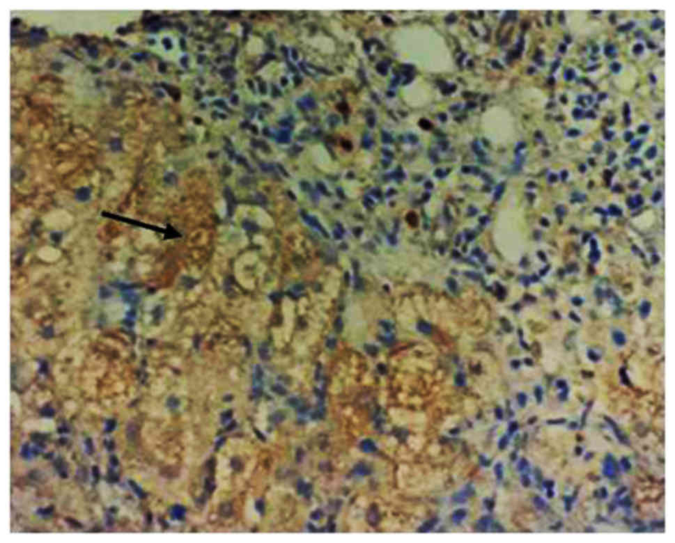
pDC analysis
The changes in the levels of pDCs in the response
group and non-responders group prior to and after treatment with
IFN-α were analyzed. The pDC markers BDCA-2 and ILT7 were detected
by IHC analysis, and they were uniformly expressed throughout the
cytoplasm of these cells (Fig. 1,
arrow). There was no significant difference in amount of pDCs
(BDCA-2 and ILT7 expressions) between the response group and the
non-responders group prior to IFN-α treatment. After IFN-α
treatment, BDCA-2 and ILT7 expression was obviously increased in
the response group compared with that in the non-responders group
(P<0.05; Fig. 2).
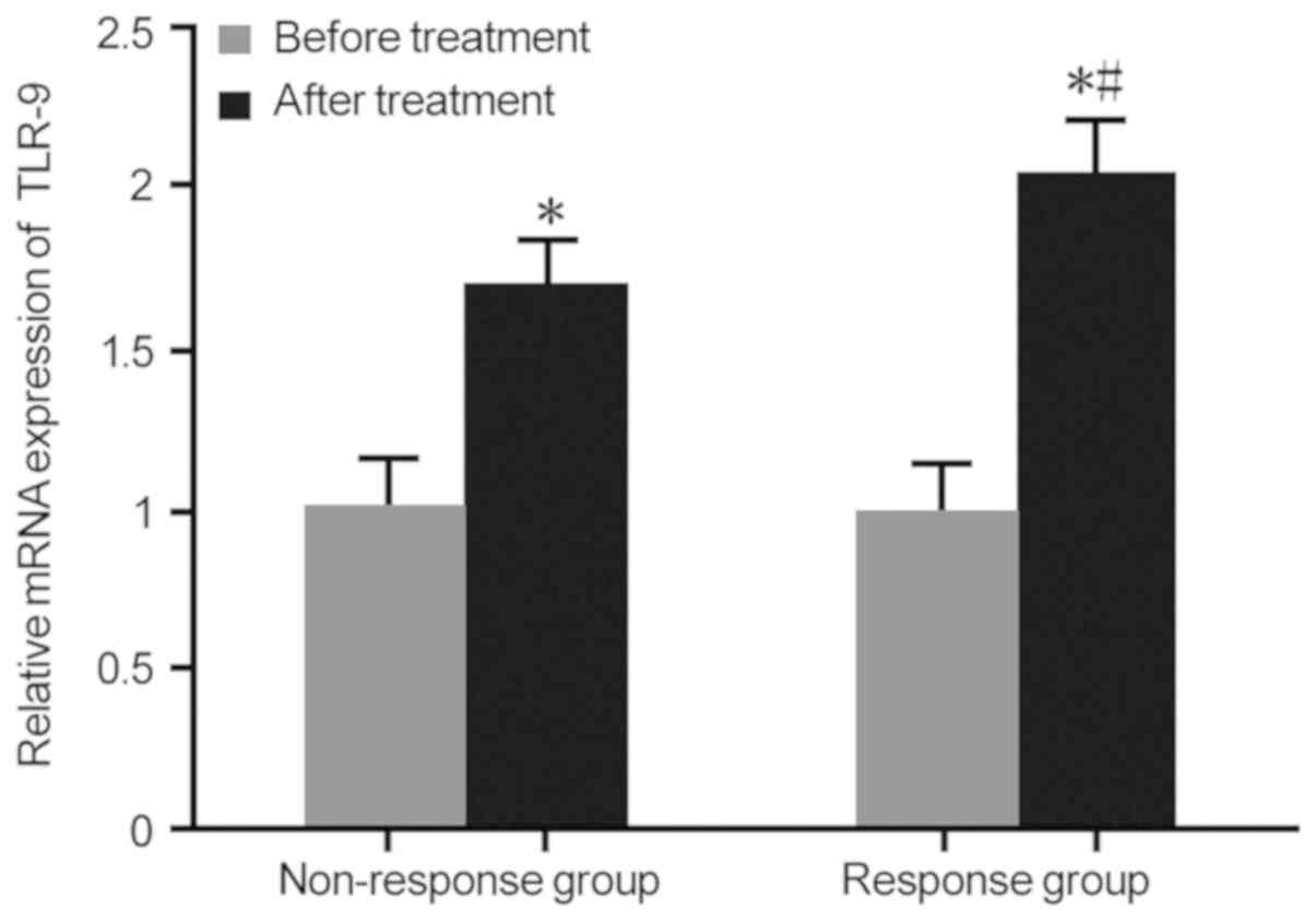
TLR-9 expression analysis
TLR-9 mRNA expression in the response group and
non-responders group prior to and after treatment with IFN-α was
assessed by RT-qPCR. There was no statistically significant
difference in TLR-9 mRNA expression between the response group and
the non-responders group prior to IFN-α treatment. After IFN-α
treatment, TLR-9 mRNA expression was markedly upregulated in the
response group compared with that in the non-responders group
(P<0.05; Fig. 3). It was then
investigated which type of cell was responsible for the increase in
the expression of TLR9 after anti-viral therapy. The expression of
TLR9 in the cytoplasm of epithelial cells in the liver biopsies of
the response group was higher than that in the non-responders group
after anti-viral therapy (Fig.
S2)
Correlation analysis of pDCs or TLR-9
expression with HBV DNA content
The correlation between pDCs or TLR-9 expression and
HBV DNA content was then analyzed. It was demonstrated that with
the increase of pDCs and TLR-9 expression, the HBV DNA content
declined. Therefore, these parameters were negatively correlated
with the HBV DNA content (P<0.05), suggesting that pDCs and
TLR-9 expressions may predict the IFN-α treatment response in
patients with HBeAg-positive CHB (Table
IV).
| Table IV.Correlation analysis of pDCs and
TLR-9 expression with HBV DNA content. |
Table IV.
Correlation analysis of pDCs and
TLR-9 expression with HBV DNA content.
| Correlation | R-value | P-value |
|---|
| pDCs vs. HBV
DNA | −0.718 | <0.05 |
| TLR-9 vs. HBV
DNA | −0.672 | <0.05 |
Discussion
pDCs are mainly produced by bone marrow
hematopoietic stem cells, which are continuously released into
peripheral blood as plasma-like cells. They specifically express
TLR-7 and TLR-9, and the type I IFN produced may directly exert
anti-viral effects. At the same time, the anti-viral ability of
natural killer (NK) and B lymphocytes is enhanced, so that pDCs
stimulate the innate as well as the acquired immune response, which
resembles a bridge between innate immunity and specific immune
response (21). It has been observed
in vitro that that administration of IFN-α promotes the
maturation of pDCs and promotes the production of IL-12, thereby
acting on CD40-activated B cells to promote plasma cells and
immunoglobulin secretion (22,23).
Therefore, pDCs are involved in the activation of T cells, B cells
and NK cells. During viral infection, pDCs differentiate into
mature DCs and regulate T-cell function. It is of great clinical
significance to explore the expression and role of pDCs in
infection and treatment of CHB patients. However, there is
currently a lack of established specific surface markers for pDCs.
Since ILT7 and BDCA-2 are selectively expressed in pDCs, which are
human pDC-specific and not expressed in any other mature DCs or
peripheral lymphocytes, they were used as specific markers for pDCs
in the present study (24,25). The present results confirmed that
after IFN-α treatment, the expression of BDCA-2 and ILT7 in the
response group was significantly higher than that in the
non-responders group, suggesting that the expression of pDCs
predicts the IFN-α treatment response in HBeAg-positive CHB
patients.
TLR-9 is only expressed on the surface of human B
cells and pDCs. The surface molecules on pDCs include BDCA-2 and
ITL7, and they may be upregulated upon activation with TLR-9. The
TLR7/9-dependent pathway appears to be a predominant mode of
nucleic acid sensing in pDCs, but is essential for TLR9-induced IFN
production by pDCs. This indirectly accelerates the maturation,
differentiation and proliferation of lymphocytes (26,27).
TLR-9 assists pDCs in chemotaxis of lymph nodes and aggregation,
which in turn assists in the exertion of the anti-viral effect
(28). The direct causal association
between pDC-derived IFN and lupus progression/severity is difficult
to establish in the human system and is to be elucidated in animal
models (29).
In the present study, the expression of TLR-9 was
analyzed in the peripheral blood of patients with HBeAg-positive
CHB treated with IFN-α, providing a reference for the safe and
effective treatment of HBV infection. The present study confirmed
that after IFN-α treatment, the mRNA expression of TLR-9 was
markedly upregulated in the response group compared with that in
the non-responders group, and was negatively correlated with the
HBV DNA content, suggesting that the expression of TLR-9 may also
predict the treatment effect of IFN-α. In line with this, previous
studies suggested that markers of fibrosis were obviously higher in
non-responders than in responders (30,31). In
the present study, TLR-9 expression was detected in peripheral
blood mononuclear cells and not in liver tissues, which is a
limitation.
The present study demonstrated changes in the levels
of pDCs and the expression of TLR-9 in patients with HBeAg-positive
CHB treated with IFN-α, and analyzed their predictive significance
regarding treatment response. Further study is required to explore
the mechanistic roles of pDCs and TLR-9 in the treatment of CHB
patients with IFN-α.
In conclusion, increased levels of pDCs and TLR-9
were negatively correlated with HBV DNA, and may thus predict the
IFN-α treatment response in patients with HBeAg-positive CHB. The
present study provided a theoretical basis for selecting more
effective anti-HBV programs for patients with CHB. However, as the
range of follow-up is very wide, further studies with close
follow-ups are required to confirm this finding in the future.
Supplementary Material
Supporting Data
Acknowledgements
Not applicable.
Funding
This work was supported by the Xiamen Science and
Technology Fund (grant no. 3502Z20144028).
Availability of data and materials
All data generated or analyzed during this study are
included in this published article.
Authors' contributions
YC, JEY, JMT, QGM and QZZ performed the experiments
and analyzed the data. YZ designed the study and wrote the
manuscript.
Ethics approval and consent to
participate
The current study was approved by the Xiamen
Hospital of Traditional Chinese Medicine and consent was obtained
for participation.
Patient consent for publication
Informed consent for the publication of data was
obtained from all patients.
Competing interests
The authors declare that they have no competing
interests.
Glossary
Abbreviations
Abbreviations:
|
HBV
|
hepatitis B virus
|
|
HBeAg
|
hepatitis B e antigen
|
|
IFN-α
|
interferon-α
|
|
pDCs
|
plasmacytoid dendritic cells
|
|
TLR-9
|
Toll-like receptor-9
|
|
ALT
|
alanine aminotransferase
|
|
BDCA-2
|
blood dendritic cell antigen 2
|
|
ILT7
|
immunoglobulin-like transcript 7
|
References
|
1
|
Tang X, Yan L, Li H, Du L, Shi Y, Huang F
and Tang H: Increased expression of phosphoenolpyruvate
carboxykinase cytoplasmic isoform by hepatitis B virus X protein
affects hepatitis B virus replication. J Med Virol. 91:258–264.
2019. View Article : Google Scholar : PubMed/NCBI
|
|
2
|
Yambasu EE, Reid A, Owiti P, Manzi M,
Murray MJS and Edwin AK: Hidden dangers-prevalence of blood borne
pathogens, hepatitis B, C, HIV and syphilis, among blood donors in
Sierra Leone in 2016: Opportunities for improvement: A
retrospective, cross-sectional study. Pan Afr Med J. 30:442018.
View Article : Google Scholar : PubMed/NCBI
|
|
3
|
Herrero-Fernández I, Rosado-Sánchez I,
Genebat M, Tarancón-Díez L, Rodríguez-Méndez MM, Pozo-Balado MM,
Lozano C, Ruiz-Mateos E, Leal M and Pacheco YM: Improved CD4 T cell
profile in HIV-infected subjects on maraviroc-containing therapy is
associated with better responsiveness to HBV vaccination. J Transl
Med. 16:2382018. View Article : Google Scholar : PubMed/NCBI
|
|
4
|
Chen M, Du D, Zheng W, Liao M, Zhang L,
Liang G and Gong M: Small hepatitis delta antigen selectively binds
to target mRNA in hepatic cells: A potential mechanism by which
hepatitis D virus down-regulates glutathione S-transferase P1 and
induces liver injury and hepatocarcinogenesis. Biochem Cell Biol.
97:130–139. 2019. View Article : Google Scholar : PubMed/NCBI
|
|
5
|
Howell J, Pedrana A, Cowie BC, Doyle J,
Getahun A, Ward J, Gane E, Cunningham C, Wallace J, Lee A, et al:
Aiming for the elimination of viral hepatitis in Australia, New
Zealand, the Pacific Islands and Territories: Where are we now and
barriers to meeting WHO targets by 2030. J Gastroenterol Hepatol.
34:40–48. 2019. View Article : Google Scholar : PubMed/NCBI
|
|
6
|
Li MR, Zheng HW, Ma SM, Liu YY, Qie LX, Li
JQ, Wang DH, Sun XL, Ren GF, Zheng YH, et al: Correlations between
serum hepatitis B surface antigen and hepatitis B core antibody
titers and liver fibrosis in treatment-naive CHB patients. J Chin
Med Assoc. 81:1052–1059. 2018. View Article : Google Scholar : PubMed/NCBI
|
|
7
|
Yang J, Yang G, He H, Ning L, Liu Z, Fu Q,
Chen H, Deng H, Wang Z and Luo K: Association of characteristics of
HBV quasispecies with hepatitis B surface antigen seroconversion
after pegylated interferon-α-2a treatment in child patients.
Antivir Ther. 23:567–574. 2018. View
Article : Google Scholar : PubMed/NCBI
|
|
8
|
Lutterkort GL, Wranke A, Hengst J,
Yurdaydin C, Stift J, Bremer B, Hardtke S, Keskin O, Idilman R,
Manns MP, et al: Viral dominance patterns in chronic hepatitis
delta determine early response to interferon alpha therapy. J Viral
Hepat. 25:1384–1394. 2018. View Article : Google Scholar : PubMed/NCBI
|
|
9
|
Cao WH, Li MH, Pan CQ, Lu Y, Zhang L, Ran
CP, Wu SL, Hua WH, Liu SA, Shen G, et al: Quantitation of
plasmacytoid dendritic cells in chronic hepatitis B patients with
HBeAg positivity during PEG-IFN and entecavir therapy. J Interferon
Cytokine Res. 38:197–205. 2018. View Article : Google Scholar : PubMed/NCBI
|
|
10
|
Karamitros T, Papatheodoridis G,
Paraskevis D, Hatzakis A, Mbisa JL, Georgopoulou U, Klenerman P and
Magiorkinis G: Impact of interferon-α receptor-1 promoter
polymorphisms on the transcriptome of the hepatitis B
virus-associated hepatocellular carcinoma. Front Immunol.
9:7772018. View Article : Google Scholar : PubMed/NCBI
|
|
11
|
Sepulveda-Toepfer JA, Pichler J, Fink K,
Sevo M, Wildburger S, Mudde-Boer LC, Taus C and Mudde GC:
TLR9-mediated activation of dendritic cells by CD32 targeting for
the generation of highly immunostimulatory vaccines. Hum Vaccin
Immunother. 15:179–188. 2019. View Article : Google Scholar : PubMed/NCBI
|
|
12
|
Tomasello E, Naciri K, Chelbi R, Bessou G,
Fries A, Gressier E, Abbas A, Pollet E, Pierre P, Lawrence T, et
al: Molecular dissection of plasmacytoid dendritic cell activation
in vivo during a viral infection. EMBO J. 37:e988362018. View Article : Google Scholar : PubMed/NCBI
|
|
13
|
Wimmers F, Subedi N, van Buuringen N,
Heister D, Vivié J, Beeren-Reinieren I, Woestenenk R, Dolstra H,
Piruska A, Jacobs JF, et al: Single-cell analysis reveals that
stochasticity and paracrine signaling control interferon-alpha
production by plasmacytoid dendritic cells. Nat Commun. 9:33172018.
View Article : Google Scholar : PubMed/NCBI
|
|
14
|
Frank MJ, Reagan PM, Bartlett NL, Gordon
LI, Friedberg JW, Czerwinski DK, Long SR, Hoppe RT, Janssen R,
Candia AF, et al: In situ vaccination with a TLR 9 agonist and
local low dose radiation induces systemic responses in untreated
indolent lymphoma. Cancer Discov. 8:1258–1269. 2018. View Article : Google Scholar : PubMed/NCBI
|
|
15
|
Atreya R, Reinisch W, Peyrin-Biroulet L,
Scaldaferri F, Admyre C, Knittel T, Kowalski J, Neurath MF and
Hawkey C: Clinical efficacy of the Toll-like receptor 9 agonist
cobitolimod using patient-reported-outcomes defined clinical
endpoints in patients with ulcerative colitis. Dig Liver Dis.
50:1019–1029. 2018. View Article : Google Scholar : PubMed/NCBI
|
|
16
|
Xu J, Lee JW, Park SK, Lee SB, Yoon YH,
Yeon SH, Rha KS, Choi JA, Song CH and Kim YM: Toll-like receptor 9
ligands increase type I interferon induced B-cell activating factor
expression in chronic rhinosinusitis with nasal polyposis. Clin
Immunol. 197:19–26. 2018. View Article : Google Scholar : PubMed/NCBI
|
|
17
|
Gibbert K, Schlaak JF, Yang D and Dittmer
U: IFN-α subtypes: Distinct biological activities in anti-viral
therapy. Br J Pharmacol. 168:1048–1058. 2013. View Article : Google Scholar : PubMed/NCBI
|
|
18
|
Hou J, Wang G, Wang F, Cheng J, Ren H,
Zhuang H, Sun J, Li L, Li J, Meng Q, et al: Guideline of prevention
and treatment for chronic hepatitis B (2015 update). J Clin Transl
Hepatol. 5:297–318. 2017. View Article : Google Scholar : PubMed/NCBI
|
|
19
|
Zhu S, Zhang H, Dong Y, Wang L, Xu Z, Liu
W, Gan Y, Tang H, Chen D, Wang F and Zhao P: Antiviral therapy in
hepatitis B virus-infected children with immune-tolerant
characteristics: A pilot open-label randomized study. J Hepatol.
68:1123–1128. 2018. View Article : Google Scholar : PubMed/NCBI
|
|
20
|
Livak KJ and Schmittgen TD: Analysis of
relative gene expression data using real-time quantitative PCR and
the 2(-Delta Delta C(T) method. Methods. 25:402–408. 2001.
View Article : Google Scholar : PubMed/NCBI
|
|
21
|
Xi Y, Troy NM, Anderson D, Pena OM, Lynch
JP, Phipps S, Bosco A and Upham JW: Critical role of plasmacytoid
dendritic cells in regulating gene expression and innate immune
responses to human rhinovirus-16. Front Immunol. 8:13512017.
View Article : Google Scholar : PubMed/NCBI
|
|
22
|
Cédile O, Jørgensen LØ, Frank I,
Wlodarczyk A and Owens T: The chemokine receptor CCR2 maintains
plasmacytoid dendritic cell homeostasis. Immunol Lett. 192:72–78.
2017. View Article : Google Scholar : PubMed/NCBI
|
|
23
|
Ainola M, Porola P, Takakubo Y, Przybyla
B, Kouri VP, Tolvanen TA, Hänninen A and Nordström DC: Activation
of plasmacytoid dendritic cells by apoptotic particles-mechanism
for the loss of immunological tolerance in Sjögren's syndrome. Clin
Exp Immunol. 191:301–310. 2018. View Article : Google Scholar : PubMed/NCBI
|
|
24
|
Ruben JM, García-Romo GS, Breman E, van
der Kooij S, Redeker A, Arens R and van Kooten C: Human
plasmacytoid dendritic cells acquire phagocytic capacity by TLR9
ligation in the presence of soluble factors produced by renal
epithelial cells. Kidney Int. 93:355–364. 2018. View Article : Google Scholar : PubMed/NCBI
|
|
25
|
Murayama G, Furusawa N, Chiba A, Yamaji K,
Tamura N and Miyake S: Enhanced IFN-α production is associated with
increased TLR7 retention in the lysosomes of palasmacytoid
dendritic cells in systemic lupus erythematosus. Arthritis Res
Ther. 19:2342017. View Article : Google Scholar : PubMed/NCBI
|
|
26
|
Torigoe M, Sakata K, Ishii A, Iwata S,
Nakayamada S and Tanaka Y: Hydroxychloroquine efficiently
suppresses inflammatory responses of human class-switched memory B
cells via Toll-like receptor 9 inhibition. Clin Immunol. 195:1–7.
2018. View Article : Google Scholar : PubMed/NCBI
|
|
27
|
Han N, Zhang Z, Jv H, Hu J, Ruan M and
Zhang C: Culture supernatants of oral cancer cells induce impaired
IFN-α production of pDCs partly through the down-regulation of
TLR-9 expression. Arch Oral Biol. 93:141–148. 2018. View Article : Google Scholar : PubMed/NCBI
|
|
28
|
White MP, Webster G, Leonard F and La
Flamme AC: Innate IFN-γ ameliorates experimental autoimmune
encephalomyelitis and promotes myeloid expansion and PDL-1
expression. Sci Rep. 8:2592018. View Article : Google Scholar : PubMed/NCBI
|
|
29
|
Reizis B, Bunin A, Ghosh HS, Lewis KL and
Sisirak V: Plasmacytoid dendritic cells: Recent progress and open
questions. Annu Rev Immunol. 29:163–83. 2011. View Article : Google Scholar : PubMed/NCBI
|
|
30
|
Lebensztejn DM, Sobaniec-Lotowska ME,
Kaczmarski M, Voelker M and Schuppan D: Matrix-derived serum
markers in monitoring liver fibrosis in children with chronic
hepatitis B treated with interferon alpha. World J Gastroenterol.
12:3338–3343. 2006. View Article : Google Scholar : PubMed/NCBI
|
|
31
|
Lebensztejn DM, Sobaniec-Łotowska ME,
Bauer M, Kaczmarski M, Voelker M and Schuppan D: Serum fibrosis
markers as predictors of an antifibrotic effect of interferon alfa
in children with chronic hepatitis B. Eur J Gastroenterol Hepatol.
17:843–848. 2005. View Article : Google Scholar : PubMed/NCBI
|