Introduction
As a papain cysteine protease and one of lysosomal
proteases, cathepsin S mainly participates in the degradation of
various tissue proteins and acts as a catalytic enzyme in the
process of proteolysis, and therefore it is the most common
endonuclease in the body (1).
Cathepsin S can not only degrade and transform intracellular
proteins under acid conditions, but can also exert physiological
effects (2), vascular
activity-regulating effects and modulatory effects on the
proliferation and differentiation of endothelial cells (3) under extracellular neutral conditions.
Moreover, it has important clinical value in the proliferation and
migration of a variety of cytokines (4).
Cathepsin S can also promote the degradation of type
I and IV collagens, as well as their laminins among the components
of extracellular matrix (5). In the
physiological process, it degrades the collagens in the above
mentioned intercellular substances mainly through the chemotaxis
and aggregation of smooth muscle cells toward the macrophages,
fibrous cap cells, tunica media and other sites, thus leading to
rupture of atherosclerotic plaques (6). Moreover, cathepsin S is able to
accelerate the production of inflammatory cytokines in the body,
further degrading elastic fibrous protein and collagen in the
atherosclerotic plaques causing attenuation and even rupture of the
atherosclerotic plaques. Finally, it can migrate to the vicinity of
the vascular intima media, and positive feedback can increase the
secretion and release of cathepsin S, thereby triggering the
formation of massive foam cells in the body and prominently
speeding up the instability of the atherosclerotic plaques
(7). Although related clinical
studies have verified that (8) the
level of cathepsin S is associated with the formation of
atherosclerosis, there are few reports on the correlation of
cathepsin S with the coronary stenosis degree and carotid thickness
in atherosclerosis patients. This study investigated these issues
and analyzed the relation of cathepsin S with blood pressure,
glucose and lipid metabolism, and vascular endothelial
function.
Patients and methods
General data
Relevant data of 120 patients with increased
cathepsin S levels, admitted and treated in The Third Xiangya
Hospital of Central South University (Changsha, China) from
February 2016 to August 2018 (increased group), and data from 120
subjects with normal cathepsin S levels, enrolled in the same time
period (normal group), were retrospectively analyzed. In the
increased group, there were 40 patients definitely diagnosed with
coronary atherosclerotic heart disease, including 23 males and 17
females, 50–70 years of age, with an average age of 63.3±2.1 years.
The course of disease was 3–21 years, with an average of 8.4±0.4
years. Forty patients were definitely diagnosed with carotid
intima-media thickening, including 21 males and 19 females, 50–70
years of age, with an average age of 63.4±2.0 years. The course of
disease was 3–20 years, with an average of 8.5±0.4 years. There
were 40 patients with hypertension, including 23 males and 17
females, 50–70 years of age, with an average age of 63.5±2.1 years,
a disease course of 5–25 years and an average course of 9.4±0.4
years. Normal group consisted of 63 males and 57 females, 50–70
years of age, with an average age of 63.4±2.0 years. The general
data of the groups are presented in Table I. The study was approved by the
Ethics Committee of The Third Xiangya Hospital of Central South
University. Signed informed consents were obtained from all
participants before the study.
 | Table I.General data. |
Table I.
General data.
| Factors | Increased group | Normal group |
|---|
| Sex |
| Male | 66 | 63 |
|
Female | 54 | 57 |
| Age (years) | 63.4±2.1 | 63.2±2.2 |
| CAD |
| Yes | 40 | 5 |
| No | 80 | 115 |
| IMT |
| Yes | 40 | 9 |
| No | 80 | 111 |
| Hypertension |
| Yes | 40 | 11 |
| No | 80 | 109 |
Methods
The level of cathepsin S was detected in all
patients using enzyme-linked immunosorbent assay (R&D Systems,
Inc.). The serum cathepsin S level and Gensini score were compared
between the healthy subjects and patients with coronary
atherosclerotic heart disease, and the correlation between serum
cathepsin S level and Gensini score was analyzed. The carotid
thickness, mean arterial pressure and indexes related to glucose
and lipid metabolism, as well as vascular endothelial function were
compared. Also, the correlation of the serum cathepsin S level with
carotid intima-media thickness (IMT), mean arterial pressure,
fasting blood glucose, total cholesterol (TC) and nitric oxide (NO)
was investigated. Fasting blood glucose and TC were measured by an
Automatic Biochemistry Analyzer (Abbott 8200; Abbott Pharmaceutical
Co. Ltd.). NO was measured by a Nitric Oxide Colorimetric Assay kit
(K262-200; AmyJet Scientific, Inc.).
Evaluation criteria
Coronary artery stenosis was evaluated on the basis
of coronary angiography. All coronary angiography results were
entered into a software (CAAS II System; Pie Medical Imaging BV)
and then analyzed by physicians with >5 years of experience in
interventional therapy or interventional therapy in the Department
of Cardiology. Patients with a degree of coronary artery stenosis
>50% were assessed as positive and diagnosed with coronary
artery disease (CAD), and those with a degree of coronary artery
stenosis <50% were assessed as negative. The degree of vascular
stenosis was evaluated with reference to the Gensini scoring system
of the American Heart Association, including 1 point (stenosis
degree of each coronary artery, <25%), 2 points (stenosis degree
of each coronary artery, 26–50%), 4 points (stenosis degree of each
coronary artery, 51–75%), 8 points (stenosis degree of each
coronary artery, 76–90%), 16 points (stenosis degree of each
coronary artery, 91–99%) and 32 points (stenosis degree of each
coronary artery, 100%). In terms of IMT, the thickness at 2 cm of
the proximal part of bilateral internal carotid arteries of all the
patients was measured using GE Vivid 7 color ultrasound diagnostic
instrument (GE Healthcare), which was regarded as the carotid IMT,
with a normal value of <1.0 mm. The indexes of glucose
metabolism included fasting blood glucose (normal reference value
for adults, 3.9–6.1 mmol/l) and fasting insulin (FINS) (normal
reference value for adults, 3.0–24.9 U/ml). The indexes of lipid
metabolism consisted of triglyceride (TG) (normal reference value
for adults, 0.56–1.71 mmol/l) and TC (normal reference value for
adults, 2.83–5.17 mmol/l). The indexes of vascular endothelial
function were endothelin-1 (ET-1) (normal reference value for
adults, 43.50–58.38 ng/l) and NO (normal reference value for
adults, 13.8–34.6 µmol/l).
Statistical analysis
Statistical Product and Service Solutions (SPSS)
20.0 software (IBM Corp.) was used. The measurement data, such as
the data of serum cathepsin S level, Gensini score, carotid
thickness, mean arterial pressure and related indexes to glucose
and lipid metabolism, as well as vascular endothelial function,
were expressed as mean ± standard deviation (mean ± SD). t-test was
performed for the comparison of the mean between two groups.
Comparison among multiple groups was carried out using one-way
ANOVA followed by a post hoc test (Tukey's honestly significant
difference). Also, Pearson's correlation analysis was applied to
analyze the correlation of serum cathepsin S level with Gensini
score, IMT, mean arterial pressure, fasting blood glucose, TC and
NO. P<0.05 was considered to indicate a statistically
significant difference.
Results
Serum cathepsin S level in healthy
subjects and patients with coronary atherosclerotic heart
disease
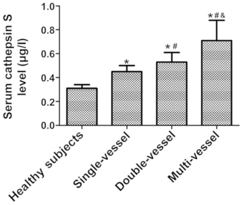
The serum cathepsin S level was 0.31±0.03 µg/l in
healthy subjects, 0.45±0.05 µg/l in patients with single-vessel
CAD, 0.53±0.08 µg/l in patients with double-vessel disease, and
0.71±0.17 µg/l in patients with multi-vessel disease. The patients
with multi-vessel CAD had a higher serum cathepsin S level than
those with double-vessel and single-vessel disease, and a higher
level than the healthy subjects (F=7.493, P<0.05) (Fig. 1).
Gensini score in healthy subjects and
patients with coronary atherosclerotic heart disease
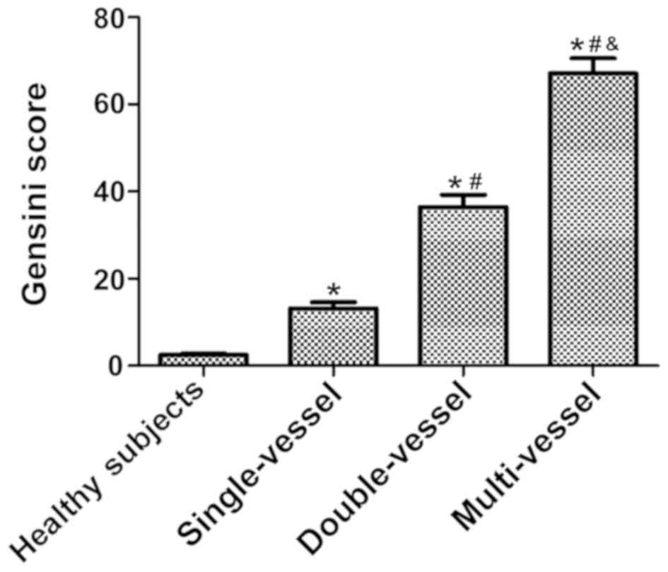
The Gensini scores were 2.5±0.3, 13.2±1.4, 36.4±2.8
and 67.1±3.5 points in healthy subjects, patients with
single-vessel CAD, patients with double-vessel disease and patients
with multi-vessel disease, respectively. The patients with
multi-vessel CAD had a higher Gensini score than those with
double-vessel and single-vessel disease, and a higher score than
the healthy subjects (F=9.201, P<0.05) (Fig. 2).
Correlation analysis between the serum
cathepsin S level and Gensini score
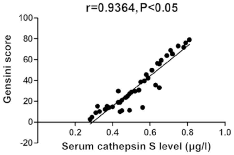
The serum cathepsin S level was positively
correlated with Gensini score (r=0.9364, P<0.05) (Fig. 3).
Comparison of carotid thickness, mean
arterial pressure and indexes related to glucose and lipid
metabolism, as well as vascular endothelial function
Patients with increased serum cathepsin S level had
greater IMT (P<0.05), higher mean arterial pressure (P<0.05),
fasting blood glucose, FINS (P<0.05), TG, TC (P<0.05) and
endothelin-1 (P<0.05), however, lower NO level (P<0.05) than
those of healthy subjects (Table
II).
| Table II.Comparison of carotid thickness, mean
arterial pressure, and indexes related to glucose and lipid
metabolism, as well as vascular endothelial function (mean ± SD)
between the two groups. |
Table II.
Comparison of carotid thickness, mean
arterial pressure, and indexes related to glucose and lipid
metabolism, as well as vascular endothelial function (mean ± SD)
between the two groups.
| Groups | IMT (mm) | Mean arterial
pressure (mmHg) | Fasting blood glucose
(mmol/l) | FINS (mU/l) | TG (mmol/l) | TC (mmol/l) | ET-1 (ng/l) | NO (µmol/l) |
|---|
| Normal group | 0.82±0.03 | 108.3±1.6 | 5.0±0.3 | 4.2±0.1 | 1.7±0.1 | 3.0±0.1 |
51.6±3.0 | 30.7±3.2 |
| Increased group | 1.78±0.11 | 143.2±2.8 | 9.7±0.7 | 9.1±0.3 | 3.4±0.2 | 5.5±0.3 | 129.5±4.5 |
8.8±1.3 |
| t value | 92.234 | 118.549 | 67.604 | 169.741 | 83.283 | 86.603 | 157.785 | 69.457 |
| P-value | <0.001 | <0.001 | <0.001 | <0.001 | <0.001 | <0.001 | <0.001 | <0.001 |
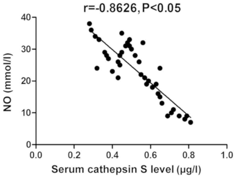
Correlation analysis of the serum
cathepsin S level with IMT, mean arterial pressure, fasting blood
glucose, TC and NO
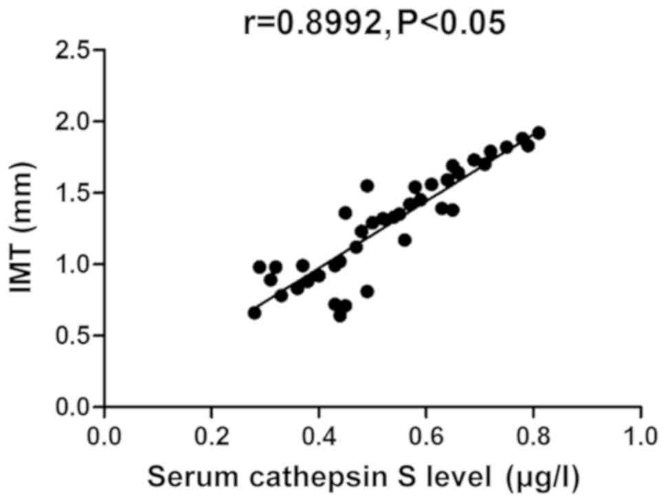
The serum cathepsin S level was positively
correlated with IMT, mean arterial pressure, fasting blood glucose
and TC levels (P<0.05), however, was negatively correlated with
NO level (P<0.05) (Figs.
4–8).
Discussion
As a papain with the richest functions in the
mammalian elastase system, cathepsin S is of great value in
numerous pathophysiological processes, such as gene transcription
and regulation through controlling the promoter of protein genes
(9). Therefore, it is considered as
the most common candidate gene for the occurrence and development
of atherosclerosis (10).
Immunohistochemical assay results have indicated that remarkable
expression of cathepsin S exists in macrophages, smooth muscle
cells of fibrous cap and intimal elastic lumina during
atherosclerotic process (11).
However, western blotting combined with elastase analysis have
suggested that the expression levels of cathepsin S and cathepsin K
in the extracts of atherosclerotic tissues are markedly elevated,
thus strengthening the dissociation activity of elastic tissues
(12). As a result, cathepsin S
plays a vital role in the occurrence and development of
atherosclerosis. Another study has identified cathepsin S as a
marker of adiposity and it has been proposed that cathepsin S
represents a molecular link between obesity and atherosclerosis
(13). In addition, large quantities
of studies have indicated that (14)
the activity of lysosomal cysteine proteases has great impact on
the occurrence and development of atherosclerosis, and cathepsin S
is also the most important lysosomal cysteine protease. Chen et
al (15) found that cathepsin S
and insulin resistance are independent of each other. At the same
time, increased blood pressure, abnormal glucose and lipid
metabolism, as well as vascular endothelial function are relevant
or independent risk factors for atherosclerosis. However, there is
no previous research report on the correlation of cathepsin S with
blood pressure change, glucose and lipid metabolism and vascular
endothelial function.
This investigation focused on the comparisons of
serum cathepsin S level and Gensini score between healthy subjects
and patients with coronary atherosclerotic heart disease, and it
was discovered that the patients with multi-vessel CAD have higher
serum cathepsin S level and Gensini score than those with
double-vessel and single-vessel disease, as well as healthy
subjects. Furthermore, correlation analysis between the serum
cathepsin S level and Gensini score revealed that the serum
cathepsin S level is positively correlated with Gensini score.
These results suggest that for the patients with coronary
atherosclerotic heart disease, the wider the extent of the lesion
is, the higher the serum cathepsin S level will be, and the
elevated serum cathepsin S level will lead to a higher Gensini
score. Also, the serum cathepsin S level, carotid thickness, mean
arterial pressure and indexes related to glucose and lipid
metabolism, as well as vascular endothelial function were compared.
The results showed that the patients with increased serum cathepsin
S level have greater IMT, higher mean arterial pressure, fasting
blood glucose, FINS, TG, TC and ET-1, however, lower NO level than
healthy subjects. Patients with elevated serum cathepsin S level
had also increased carotid IMT, raised levels of blood pressure,
blood glucose and blood lipid and impaired vascular endothelial
function. Finally, correlation analysis of the serum cathepsin S
level with IMT, mean arterial pressure, fasting blood glucose, TC
and NO demonstrated that the serum cathepsin S level is positively
correlated with IMT, mean arterial pressure, fasting blood glucose
and TC levels, and negatively correlated with NO level, further
suggesting that the serum cathepsin S level is not only related to
the degree of coronary artery stenosis in the patients with
coronary atherosclerotic heart disease, but also positively
correlated with IMT, mean arterial pressure, fasting blood glucose
and TC levels, and negatively associated with NO level, a cytokine
related to the vascular endothelial function.
Serum cathepsin S can reduce the adhesiveness of
extracellular matrix, promote the migration of atherosclerotic
factors in the coronary tunica intima toward the site below the
tunica intima (16), and trigger
atherosclerosis-induced thickening of tunica intima of coronary
artery and carotid artery, ultimately aggravating the vascular
stenosis induced by atherosclerotic plaque, and promoting the
disease progression (17).
Additionally, serum cathepsin S is able to reduce the stability of
collagen fiber and fibrous cap in the atherosclerotic plaque,
thereby enhancing the instability of atherosclerotic plaque
(18). As the most important
protease for degrading the extracellular matrix during
atherosclerosis formation, serum cathepsin S is also capable of
accelerating the migration of monocytes to subintimal sites through
the arterial intima (19), further
causing the thickening of arterial intima, promoting the formation
of fibrous plaque, and aggravating the atherosclerotic lesions
(20). At the same time, it was also
indicated in this research that the patients with elevated serum
cathepsin S level also have increased blood pressure, and they are
prone to abnormalities in glucose and lipid metabolism and vascular
endothelial function. Differently, some studies have indicated that
high levels of cathepsin K and L are closely linked with the
presence of CAD, and are both independent biomarkers for CHD
(21,22). The present study also has some
limitations. The main limitation is the lack of in vitro or
in vivo experiments. Cathepsin S-deficient animals or cells
should be employed to verify our results.
In conclusion, the serum cathepsin S level is
significantly correlated with the coronary stenosis degree, carotid
thickness, blood pressure, glucose and lipid metabolism and
vascular endothelial function. With the elevation of serum
cathepsin S level, the coronary stenosis becomes more severe, the
carotid artery is thicker, the blood pressure is higher, and the
glucose and lipid metabolism and vascular endothelial function are
significantly abnormal.
Acknowledgements
Not applicable.
Funding
Not funding was received.
Availability of data and materials
All data generated or analyzed during this study are
included in this published article.
Authors' contributions
SH and YC designed the study and performed the
experiments. SH and YC collected the patients' data, analyzed the
data and prepared the manuscript. Both authors read and approved
the final manuscript.
Ethics approval and consent to
participate
The study was approved by the Ethics Committee of
The Third Xiangya Hospital of Central South University (Changsha,
China). Signed informed consents were obtained from all
participants before the study.
Patient consent for publication
Not applicable.
Competing interests
The authors declare that they have no competing
interests.
References
|
1
|
Zhou PP, Zhang WY, Li ZF, Chen YR, Kang XC
and Jiang YX: Association between SNPs in the promoter region in
cathepsin S and risk of asthma in Chinese Han population. Eur Rev
Med Pharmacol Sci. 20:2070–2076. 2016.PubMed/NCBI
|
|
2
|
Ignatov M, Liu C, Alekseenko A, Sun Z,
Padhorny D, Kotelnikov S, Kazennov A, Grebenkin I, Kholodov Y,
Kolosvari I, et al: Monte Carlo on the manifold and MD refinement
for binding pose prediction of protein-ligand complexes: 2017 D3R
Grand Challenge. J Comput Aided Mol Des. 33:119–127. 2019.
View Article : Google Scholar : PubMed/NCBI
|
|
3
|
Xiao L, Han X, Wang XE, Li Q and Chen Y,
Cui Y and Chen Y: Cathepsin S in the spinal microglia facilitates
morphine-induced antinociceptive tolerance in rats. Neurosci Lett.
690:225–231. 2019. View Article : Google Scholar : PubMed/NCBI
|
|
4
|
Luo L, Zhu M and Zhou J: Association
between CTSS gene polymorphism and the risk of acute
atherosclerotic cerebral infarction in Chinese population: A
case-control study. Biosci Rep. 38:BSR201805862018. View Article : Google Scholar : PubMed/NCBI
|
|
5
|
Lee BC, Kang I, Lee SE, Lee JY, Shin N,
Kim JJ, Choi SW and Kang KS: Human umbilical cord blood plasma
alleviates age-related olfactory dysfunction by attenuating
peripheral TNF-α expression. BMB Rep. 52:259–264. 2019. View Article : Google Scholar : PubMed/NCBI
|
|
6
|
Chen CY, Chen CY, Liu CC and Chen CP:
Omega-3 polyunsaturated fatty acids reduce preterm labor by
inhibiting trophoblast cathepsin S and inflammasome activation.
Clin Sci (Lond). 132:2221–2239. 2018.PubMed/NCBI
|
|
7
|
Wuopio J, Hilden J, Bring C, Kastrup J,
Sajadieh A, Jensen GB, Kjøller E, Kolmos HJ, Larsson A, Jakobsen
JC, et al: Cathepsin B and S as markers for cardiovascular risk and
all-cause mortality in patients with stable coronary heart disease
during 10 years: A CLARICOR trial sub-study. Atherosclerosis.
278:97–102. 2018. View Article : Google Scholar : PubMed/NCBI
|
|
8
|
Poulsen CB, Al-Mashhadi AL, von
Wachenfeldt K, Bentzon JF, Nielsen LB, Al-Mashhadi RH, Thygesen J,
Tolbod L, Larsen JR, Frøkiær J, et al: Treatment with a human
recombinant monoclonal IgG antibody against oxidized LDL in
atherosclerosis-prone pigs reduces cathepsin S in coronary lesions.
Int J Cardiol. 215:506–515. 2016. View Article : Google Scholar : PubMed/NCBI
|
|
9
|
Bonfante R, Napimoga MH, Macedo CG,
Abdalla HB, Pieroni V and Clemente-Napimoga JT: The P2X7 receptor,
cathepsin S and fractalkine in the trigeminal subnucleus caudalis
signal persistent hypernociception in temporomandibular rat joints.
Neuroscience. 391:120–130. 2018. View Article : Google Scholar : PubMed/NCBI
|
|
10
|
Ji C, Tang M, Harrison J, Paciorkowski A
and Johnson GVW: Nuclear transglutaminase 2 directly regulates
expression of cathepsin S in rat cortical neurons. Eur J Neurosci.
48:3043–3051. 2018. View Article : Google Scholar : PubMed/NCBI
|
|
11
|
He X, Man VH, Ji B, Xie XQ and Wang J:
Calculate protein-ligand binding affinities with the extended
linear interaction energy method: Application on the Cathepsin S
set in the D3R Grand Challenge 3. J Comput Aided Mol Des.
33:105–117. 2019. View Article : Google Scholar : PubMed/NCBI
|
|
12
|
Chaput L, Selwa E, Elisée E and Iorga BI:
Blinded evaluation of cathepsin S inhibitors from the D3RGC3
dataset using molecular docking and free energy calculations. J
Comput Aided Mol Des. 33:93–103. 2019. View Article : Google Scholar : PubMed/NCBI
|
|
13
|
Taleb S, Lacasa D, Bastard JP, Poitou C,
Cancello R, Pelloux V, Viguerie N, Benis A, Zucker JD, Bouillot JL,
et al: Cathepsin S, a novel biomarker of adiposity: Relevance to
atherogenesis. FASEB J. 19:1540–1542. 2005. View Article : Google Scholar : PubMed/NCBI
|
|
14
|
Kubo K, Kawato Y, Nakamura K, Nakajima Y,
Nakagawa TY, Hanaoka K, Oshima S, Fukahori H, Inami M, Morokata T,
et al: Effective suppression of donor specific antibody production
by cathepsin S inhibitors in a mouse transplantation model. Eur J
Pharmacol. 838:145–152. 2018. View Article : Google Scholar : PubMed/NCBI
|
|
15
|
Chen RP, Ren A and Ye SD: Correlation
between serum cathepsin S and insulin resistance in type 2
diabetes. Exp Ther Med. 6:1237–1242. 2013. View Article : Google Scholar : PubMed/NCBI
|
|
16
|
Gautam J, Banskota S, Lee H, Lee YJ, Jeon
YH, Kim JA and Jeong BS: Down-regulation of cathepsin S and matrix
metalloproteinase-9 via Src, a non-receptor tyrosine kinase,
suppresses triple-negative breast cancer growth and metastasis. Exp
Mol Med. 50:1182018. View Article : Google Scholar : PubMed/NCBI
|
|
17
|
Janga SR Sr, Shah M, Ju Y, Meng Z, Edman
MC and Hamm-Alvarez SF: Longitudinal analysis of tear cathepsin S
activity levels in male non-obese diabetic mice suggests its
potential as an early stage biomarker of Sjögren's Syndrome.
Biomarkers. 24:91–102. 2019. View Article : Google Scholar : PubMed/NCBI
|
|
18
|
Seo SU, Min KJ, Woo SM and Kwon TK:
Z-FL-COCHO, a cathepsin S inhibitor, enhances oxaliplatin-mediated
apoptosis through the induction of endoplasmic reticulum stress.
Exp Mol Med. 50:1072018. View Article : Google Scholar : PubMed/NCBI
|
|
19
|
Nguyen DD, Cang Z, Wu K, Wang M, Cao Y and
Wei GW: Mathematical deep learning for pose and binding affinity
prediction and ranking in D3R Grand Challenges. J Comput Aided Mol
Des. 33:71–82. 2019. View Article : Google Scholar : PubMed/NCBI
|
|
20
|
Altieri A, Piyadasa H, Recksiedler B,
Spicer V and Mookherjee N: Cytokines IL-17, TNF and IFN-γ alter the
expression of antimicrobial peptides and proteins disparately: A
targeted proteomics analysis using SOMAscan technology. Vaccines
(Basel). 6:512018. View Article : Google Scholar
|
|
21
|
Cheng XW, Kikuchi R, Ishii H, Yoshikawa D,
Hu L, Takahashi R, Shibata R, Ikeda N, Kuzuya M, Okumura K, et al:
Circulating cathepsin K as a potential novel biomarker of coronary
artery disease. Atherosclerosis. 228:211–216. 2013. View Article : Google Scholar : PubMed/NCBI
|
|
22
|
Liu Y, Li X, Peng D, Tan Z, Liu H, Qing Y,
Xue Y and Shi GP: Usefulness of serum cathepsin L as an independent
biomarker in patients with coronary heart disease. Am J Cardiol.
103:476–481. 2009. View Article : Google Scholar : PubMed/NCBI
|