Introduction
Osteoarthritis (OA) is a degenerative joint disease
involving cartilage and surrounding tissues. The disease usually
progresses slowly, but may eventually result in joint failure,
degeneration, pain and even disability (1). It is characterised by the focal area of
articular cartilage loss in synovial joints and its related
symptoms such as osteophyte formation, subchondral bone changes and
synovitis (2). The risk of knee and
hip diseases is high, followed by widespread lower limb, hand OA
and hip diseases (3). The main
symptoms are pain, stiffness, joint deformity and cracking
(4). Women are more affected by OA
than men, and the incidence of OA increases with age (5). So it is considered the most frequent
chronic joint disease (6). In
addition, OA is the leading cause of disability, and its incidence
is increasing (7). In most cases,
joint degeneration occurs, but the risk of OA increases with age,
joint overload, joint abnormalities and collisions (8). In addition, risk factors for the
disease include bone marrow edema, synovitis and joint effusion
(9). Therefore, OA, as a common
complex disease, is a prominent public health burden (10). Studies have shown that exercise
therapy can effectively relieve pain in patients with OA of the hip
or knee joint, but the cost is high (11). Non-surgical treatment is usually the
best choice, mainly based on everyday life adaptation, weight loss
and exercise, combined with drug therapy (12). In addition, conservative OA-specific
therapy can also improve pain and function, and reduce the risks of
surgery in patients with hip or knee OA (13). In addition to surgical treatment of
severe OA, traditional treatments include non-steroidal
anti-inflammatory drugs to relieve pain symptoms, anesthetic and
non-anesthetic (limp) analgesics and physiotherapy (14). Viscoelastic supplementation with
hypertonic acid (HA) injection is often used for local treatment of
OA (15). OA has a potential
inflammatory phenomenon which causes loss of chondrocytes, thus
reducing the cartilage layer at the joint. Compounds with
anti-inflammatory properties are potential therapeutic agents for
OA (16). Some studies have shown
that etoxib has a positive effect on OA inflammation, and shows
good tolerance and low incidence of side effects (17). Collagen hydrolysate is a potential
therapeutic agent for OA and osteoporosis (18). However, these drugs can only improve
mild OA and have no good effect on serious diseases requiring
surgical treatment. Therefore, how to reduce the clinical symptoms
of severe OA patients by non-surgical methods is of particular
importance.
Herein, we propose a comprehensive strategy for
exploring the probable pathogenic process of OA based on functional
dysfunction module. In this study, we identified that STAT3 may
take part in these dysfunction modules and play an important role
in mediating the NF-κB signaling pathway, thereby promoting OA. In
conclusion, our integrated strategy based on functional modules not
only helps to explore the hypothetical molecular mechanism of OA,
but also provides rich resources and guidance for biologists to
further design experiments.
Materials and methods
Differential expression analysis
We collected expression microarray data sets of
OA-related disease samples from NCBI Gene Expression Omnibus (GEO)
database (19), number GSE55235.
Difference analysis was performed on the collected disease samples
(patients with normal-OA) using the R language limma package
(20).
The study was approved by the Ethics Committee of
Second Hospital of Shanxi Medical University (Taiyuan, China).
Co-expression analysis
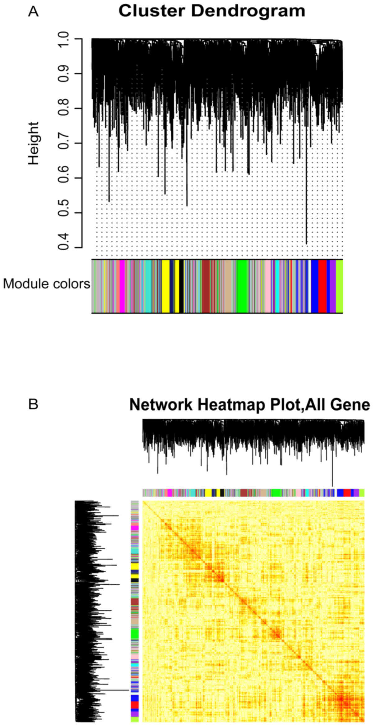
In order to study the co-expression of OA-related
genes, we used weighted gene co-expression network analysis (WGCNA)
(21) to analyze the RNA expression
matrix of OA-related genes and to find the co-expression gene
module. The weighted value of correlation coefficient, i.e., the N
power of gene correlation coefficient, was utilized to calculate
the correlation coefficient (Person coefficient) between any two
genes. The connection between genes in the network obeys scale-free
networks, which make the algorithm more biologically meaningful.
Then, a hierarchical clustering tree was built by the correlation
coefficient between genes. Different branches of the clustering
tree represent separate gene modules, and different colors
represent different modules. Finally, 16 significant OA-related
gene co-expression modules were extracted.
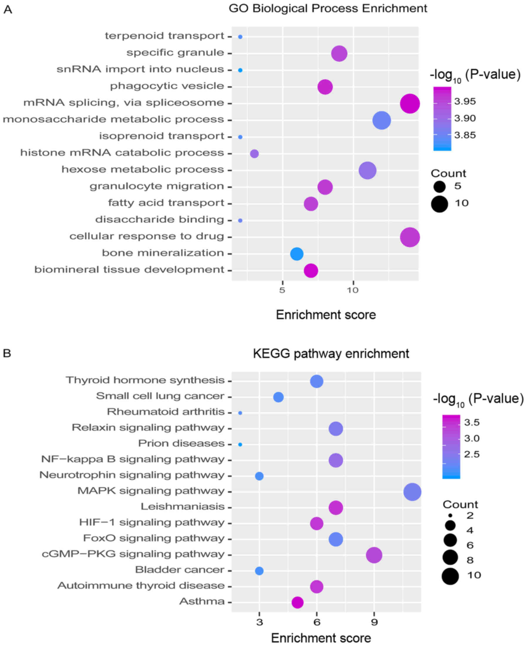
Functional pathway enrichment analysis
and identification of dysfunctional modules
Exploring the function and signaling pathway of
genes is often an effective wany to study the molecular mechanism
of diseases. R language Cluster Profiler package (22) was used to analyze gene enrichment in
16 modules (P-value cutoff = 0.01, q-value cutoff = 0.01) and KEGG
pathway (P-value cutoff = 0.05, q value Cutoff = 0.2). Then, the
functions and pathways related to the process of OA were screened
and bubble maps were drawn.
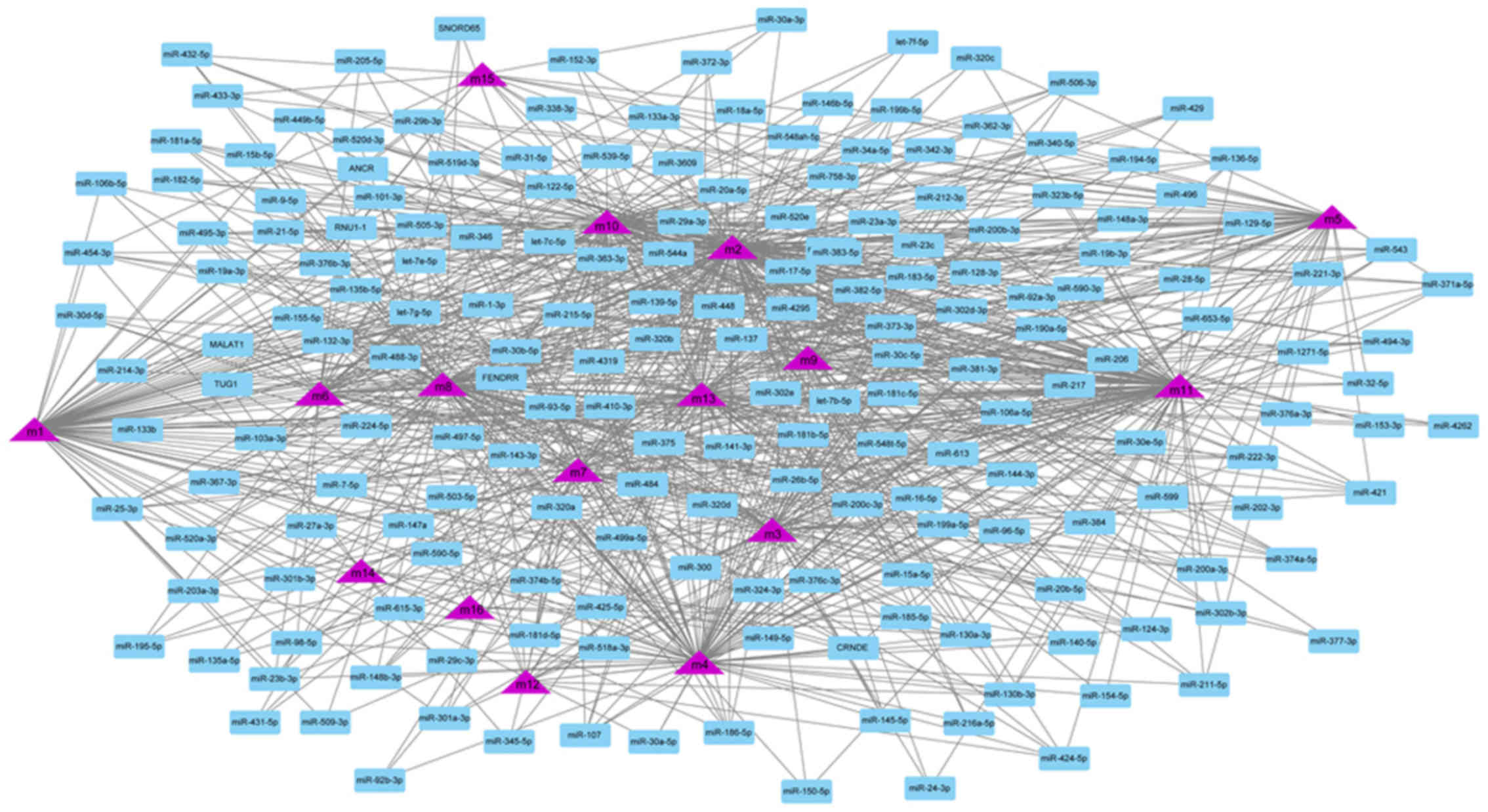
Regulator analysis
Pivot regulator is defined as a regulator, which
substantially regulates the dysfunction module of arthritis. Gene
transcription and post-transcriptional regulation are often driven
by non-coding genes (ncRNA) and transcription factors (TF). The
transcription factor target data was downloaded from TRRUST V2
database (23), and 71 interaction
pairs of 59 transcription factors were obtained. Then, ncRNA-RNA
(protein) data were downloaded from RAID 2.0 database (24), and 1,661 interaction pairs involving
842 ncRNAs were obtained. Pivot analysis based on the interaction
data was carried out to identify the regulatory effects of these
transcription factors and ncRNA on the modules. Pivot analysis
refers to search for at least two interacting drivers with the
module in the target pair and calculating the significance of the
interaction between the driver and the module according to the
hypergeometric test. TF and ncRNA with P<0.01 are the pivots of
the significant regulatory module. Finally, the core pivots were
recognized by statistical analysis.
Results
Identification of time-series
expression disorders in OA
In order to further explore the occurrence and
progress of OA, data sets of gene expression profiles related to OA
we downloaded from GEO database. Built on the differential analysis
of genes, 3,239 differential genes were obtained to identify the
key genes that play a continuous role in the regulation of OA in
the disease process (Table SI).
Identification of staging related
modules for functional OA
Based on WGCNA network, 16 co-expression modules
(Fig. 1A and B) were generated for
the analysis of persistent dysfunction genes. The key genes of each
module were identified based on functional impairment modules,
which showed significant clustering phenomena in the samples. These
functional modules may participate in different functions and
pathways, thus representing diverse regulatory mechanisms to
mediate the occurrence and development of OA dysfunction.
Functions and pathways of genes of
interest
Studying the function and pathway of gene
involvement is an essential means to identify the mediating
pathogenesis. In order to study possible dysfunction caused by
module gene disorder, the enrichment was analyzed of function and
pathway of each module. The results showed that most of the
functional modules added value in OA-related functions and
pathways. GO function and KEGG pathway enrichment analysis on 16
functional modules were performed. A total of 10,759 functions and
263 KEGG pathway enrichment results were obtained. These include
1,576 molecular functions (MF), 852 cell components (CC) and 8,331
biological processes (BP) involving genes (Fig. 2 and Table SII).
It is noteworthy that the AMPK/NF-κB signaling
pathway in which they are substantially involved may be identified
as the core signaling pathway to accelerate the disease progression
of OA. As noted above, we found that the NF-κB signaling pathway
may be closely related to the acceleration of OA.
TF and ncRNA driving OA
Although the regulation of single or several
transcription factors and ncRNA on OA have been studied by
biologists, few studies have focused on their comprehensive
regulation of dysfunctional modules. According to the number of
regulatory module genes and the significance, the potential
regulatory effect of 901 pivot regulators on the module were
determined. These include 842 ncRNA and 59 transcription factors,
involving 1,661 ncRNA-module interaction pairs and 71 TF-module
target pairs. Statistical analysis of the predicted results showed
that miR-132-3p targeted as many as 11 dysfunction modules,
respectively, and had a significant regulatory effect on OA
(Fig. 3). In addition, miR-130a-3p
and miR-590-3p regulated 10 dysfunction modules, respectively, and
were identified as pivot regulators. Other ncRNAs also regulate
multiple dysfunction modules to various degrees, and have potential
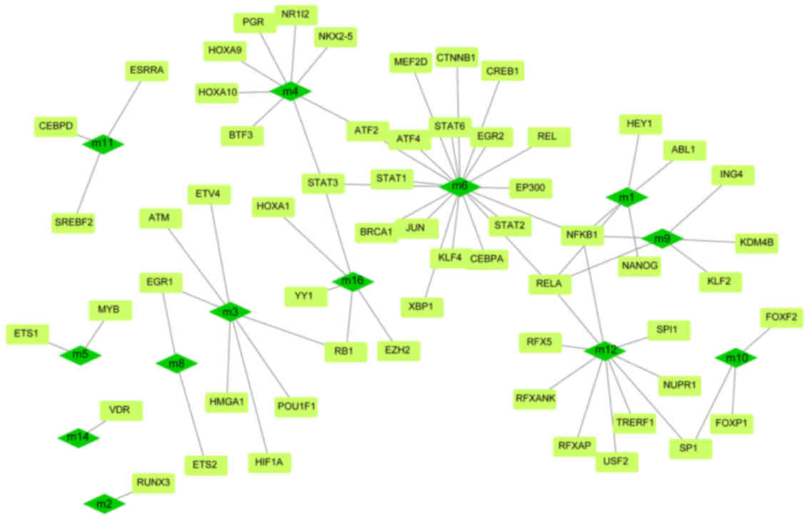
effects on OA. The regulatory effect of transcription factors on
diseases could be overlooked. Depending on statistics, NFKB1 and
RELA have significant regulatory effects on four dysfunction
modules, while STAT3 plays a role on three modules (Fig. 4). These transcription factors may
mediate dysfunction modules to regulate the occurrence and progress
of OA, and play a key role in the pathogenesis of OA. Other TFs
also have a definite regulatory effect on functional modules, which
may affect the progress of the disease. A thorough study of the
regulatory role of pivot regulators on dysfunction modules will
allow us to understand the underlying mechanisms of diseases. These
pivot regulators can be used as candidates for further experimental
studies.
Discussion
OA is one of the most frequent chronic diseases.
With the increase in life expectancy, the incidence of OA will
continue to rise (25). The
pathogenesis of OA involves many factors, including mechanics,
effects of aging on the composition and structure of the cartilage
matrix, and genetic factors (26).
Among them, serum microRNA level has an important regulatory
relationship with the development of severe OA of the knee joint
and hip joint (27). Rousseau et
al (28) found that serum
periosteal protein was involved in the prevalence and development
risk of knee OA in women. In order to fully explore the core
pathways and regulators of OA, we first integrated related
differential genes for differential analysis, and finally obtained
3,239 differential genes. The key gene statistics found that
proteins play an important role in promoting the operative
mechanism of OA. In view of the early stage of OA, including the
effects of cell proliferation and chondrocytes in the synthesis of
inflammatory mediators such as matrix proteins, proteases, growth
factors and cytokines (26). Studies
have revealed that in the late stage of OA, bone tissue blood flow
and oxygen content significantly decreased. It also has a negative
effect on bone cells, induces them to release protein (cytokines),
promotes bone remodeling and cartilage destruction (29). We observed the co-expression behavior
of the differentially expressed genes in the disease samples. From
this, we obtained 16 co-expression modules. The genes contained in
the modules were reckoned to have co-expression. Subsequently, in
view of the consequences of enrichment analysis, we found that
genes in 11 functional modules of OA mainly participate in the
functional pathways of NF-κB signaling pathway. These include
maintenance of protein localization in organelle, epidermal growth
factor-activated receptor trans activation by G-protein coupled
receptor signaling pathway and chondrocyte proliferation. The NF-κB
signaling pathway is thought to be the core signaling pathway to
promote the progression of OA. Through the review of Saito and
Tanaka (30) we found two kinds of
multifunctional signaling pathways in the development of OA: Notch
and NF-κB in activated B cells, and determined that articular
chondrocytes regulate the progress of OA through these pathways.
miR-146 promotes the proliferation and inhibit apoptosis of OA
chondrocytes by inhibiting the expression of TRAF6 and the
inhibition of NF-κB signaling pathway (31). In addition, high expression of Sam68
can promote the activation of TNF-α-induced chondrocyte NF-κB
signaling, the expression of decomposition genes and cell
apoptosis, which provides a possible target for the
pathophysiological study and treatment of OA (32).
To elucidate the transcriptional regulatory factors
of OA, pivot regulators were analyzed based on transcriptional and
post-transcriptional regulatory relationships. The results showed
that microRNAs mainly composed of microRNAs such as miR-132-3p,
miR-130a-3p and miR-590-3p played a major role in the regulation of
OA. The results showed that miR-132-3p promoted cartilage formation
and differentiation of rMSCs, possibly through targeting ADAMTS-5,
which provided a new perspective for cartilage differentiation and
pathology of OA (33). In addition,
miR-130a plays an important role in regulating the expression of
TNF-alpha in human chondrocytes, and it will be a novel therapeutic
target for OA (34). Relevant
studies have shown that elevated concentration of melatonin may
lead to the inhibition of the expression of microRNA-590-3p and
upregulation of the target gene of human osteoblast apoptosis
(35). Building on our observations,
we found that microRNAs that regulate multiple modules target genes
and pathways related to OA, thus providing a comprehensive
understanding of the molecular network underlying the pathogenesis
of OA. Transcription factors such as NFKB1, RELA and STAT3 are key
mediators in the process of OA-related potential diseases. Their
interactions can regulate inflammation and metabolism in cells, and
drive inflammatory cytokines and immune responses in OA
microenvironment. In the process of OA, these three transcription
factors mediate four and three dysfunction modules respectively,
and play an important role in the pathogenesis of OA. Recent
studies have demonstrated that upregulation of microRNA-9 or
downregulation of NF-κB1 can promote cell proliferation and inhibit
cell apoptosis, and downregulation of microRNA-9 can directly bind
to NF-κB1, promote the proliferation and anti-apoptosis of knee OA
chondrocytes (36). In addition,
in vitro studies have shown that Rela is a fundamental
subunit mediating the signal transduction of NF-κB, involved in
cartilage formation and differentiation, cell survival and
production of catabolic enzymes (37). These pivot regulators together
mediate functional dysfunction modules, play an overall supervisory
role, and represent the accelerating process of OA disease.
In conclusion, the study of gene involvement in
signaling pathways will allow us to analyze the progress of
disease. In view of the differential genes, we obtained, STAT3
speeds up the progression of OA through the NF-κB signaling
pathway. Activation of nuclear factor NF-κB protein triggers the
expression of a series of genes, leading to joint destruction and
the occurrence and development of OA (OA) (38). ACY-1215, an inhibitor, mainly induces
STAT3 in OA chondrocytes to downregulate the expression of
matrix-degrading proteinase through NF-κB signaling pathway, thus
improving cartilage degradation and exerting effective cartilage
protection (39). In addition,
chondroitin sulfate (CS) is a slow-acting disease regulator in the
treatment of OA. Its beneficial effect is the anti-inflammatory
properties caused by the inhibition of the signal transduction
pathway of NF-κB or STAT3 (40). In
general, functional module-based approaches cannot only explore the
pathogenesis and development of diseases comprehensively and
deeply, but also provide abundant resources for potential
candidates of TF and pivot ncRNAs, and predict their potential
therapeutic methods and mechanisms.
Acknowledgements
Not applicable.
Funding
No funding was received.
Availability of data and materials
The datasets used and/or analyzed during the current
study are available from the corresponding author on reasonable
request.
Authors' contributions
FW wrote the manuscript, interpreted and analyzed
the data. ZG designed the study and performed the experiments. YY
was responsible for the analysis and discussion of the data. All
authors read and approved the final manuscript.
Ethics approval and consent to
participate
The study was approved by the Ethics Committee of
Second Hospital of Shanxi Medical University (Taiyuan, China).
Patient consent for publication
Not applicable.
Competing interests
The authors declare that they have no competing
interests.
References
|
1
|
Litwic A, Edwards MH, Dennison EM and
Cooper C: Epidemiology and burden of osteoarthritis. Br Med Bull.
105:185–199. 2013. View Article : Google Scholar : PubMed/NCBI
|
|
2
|
Dieppe PA and Lohmander LS: Pathogenesis
and management of pain in osteoarthritis. Lancet. 365:965–973.
2005. View Article : Google Scholar : PubMed/NCBI
|
|
3
|
Prieto-Alhambra D, Judge A, Javaid MK,
Cooper C, Diez-Perez A and Arden NK: Incidence and risk factors for
clinically diagnosed knee, hip and hand osteoarthritis: Influences
of age, gender and osteoarthritis affecting other joints. Ann Rheum
Dis. 73:1659–1664. 2014. View Article : Google Scholar : PubMed/NCBI
|
|
4
|
Pérez Martín Á: [Symptoms. Localizations:
Knee, hip, hands, spine, other localizations]. Aten Primaria. 46
(Suppl 1):11–17. 2014.PubMed/NCBI
|
|
5
|
Flugsrud GB, Nordsletten L, Reinholt FP,
Risberg MA, Rydevik K and Uhlig T: Osteoarthritis. Tidsskr Nor
Laegeforen. 130:2136–2140. 2010.(In Norwegian). View Article : Google Scholar : PubMed/NCBI
|
|
6
|
Bijlsma JW, Berenbaum F and Lafeber FP:
Osteoarthritis: An update with relevance for clinical practice.
Lancet. 377:2115–2126. 2011. View Article : Google Scholar : PubMed/NCBI
|
|
7
|
Thomas AC, Hubbard-Turner T, Wikstrom EA
and Palmieri-Smith RM: Epidemiology of posttraumatic
osteoarthritis. J Athl Train. 52:491–496. 2017. View Article : Google Scholar : PubMed/NCBI
|
|
8
|
Buckwalter JA and Martin JA:
Osteoarthritis. Adv Drug Deliv Rev. 58:150–167. 2006. View Article : Google Scholar : PubMed/NCBI
|
|
9
|
Felson DT: An update on the pathogenesis
and epidemiology of osteoarthritis. Radiol Clin North Am. 42:1–9.
2004. View Article : Google Scholar : PubMed/NCBI
|
|
10
|
Steinberg J and Zeggini E: Functional
genomics in osteoarthritis: Past, present, and future. J Orthop
Res. 34:1105–1110. 2016. View Article : Google Scholar : PubMed/NCBI
|
|
11
|
Kloek CJ, Bossen D, Veenhof C, van Dongen
JM, Dekker J and de Bakker DH: Effectiveness and cost-effectiveness
of a blended exercise intervention for patients with hip and/or
knee osteoarthritis: Study protocol of a randomized controlled
trial. BMC Musculoskelet Disord. 15:2692014. View Article : Google Scholar : PubMed/NCBI
|
|
12
|
Grazina R, Andrade R, Bastos R, Costa D,
Pereira R, Marinhas J, Maestro A and Espregueira-Mendes J: Clinical
management in early OA. Adv Exp Med Biol. 1059:111–135. 2018.
View Article : Google Scholar : PubMed/NCBI
|
|
13
|
Teoh LSG, Eyles JP, Makovey J, Williams M,
Kwoh CK and Hunter DJ: Observational study of the impact of an
individualized multidisciplinary chronic care program for hip and
knee osteoarthritis treatment on willingness for surgery. Int J
Rheum Dis. 20:1383–1392. 2017. View Article : Google Scholar : PubMed/NCBI
|
|
14
|
Adatia A, Rainsford KD and Kean WF:
Osteoarthritis of the knee and hip. Part I: Aetiology and
pathogenesis as a basis for pharmacotherapy. J Pharm Pharmacol.
64:617–625. 2012. View Article : Google Scholar : PubMed/NCBI
|
|
15
|
Legré-Boyer V: Viscosupplementation:
Techniques, indications, results. Orthop Traumatol Surg Res. 101
(Suppl):S101–S108. 2015. View Article : Google Scholar : PubMed/NCBI
|
|
16
|
Chin KY: The spice for joint inflammation:
Anti-inflammatory role of curcumin in treating osteoarthritis. Drug
Des Devel Ther. 10:3029–3042. 2016. View Article : Google Scholar : PubMed/NCBI
|
|
17
|
Sivordova LE, Zavodovsky BV, Polyakova JV
and Akhverdyan YR: Evidence of feasibility etoricoxib therapy in
osteoarthritis in elderly patients. Adv Gerontol. 29:286–290.
2016.(In Russian). PubMed/NCBI
|
|
18
|
Moskowitz RW: Role of collagen hydrolysate
in bone and joint disease. Semin Arthritis Rheum. 30:87–99. 2000.
View Article : Google Scholar : PubMed/NCBI
|
|
19
|
Barrett T, Wilhite SE, Ledoux P,
Evangelista C, Kim IF, Tomashevsky M, Marshall KA, Phillippy KH,
Sherman PM, Holko M, et al: NCBI GEO: Archive for functional
genomics data sets - update. Nucleic Acids Res. 41(D1): D991–D995.
2013. View Article : Google Scholar : PubMed/NCBI
|
|
20
|
Ritchie ME, Phipson B, Wu D, Hu Y, Law CW,
Shi W and Smyth GK: limma powers differential expression analyses
for RNA-sequencing and microarray studies. Nucleic Acids Res.
43:e472015. View Article : Google Scholar : PubMed/NCBI
|
|
21
|
Langfelder P and Horvath S: WGCNA: An R
package for weighted correlation network analysis. BMC
Bioinformatics. 9:5592008. View Article : Google Scholar : PubMed/NCBI
|
|
22
|
Yu G, Wang LG, Han Y and He QY:
clusterProfiler: an R package for comparing biological themes among
gene clusters. OMICS. 16:284–287. 2012. View Article : Google Scholar : PubMed/NCBI
|
|
23
|
Han H, Cho JW, Lee S, Yun A, Kim H, Bae D,
Yang S, Kim CY, Lee M, Kim E, et al: TRRUST v2: An expanded
reference database of human and mouse transcriptional regulatory
interactions. Nucleic Acids Res. 46D:D380–D386. 2018. View Article : Google Scholar
|
|
24
|
Yi Y, Zhao Y, Li C, Zhang L, Huang H, Li
Y, Liu L, Hou P, Cui T, Tan P, et al: RAID v2.0: An updated
resource of RNA-associated interactions across organisms. Nucleic
Acids Res. 45D:D115–D118. 2017. View Article : Google Scholar
|
|
25
|
Pereira D, Ramos E and Branco J:
Osteoarthritis. Osteoarthritis Acta Med Port. 28:99–106. 2015.
View Article : Google Scholar : PubMed/NCBI
|
|
26
|
Goldring MB and Goldring SR:
Osteoarthritis. J Cell Physiol. 213:626–634. 2007. View Article : Google Scholar : PubMed/NCBI
|
|
27
|
Beyer C, Zampetaki A, Lin NY, Kleyer A,
Perricone C, Iagnocco A, Distler A, Langley SR, Gelse K, Sesselmann
S, et al: Signature of circulating microRNAs in osteoarthritis. Ann
Rheum Dis. 74:e182015. View Article : Google Scholar : PubMed/NCBI
|
|
28
|
Rousseau JC, Sornay-Rendu E, Bertholon C,
Garnero P and Chapurlat R: Serum periostin is associated with
prevalent knee osteoarthritis and disease incidence/progression in
women: The OFELY study. Osteoarthritis Cartilage. 23:1736–1742.
2015. View Article : Google Scholar : PubMed/NCBI
|
|
29
|
Racine J and Aaron RK: Pathogenesis and
epidemiology of osteoarthritis. R I Med J. 96:19–22. 2013.
|
|
30
|
Saito T and Tanaka S: Molecular mechanisms
underlying osteoarthritis development: Notch and NF-κB. Arthritis
Res Ther. 19:942017. View Article : Google Scholar : PubMed/NCBI
|
|
31
|
Zhong JH, Li J, Liu CF, Liu N, Bian RX,
Zhao SM, Yan SY and Zhang YB: Effects of microRNA-146a on the
proliferation and apoptosis of human osteoarthritis chondrocytes by
targeting TRAF6 through the NF-κB signalling pathway. Biosci Rep.
37:372017. View Article : Google Scholar
|
|
32
|
Xu L, Sun C, Zhang S, Xu X, Zhai L, Wang
Y, Wang S, Liu Z, Cheng H, Xiao M, et al: Sam68 promotes NF-κB
activation and apoptosis signaling in articular chondrocytes during
osteoarthritis. Inflamm Res. 64:895–902. 2015. View Article : Google Scholar : PubMed/NCBI
|
|
33
|
Zhou X, Luo D, Sun H, Qi Y, Xu W, Jin X,
Li C, Lin Z and Li G: miR-132-3p regulates ADAMTS-5 expression and
promotes chondrogenic differentiation of rat mesenchymal stem
cells. J Cell Biochem. 119:2579–2587. 2018. View Article : Google Scholar : PubMed/NCBI
|
|
34
|
Li ZC, Han N, Li X, Li G, Liu YZ, Sun GX,
Wang Y, Chen GT and Li GF: Decreased expression of microRNA-130a
correlates with TNF-α in the development of osteoarthritis. Int J
Clin Exp Pathol. 8:2555–2564. 2015.PubMed/NCBI
|
|
35
|
Meng X, Zhu Y, Tao L, Zhao S and Qiu S:
miR-590-3p mediates melatonin-induced cell apoptosis by targeting
septin 7 in the human osteoblast cell line hFOB 1.19. Mol Med Rep.
17:7202–7208. 2018.PubMed/NCBI
|
|
36
|
Gu R, Liu N, Luo S, Huang W, Zha Z and
Yang J: MicroRNA-9 regulates the development of knee osteoarthritis
through the NF-kappaB1 pathway in chondrocytes. Medicine
(Baltimore). 95:e43152016. View Article : Google Scholar : PubMed/NCBI
|
|
37
|
Kobayashi H, Chang SH, Mori D, Itoh S,
Hirata M, Hosaka Y, Taniguchi Y, Okada K, Mori Y, Yano F, et al:
Biphasic regulation of chondrocytes by Rela through induction of
anti-apoptotic and catabolic target genes. Nat Commun. 7:133362016.
View Article : Google Scholar : PubMed/NCBI
|
|
38
|
Rigoglou S and Papavassiliou AG: The NF-κB
signalling pathway in osteoarthritis. Int J Biochem Cell Biol.
45:2580–2584. 2013. View Article : Google Scholar : PubMed/NCBI
|
|
39
|
Cheng C, Shan W, Huang W, Ding Z, Cui G,
Liu F, Lu W, Xu J, He W and Yin Z: ACY-1215 exhibits
anti-inflammatory and chondroprotective effects in human
osteoarthritis chondrocytes via inhibition of STAT3 and NF-κB
signaling pathways. Biomed Pharmacother. 109:2464–2471. 2019.
View Article : Google Scholar : PubMed/NCBI
|
|
40
|
Andrés RM, Payá M, Montesinos MC, Ubeda A,
Navalón P, Herrero M, Vergés J and Terencio MC: Potential
antipsoriatic effect of chondroitin sulfate through inhibition of
NF-κB and STAT3 in human keratinocytes. Pharmacol Res. 70:20–26.
2013. View Article : Google Scholar : PubMed/NCBI
|