|
1
|
Kuan EC, Yoo F, Chyu J, Bergsneider M and
Wang MB: Treatment outcomes of Rathke's cleft cysts managed with
marsupialization. J Neurol Surg B Skull Base. 78:112–115. 2017.
View Article : Google Scholar : PubMed/NCBI
|
|
2
|
Lin M, Wedemeyer MA, Bradley D, Donoho DA,
Fredrickson VL, Weiss MH, Carmichael JD and Zada G: Long-term
surgical outcomes following transsphenoidal surgery in patients
with Rathke's cleft cysts. J Neurosurg. 130:831–837. 2018.
View Article : Google Scholar : PubMed/NCBI
|
|
3
|
Kim JE, Kim JH, Kim OL, Paek SH, Kim DG,
Chi JG and Jung HW: Surgical treatment of symptomatic Rathke cleft
cysts: Clinical features and results with special attention to
recurrence. J Neurosurg. 100:33–40. 2004. View Article : Google Scholar : PubMed/NCBI
|
|
4
|
Jahangiri A, Molinaro AM, Tarapore PE,
Blevins L Jr, Auguste KI, Gupta N, Kunwar S and Aghi MK: Rathke
cleft cysts in pediatric patients: Presentation, surgical
management and postoperative outcomes. Neurosurg Focus. 31:E32011.
View Article : Google Scholar : PubMed/NCBI
|
|
5
|
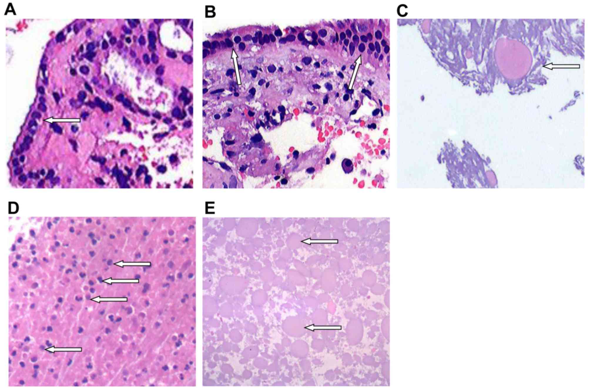
Mrelashvili A, Braksick SA, Murphy LL,
Morparia NP, Neena N and Neeraj K: Chemical meningitis: A rare
presentation of Rathke's cleft cyst. J Clin Neurosci. 21:692–694.
2014. View Article : Google Scholar : PubMed/NCBI
|
|
6
|
Naama O, Gazzaz M, Boulahroud O and
Elmoustarchid B: Infection of a Rathke cleft cyst: A rare cause of
pituitary abscess. Surg Infect (Larchmt). 15:358–360. 2014.
View Article : Google Scholar : PubMed/NCBI
|
|
7
|
Jimbo H, Ichikawa M, Fukami S, Otsuka K,
Tsurukiri J, Sunaga S and Lkeda Y: Rapid De Novo aneurysm formation
after Rathke cleft cyst rupture. World Neurosurg.
88:690.e11–690.e16. 2016. View Article : Google Scholar
|
|
8
|
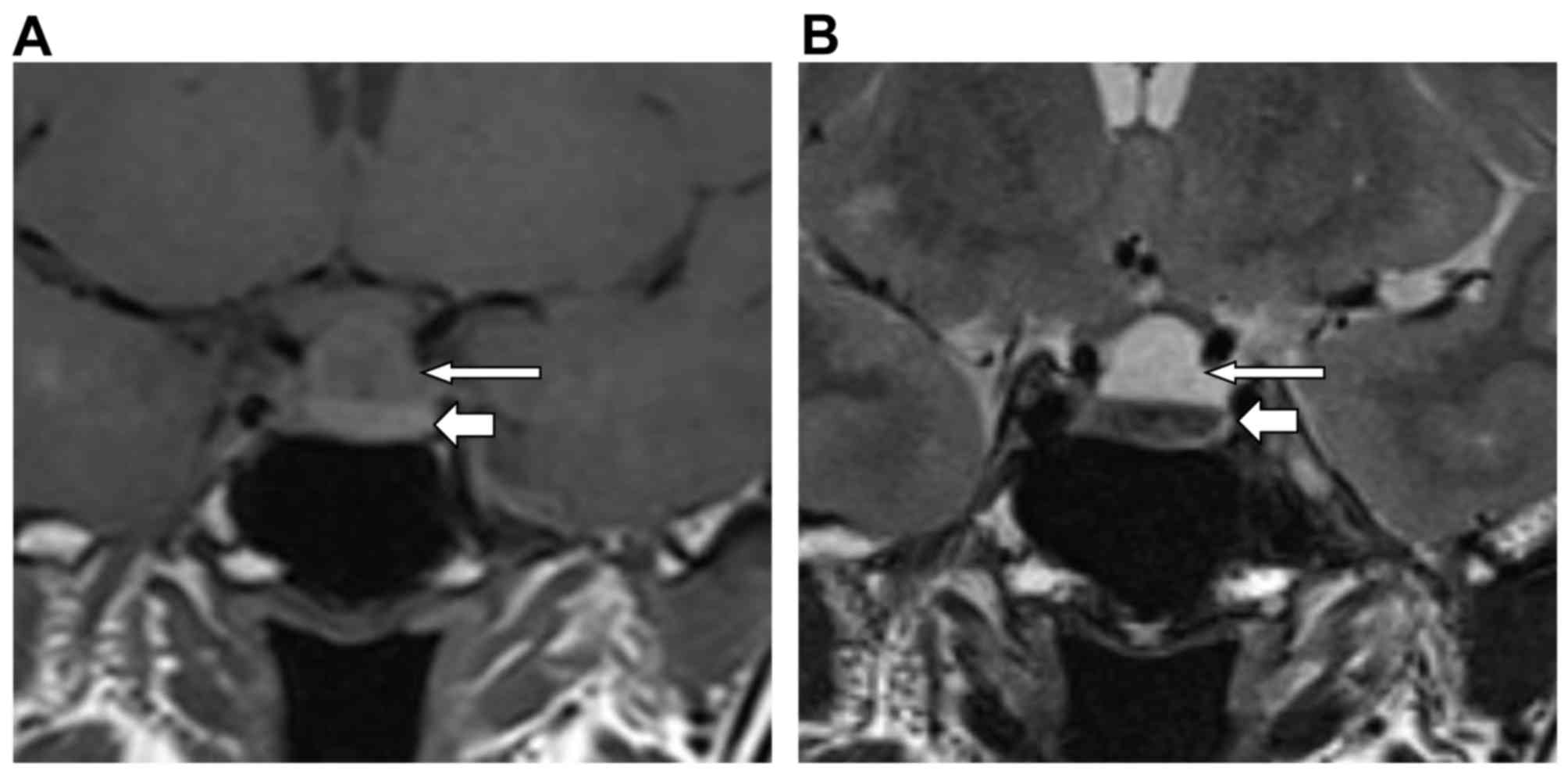
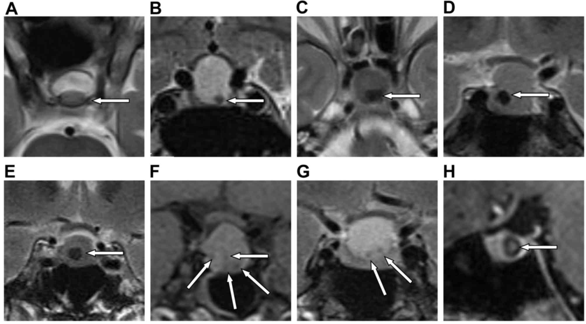
Han X and Zhao DJ: Imaging diagnosis of
Rathke's cleft cyst. J Med Imag. 20:782–784. 2010.
|
|
9
|
Wang SS, Xiao DY, Yu YH, Jing JJ, Zhao L
and Wang RM: Diagnostic significance of intracystic nodules on MRI
in Rathke's cleft cyst. Int J Endocrinol. 2012:9587322012.
View Article : Google Scholar : PubMed/NCBI
|
|
10
|
Binning MJ, Gottfried ON, Osborn AG and
Couldwell WT: Rathke cleft cyst intracystic nodule: A
characteristic magnetic resonance imaging finding. J Neurosurg.
103:837–840. 2005. View Article : Google Scholar : PubMed/NCBI
|
|
11
|
Byun WM, Kim OL and Kim D: MR imaging
findings of Rathke's cleft cysts: Significance of intracystic
nodules. AJNR Am J Neuroradiol. 21:485–488. 2000.PubMed/NCBI
|
|
12
|
QI C and Wang N: Value of intracystic
nodules on MRI to diagnosis of Rathke's cleft cyst. Chin J Clin
Neurosurg. 19:212–214. 2014.
|
|
13
|
Kilday JP, Laughlin S, Urbach S, Bouffet E
and Bartels U: Diabetes insipidus in pediatric germinomas of the
suprasellar region: Characteristic features and significance of the
pituitary bright spot. J Neurooncol. 121:167–175. 2015. View Article : Google Scholar : PubMed/NCBI
|
|
14
|
Saeki N, Hoshi S, Sunada S, Sunami K,
Murai H, Kubota M, Tatsuno I, Iuchi T and Yamaura A: Correlation of
high signal intensity of the pituitary stalk in macroadenoma and
postoperative diabetes insipidus. AJNRAm J Neuroradiol. 23:822–827.
2002.
|
|
15
|
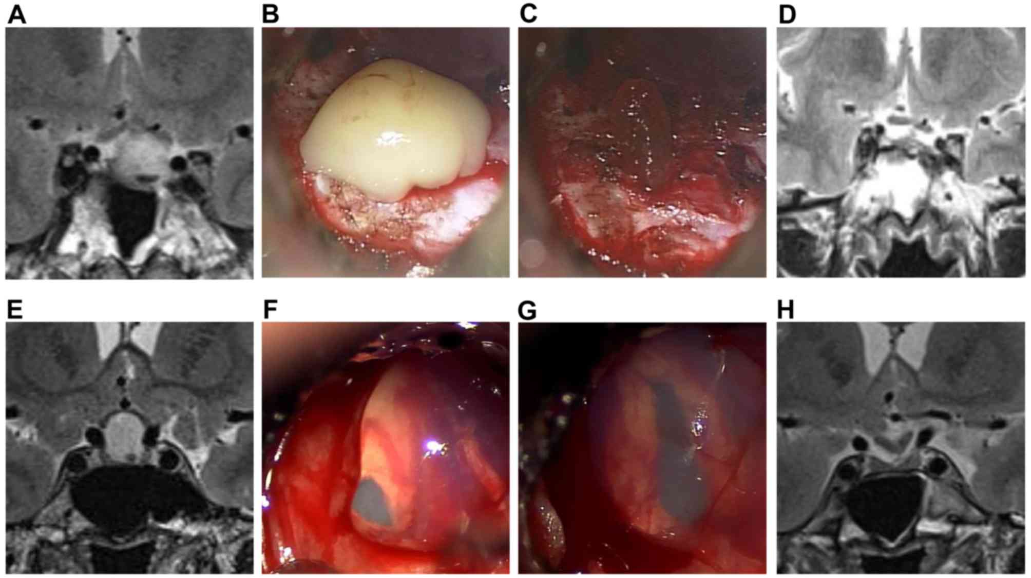
Benveniste RJ, King WA, Walsh J, Lee JS,
Naidich TP and Post KD: Surgery for Rathke cleft cysts: Technical
considerations and outcomes. J Neurosurg. 101:577–584. 2004.
View Article : Google Scholar : PubMed/NCBI
|
|
16
|
Côté M, Salzman KL, Sorour M and Couldwell
WT: Normal dimensions of the posterior pituitary bright spot on
magnetic resonance imaging. J Neurosurg. 120:357–362. 2014.
View Article : Google Scholar : PubMed/NCBI
|
|
17
|
Schreckinger M, Szerlip N and Mittal S:
Diabetes insipidus following resection of pituitary tumors. Clin
Neurol Neurosurg. 115:121–126. 2013. View Article : Google Scholar : PubMed/NCBI
|
|
18
|
Wang S, Lin K, Xiao D, Zhao L, Qin Y and
Wei L: MR imaging analysis of posterior pituitary in patients with
pituitary adenoma. Int J Clin Exp Med. 8:7634–7640. 2015.PubMed/NCBI
|
|
19
|
Klyn V, Dekeyzer S, Van Eetvelde R, Roels
P, Vergauwen O, Devolder P, Wiesmann M, Achten E and Nikoubashman
O: Presence of the posterior pituitary bright spot sign on MRI in
the general population: A comparison between 1.5 and 3T MRI and
between 2D-T1 spin-echo- and 3D-T1 gradient-echo sequences.
Pituitary. 21:379–383. 2018. View Article : Google Scholar : PubMed/NCBI
|
|
20
|
Ogawa Y, Watanabe M and Tominaga T:
Intraparenchymal infiltration of Rathke's cleft cysts manifesting
as severe neurological deficits and hypopituitarism: 2 Case
reports. BMC Res Notes. 9:2252016. View Article : Google Scholar : PubMed/NCBI
|
|
21
|
Locatelli D, Pozzi F, Agresta G, Padovan
S, Karligkiltis A and Castelnuovo P: Extended endoscopic endonasal
approach for suprasellar craniopharyngioma. J Neurol Surg B Skull
Base. 79:S196–S198. 2018. View Article : Google Scholar : PubMed/NCBI
|
|
22
|
Abbas M, Khairy S, AlWohaibi M, Aloraidi A
and AlQurrashi WW: Bilateral temporal extradural hematoma on top of
bilateral temporal arachnoid cyst: First case report and extensive
literature review. World Neurosurg. 115:134–137. 2018. View Article : Google Scholar : PubMed/NCBI
|
|
23
|
Ifergan H, Cazeneuve N, Merenda P and
Magni C: MR imaging features of a pituitary abscess: A case report.
Ann Endocrinol (Paris). 80:62–63. 2019. View Article : Google Scholar : PubMed/NCBI
|