Introduction
Myocardial ischemia/reperfusion (I/R) injury
exacerbates tissue injuries during reperfusion following prolonged
myocardial ischemia and is a significant clinical problem
associated with procedures such as angioplasty, thrombolysis and
coronary bypass surgery (1–3). The underlying mechanism of myocardial
I/R injury is complicated and unclear, and this may be associated
with a number of factors, including overproduction of reactive
oxygen species (ROS), apoptosis, mitochondrial dysfunction,
intracellular calcium overload and inflammation (4–6).
Although an emerging number of innovative approaches to protect
cardiac tissue against myocardial I/R injury are under preclinical
and clinical investigation; at present, there are no effective
therapeutic strategies to reduce or protect against I/R injury
(7,8). Therefore, compounds with antioxidant or
anti-inflammatory properties may prove valuable for treating
myocardial I/R injury.
Salidroside is isolated from Rhodiola rosea,
which is used as a herbal medicine used to mitigate high altitude
sickness and protect erythrocytes against oxidative stress injury
(9). Salidroside has been reported
to possess a wide range of pharmacological properties, including
anti-inflammatory, antioxidative, anti-asthmatic and
cardioprotective effects (10,11). It
has previously been reported that salidroside may possess
therapeutic value against myocardial I/R injury (12,13).
Chang et al (14)
demonstrated that salidroside protects against myocardial injury
under I/R conditions by regulating energy metabolism homeostasis
and inflammation. A previous study also demonstrated that
salidroside exhibited its cardioprotective effects against
myocardial I/R injury in rats by inhibiting apoptosis and
inflammation (15). However, the
underlying protective molecular mechanisms of salidroside during
H/R injury remains unknown.
MicroRNAs (miRs) are a class of endogenous, small
non-coding single-stranded RNAs ~22 nucleotides in length, which
participate in the regulation of multiple physiological and
pathophysiological processes (16).
miR-21 expression is endogenously high in cardiomyocytes, vascular
endothelial cells, cardiac fibroblasts and vascular smooth muscle
cells (17). Although, the
physiological functions of miR-21 in cardiovascular diseases have
not been fully determined, the contribution of miR-21 in various
cardiovascular diseases, including heart failure, myocardial
infarction, myocardial fibrosis and atherosclerosis are being
uncovered (18–20). Studies have shown that miR-21 is
involved in numerous pathophysiological processes associated with
myocardial I/R injury (21–23). MiR-21 effectively reduced the level
of myocardial apoptosis and the release of inflammatory factors
induced by myocardial I/R injury in rats (21). In addition, miR-21 expression was
decreased in myocardial I/R injury and restoring miR-21 expression
levels attenuated myocardial I/R injury (21,24,25).
Therefore, miR-21 may serve as a novel biomarker of myocardial I/R
injury and may be a promising therapeutic target.
In the present study, the protective effects of
salidroside on oxidative stress and inflammatory injuries in an
in vitro model of myocardial I/R injury was examined. The
key targets and signaling pathways associated with salidroside
during I/R were explored to elucidate the mechanism by which miR-21
mediated the cardioprotective effects of salidroside. The results
of the present study may improve understanding of the
pharmacological mechanisms of salidroside and may also provide
additional evidence of the clinical value of combining traditional
Chinese medicines treatment with agents which upregulate the
effects of miR-21 or its downstream targets for preventing and
treating myocardial I/R injury.
Materials and methods
Cell culture and hypoxia/reoxygenation
(H/R) model
H9c2 rat derived cardiomyocytes (American Type
Culture Collection; ATCC) were cultured in high glucose Dulbecco's
modified Eagle's medium (DMEM; HyClone; GE Healthcare Life
Sciences) supplemented with 10% heat-inactivated fetal bovine
syndrome (Gibco; Thermo Fisher Scientific, Inc.) with a
penicillin-streptomycin solution (100×; Beyotime Institute of
Biotechnology). Cells were seeded in a humidified atmosphere
containing 5% CO2 at 37°C (Thermo Fisher Scientific,
Inc.). To mimic myocardial I/R injury in vitro, the H9c2
cells were exposed to an H/R environment as described previously
(26). H9c2 cells at 80–90%
confluence were incubated with serum-free DMEM and placed in an
anaerobic chamber that was supplied with a gas mixture containing
94% N2, 5% CO2, and 1% O2 for 6 h
to induce hypoxia. Subsequently, the cells were provided with the
normal medium and placed in an incubator with 95% O2 and
5% CO2 for 12 h to allow reoxygenation. The control
plates were incubated with 95% O2/5% CO2 at
37°C for 18 h. In the drug treatment experiments, H9c2 cells were
pretreated with salidroside (10 µM) for 1 h and were maintained
under hypoxia for 6 h, followed by reoxygenation for 12 h. In the
transfected cells, H9c2 cells were transfected with miR-21
inhibitor or negative control inhibitors for 6 h before salidroside
(10 µM) pre-treatment for 1 h. Subsequently, the H9c2 cells were
exposed to the H/R environment as described above.
Transfection of a miR-21
inhibitor
A miR-21 inhibitor and the negative control (NC)
inhibitor were purchased from Shanghai GenePharma Co., Ltd with the
following sequences: miR-21 inhibitor,
5′-UCAACAUCAGUCUGAUAAGCUA-3′; Negative control,
5′-CAGUACUUUUGUGUAGUACAA-3′. Cells were transfected with the miR-21
inhibitor (100 nM) or NC inhibitor (100 nM) using
Lipofectamine® RNAi Max (Thermo Fisher Scientific, Inc.)
with Opti-MEM Reduced Serum Medium (Gibco; Thermo Fisher
Scientific, Inc.) according to the manufacturer's protocols. A
total of 6 h after transfection, the medium was replaced with fresh
DMEM. The transfection efficiency was determined using reverse
transcription-quantitative (RT-q)PCR 48 h after transfection.
RT-qPCR
Total RNA from cells was extracted using
TRIzol® reagent (Invitrogen; Thermo Fisher Scientific,
Inc) according to the manufacturer's protocol. The cDNA was reverse
transcribed using Prime Script™ RT master mix according to the
manufacturer's protocol (Takara Biotechnology Co., Ltd.). The
temperature protocol was: 37°C for 15 min; followed by 85°C for 5
sec and 4°C for 20 sec. RT-qPCR was performed on an ABI 7300
thermocycler (Applied Biosystems; Thermo Fisher Scientific, Inc.)
and the expression of miR-21 was quantified using SYBR Green I
fluorescent dye (Takara Biotechnology Co., Ltd.). The miR-21
expression levels were normalized to the expression of U6. The
thermocycling conditions were: 95°C for 2 min; followed by 40
cycles of 95°C for 15 sec and 58°C for 20 sec. The expression
levels of interleukin (IL)-6, IL-1β and tumor necrosis factor
(TNF)-α were normalized to the expression of GAPDH. The
thermocycling conditions were: 95°C for 2 min; followed by 40
cycles of 95°C for 5 sec, 56°C for 20 sec; and 72°C for 25 sec,
65°C for 5 sec, and 95°C for 50 sec. The relative levels of miR-21
or IL-6, IL-1β and TNF-α was calculated using the comparative
2−ΔΔCq method (27)
normalized to U6 or GAPDH, respectively. The sequences of the
primers used were presented in Table
I.
 | Table I.Primer sequences. |
Table I.
Primer sequences.
| Genes | Sequence 5′-3′ |
|---|
| miR-21,
forward |
GCCGCTAGCTTATCAGACTGATGT |
| miR-21,
reverse |
GTGCAGGGTCCGAGGT |
| U6, forward |
CTCGCTTCGGCAGCACA |
| U6, reverse |
AACGCTTCACGAATTTGCGT |
| IL-6, forward |
GCCAGAGTCATTCAGAGCAAT |
| IL-6, reverse |
CTTGGTCCTTAGCCACTCCT |
| TNF-α, forward |
CACCACGCTCTTCTGTCTACTG |
| TNF-α, reverse |
GCTACGGGCTTGTCACTCG |
| GAPDH, forward |
CGCTAACATCAAATGGGGTG |
| GAPDH, reverse |
TTGCTGACAATCTTGAGGGAG |
MTT assay
Cell viability was measured using an MTT Cell
Proliferation and Cytotoxicity assay kit (Beyotime Institute of
Biotechnology) according to manufacturer's protocols. Briefly, H9c2
cells were cultured in 96-well plates at a density of
1×10⁴ cells/well. Following the treatment as described
above, MTT reagent at a final concentration of 0.5 mg/ml (10 µl)
was added to each well and incubated at 37°C for 4 h. The formazan
crystals were subsequently dissolved using DMSO (100 µl/well). The
absorbance was measured at 570 nm using a microplate
spectrophotometer (BioTek China).
Measurement of lactate dehydrogenase
(LDH) release, malondialdehyde (MDA) levels and superoxide
dismutase (SOD) and glutathione peroxidase (GSH-Px) activity
Following treatment as described above, the cells
were collected and centrifuged at 3,000 × g for 10 min at 4°C, and
the supernatant was stored at −80°C. The LDH release levels, MDA
levels and SOD and GSH-Px activity were detected using a LDH
cytotoxicity kit (Beyotime Institute of Biotechnology), and MDA
assay kit (Nanjing Jiancheng Institute of Bioengineering
Institute), an SOD assay kit (Beijing Solarbio Science &
Technology Co., Ltd.) and a GSH-Px assay kit (Beijing Solarbio
Science & Technology Co., Ltd.), respectively. All assays were
performed strictly according to the manufacturer's protocol.
Detection of apoptosis
Cell apoptosis was measured using an Annexin
V-fluorescein isothiocyanate (FITC)/propidium iodide (PI) staining
kit (BD Bioscience; Becton, Dickinson and Company) according to
manufacturer's protocol. Following treatment, H9c2 cells
(1×10⁶) were collected, washed with cold PBS and
resuspended in 500 µl 1× binding buffer followed by Annexin V-FITC
(5 µl) and PI (10 µl). The mixture was placed in the dark for 15
min at room temperature and analyzed using a flow cytometer
(Beckman Coulter) and Kaluza software 2.1.1 (Beckman Coulter,
Inc.). Each experiment was performed at least three times.
Measurement of caspase-3 activity
The activity of caspase-3 was determined using a
caspase-3 colorimetric assay kit (RayBiotech, Inc.) according to
the manufacturer's protocol. H9c2 cells were seeded in 6-well
plates at a density of 5×10⁵ cells/well and treated as
mentioned above. Subsequently, the cells were collected and lysed
using a cell lysis buffer included in the kit followed by
centrifuging at 10,000 × g for 5 min at 4°C. The protein
concentration was measured using a bicinchoninic acid assay kit
(Beyotime Institute of Biotechnology). The caspase-3 substrate
Ac-DEVD-pNA (0.2 mM) was added to the lysis buffer and incubated at
37°C for 2 h. The relative fluorescence of each well was measured
using a fluorescence plate reader at 450 nm within 30 min.
Measurement of intracellular ROS
Intracellular ROS generation was monitored using the
Reactive Oxygen Species assay kit (Beyotime Institute of
Biotechnology) which uses the fluorescent probe DCFH-DA to detect
ROS. DCFH-DA is converted to DCFH by intracellular esterases, which
are subsequently converted to the fluorescent DCF by ROS. After
treatment, H9c2 cells were incubated with DCFH-DA (10 µM) at 37°C
for 20 min in the dark. Subsequently, the cells were washed twice
with PBS and analyzed using a flow cytometer (Beckman Coulter,
Inc.) with excitation at 488 nm and emission at 525 nm. The data
were viewed in FlowJo 10 (Tree Star, Inc.).
ELISA
The IL-6 (cat. no. M6000B), IL-1β (cat. no. RLB00)
and TNF-α (cat. no. RTA00) levels in the culture supernatant were
analyzed using commercially available high sensitivity ELISA kits
(R&D Systems Europe, Ltd.) according to manufacturer's
protocol. A microplate reader measured the optical density at 450
nm and the data were used to calculate the concentrations of
various cytokines based on the standard curves. The values were
determined in three independent experiments.
Statistical analysis
All data are expressed as the mean ± standard
deviation of at least three independent experiments and statistical
analyses were performed using Graphpad 6.0 (GraphPad Software,
Inc.). The statistical significance of differences between multiple
groups was estimated using a one-way analysis of variance followed
by a Dunnett's post-hoc test. P<0.05 was considered to indicate
a statistically significant difference.
Results
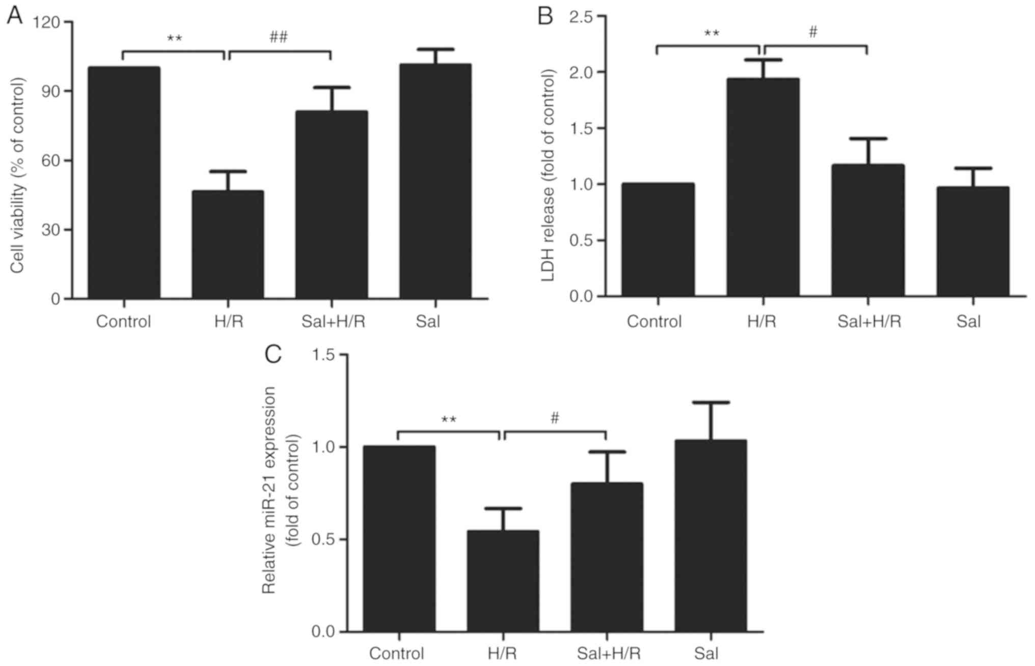
Salidroside inhibits H/R injury in
H9c2 cell
The potential effects of salidroside on H/R-induced
H9c2 cell injury were first determined. The result of the MTT assay
demonstrated that H/R treatment significantly reduced H9c2 cell
viability, which was reduced by salidroside pretreatment
(P<0.01; Fig. 1A). Salidroside
reduced H/R-induced H9c2 cell death, which was further confirmed by
the LDH assay. H/R resulted in a significant increase in LDH
release in H9c2 cells (P<0.01), whereas salidroside pretreatment
significantly attenuated the increase in LDH release (P<0.05;
Fig. 1B). Furthermore, the effects
of salidroside on miR-21 expression levels in the presence or
absence of H/R were determined. RT-qPCR results showed that,
compared with the control group, the expression of miR-21 was
significantly decreased in the H/R group (P<0.01), whereas, in
comparison with the H/R group, the expression of miR-21 in the
salidroside + H/R co-treatment group was significantly increased
(P<0.05; Fig. 1C). Together,
these results suggest that salidroside reduces or prevents
H/R-induced injury in H9c2 cells and miR-21 expression was
increased.
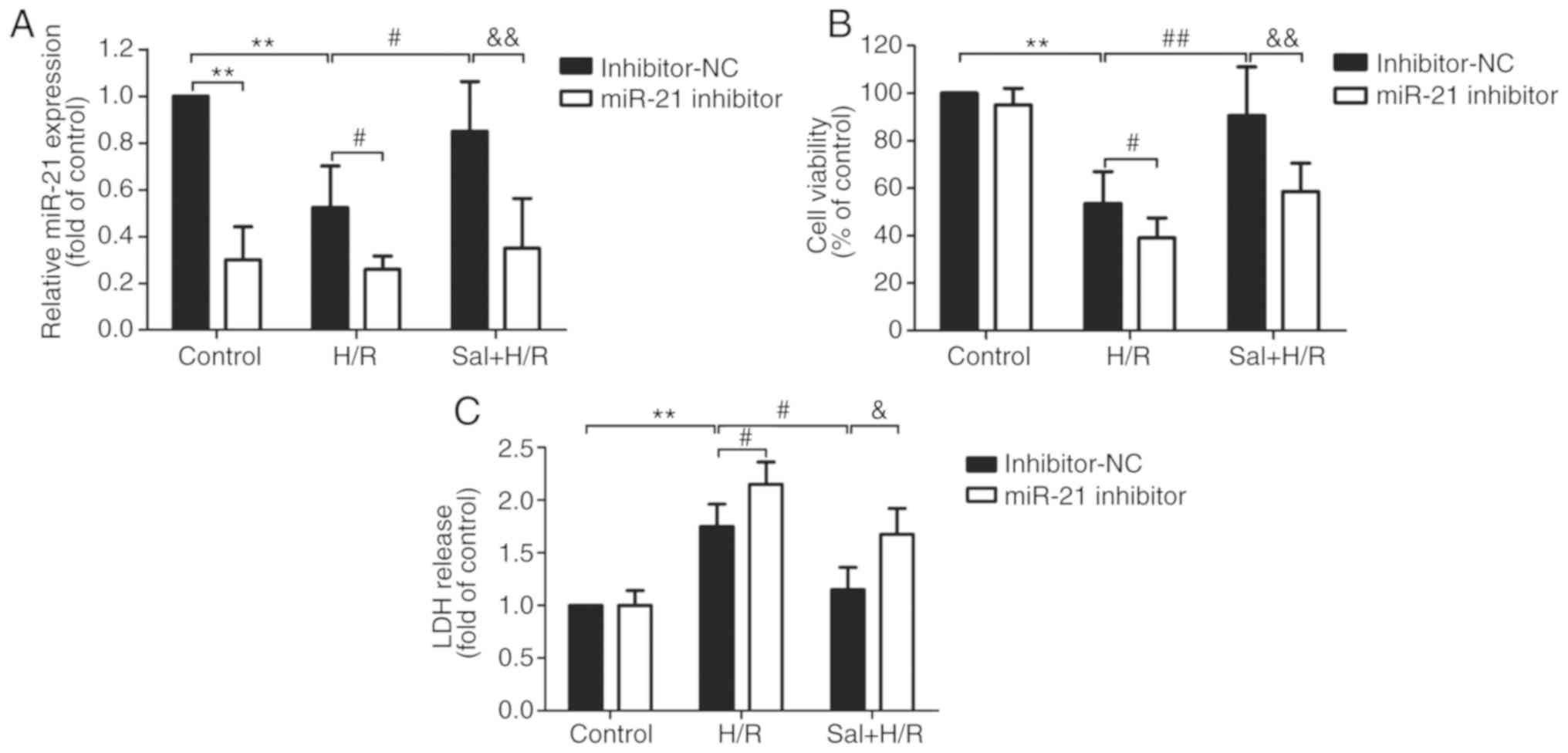
miR-21 inhibitor suppresses the
salidroside-induced protective effects in H9c2 cells
To determine whether miR-21 conferred
cardioprotective effects when salidroside was used to protect
against H/R injury, H9c2 cells were transfected with miR-21
inhibitor or inhibitor-NC and the transfection efficiency was
confirmed by RT-qPCR. The results demonstrated that compared with
the inhibitor-NC transfection group, the expression levels of
miR-21 were significantly decreased in the cells transfected with
miR-21 inhibitor following H/R or salidroside + H/R (P<0.05).
There were no significant differences in the cells transfected with
miR-21 inhibitor + control, miR-21 inhibitor + H/R and miR-21
inhibitor + salidroside + H/R group (Fig. 2A). Therefore, transfection with the
miR-21 inhibitor further aggravated H/R-induced cell viability and
reversed the protective effects of salidroside on cell viability in
H9c2 cells (Fig. 2B). In addition,
measurement of LDH release showed that transfection with a miR-21
inhibitor significantly increased the release of LDH compared with
transfection with inhibitor-NC in H9c2 cells pretreated with H/R
alone or salidroside + H/R group (Fig.
2C). These results suggest that miR-21 mediates the protective
effects of salidroside against H/R injury in H9c2 cells.
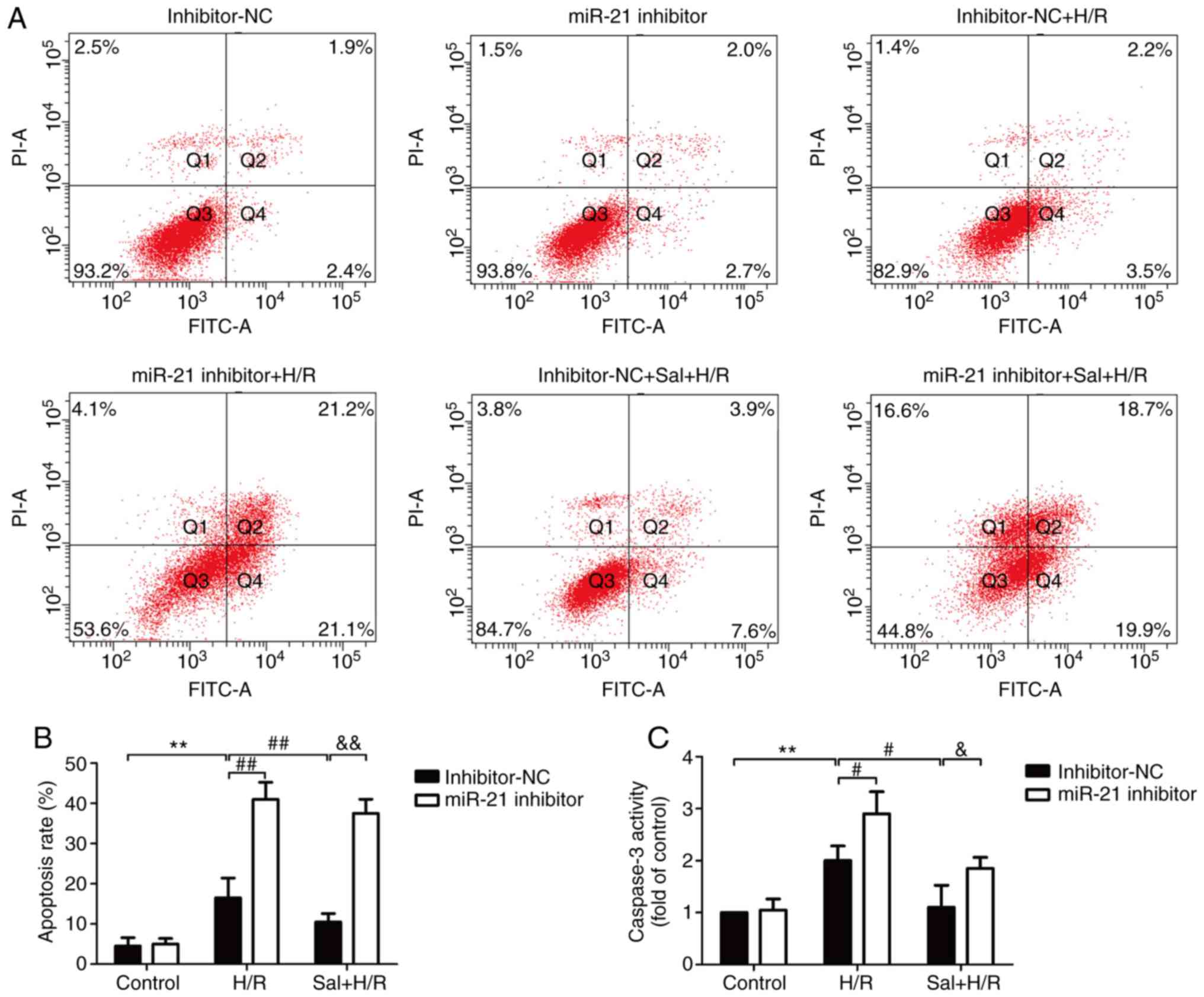
miR-21 inhibitor reduces the
salidroside-induced inhibition of apoptosis
The effects of salidroside on apoptosis in
H/R-treated H9c2 cells and the role of miR-21 in this process were
determined. Flow cytometry results showed that salidroside
pretreatment reversed the H/R-induced increase in apoptosis in the
cells transfected with the inhibitor-NC, whereas this effect was
blocked by transfection with the miR-21 inhibitor (Fig. 3A and B). Notably, the miR-21
inhibitor also further exacerbated H/R-induced apoptosis and no
significant difference in apoptosis rate was observed between the
H/R and the salidroside + H/R groups when transfected with miR-21.
In the H9c2 cells transfected with the inhibitor-NC, compared with
the H/R group, the caspase-3 activity was significantly reduced
compared with the salidroside + H/R treated cells (P<0.05;
Fig. 3C). However, the protective
effect of salidroside was reversed by transfection with the miR-21
inhibitor. These data suggest that salidroside inhibited
H/R-induced apoptosis by increasing miR-21 expression in H9c2
cells.
Salidroside decreases oxidative stress
via miR-21 in H9c2 cells
The effect of salidroside on oxidative stress in
H/R-treated H9c2 cells and the role of miR-21 in this process were
determined. As shown in Fig. 4, ROS
generation (Fig. 4A and B) and MDA
levels (Fig. 4C) were significantly
increased in the H/R-treated cells compared with the control cells
(P<0.01), and salidroside pretreatment significantly decreased
the levels of these molecules (P<0.05). However, miR-21
downregulation, by transfecting cells with the miR-21 inhibitor,
abrogated the protective effects of salidroside on oxidative stress
in H/R-treated H9c2 cells. Notably, in the miR-21 inhibitor group,
salidroside had no effect on the generation of ROS (Fig. 4A and B) and MDA levels (Fig. 4C) under H/R compared with H/R alone
group. The enzymatic activities of SOD (Fig. 4D) and GSH-Px (Fig. 4E) were significantly reduced in the
cells treated with H/R compared with the control group (P<0.01).
The observed decreases were reversed when cells were treated with
salidroside, and the protective effects were abrogated by
transfection with the miR-21 inhibitor in the salidroside + H/R
treated cells. Similarly, salidroside did not significantly alter
the activity of SOD and GSH-Px when the cells were transfected with
the miR-21 inhibitor compared with H/R alone group. Thus, miR-21
contributed to the protection of salidroside against H/R-induced
oxidative stress injury in H9c2 cells.
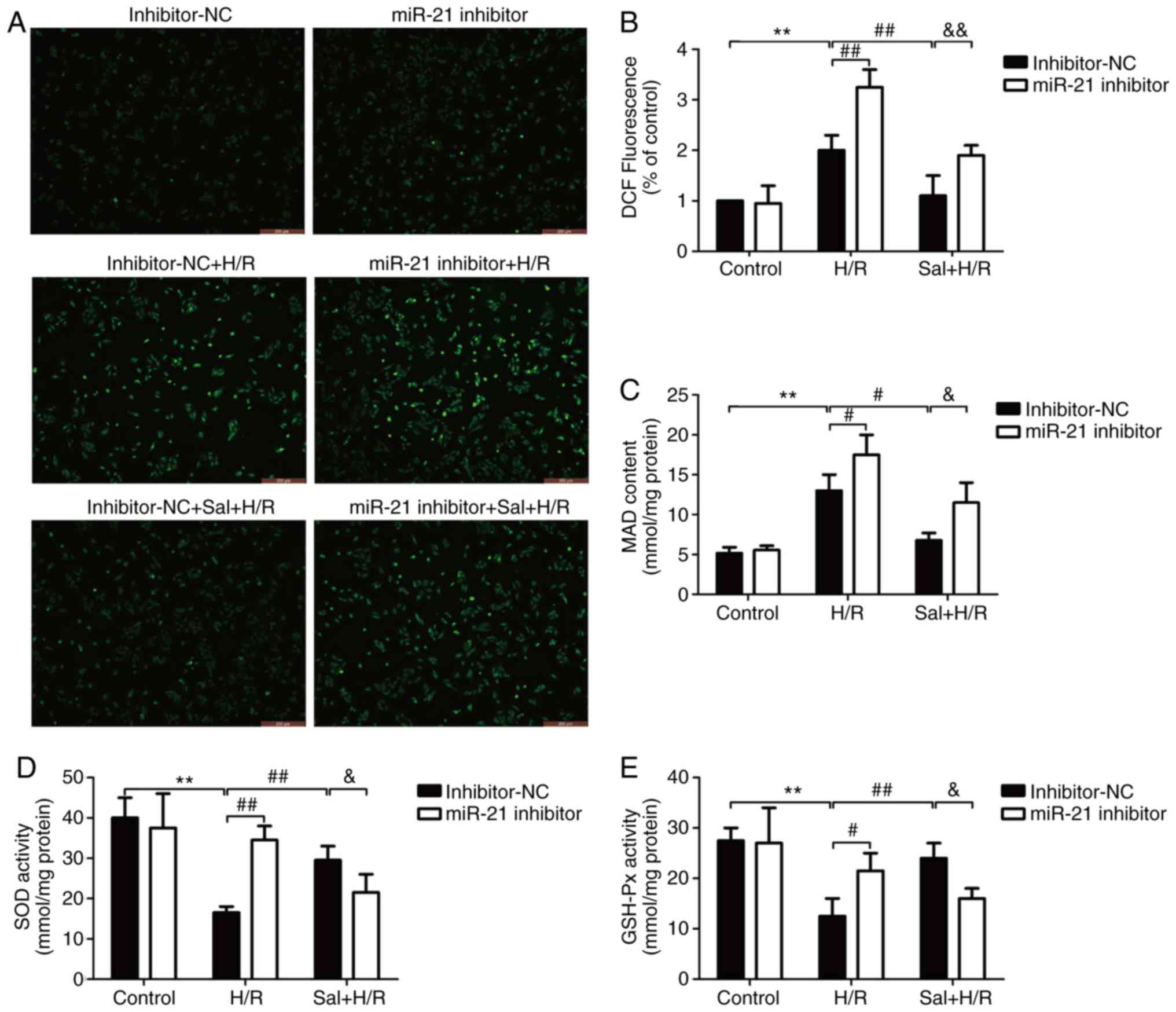 | Figure 4.Effect of the miR-21 inhibitor on
oxidative stress in H9c2 cells subjected to H/R injury in the
presence or absence of Sal. H9c2 cells were transfected with either
a miR-21 inhibitor or inhibitor-NC. Following transfection, cells
were pretreated with Sal, and were subsequently maintained under
hypoxia for 6 h, followed by 12 h of reoxygenation. (A) Production
of ROS was observed using a DCFH-DA fluorescent probe under a
fluorescence microscope. Scale bar, 100 µm. (B) ROS levels were
determined by flow cytometry. (C) MDA levels were analyzed using an
MDA assay kit. (D) SOD activity was measured using an SOD assay
kit. (E) GHS-Px activity was determined using a GSH-Px assay kit.
Data are presented as the mean ± standard deviation of three
experiments. **P<0.01 vs. the control group;
#P<0.05 and ##P<0.01 vs. the H/R group;
&P<0.05 and &&P<0.01 vs.
the Sal + H/R control group. H/R, hypoxia/reoxygenation; miR-21,
microRNA-21; MDA, malondialdehyde; SOD, superoxide dismutase;
GSH-Px, glutathione peroxidase; NC, negative control; Sal,
salidroside. |
miR-21 inhibitor abrogates the
protective effects of salidroside to the inflammatory response
The effect of salidroside on the inflammatory
response and the role of miR-21 in these processes were determined.
The levels of the inflammatory markers including IL-6, IL-1β, and
TNF-α in H9c2 cells or culture supernatant were measured by RT-qPCR
and ELISA. As shown in Fig. 5, ELISA
analysis showed that H/R treatment significantly upregulated the
mRNA expression levels of IL-6 (P<0.01; Fig. 5A), IL-1β (P<0.01; Fig. 5B) and TNF-α (P<0.01; Fig. 5C), and these effects were reversed by
salidroside in cells transfected with the inhibitor-NC.
Transfection with miR-21 inhibitor prevented the
salidroside-induced downregulation of IL-6, IL-1β and TNF-α levels.
In addition, the mRNA expression levels of IL-6 (Fig. 5D), IL-1β (Fig. 5E) and TNF-α (Fig. 5F) were also increased in the H/R
group compared with the control group, whereas the expression of
the inflammatory markers were decreased in the salidroside + H/R
group compared with the H/R group. Transfection with the miR-21
inhibitor significantly attenuated the effects of the
salidroside-induced decrease in IL-6, IL-1β and TNF-α mRNA
expression (P<0.05). Additionally, transfection with the miR-21
inhibitor further exacerbated the H/R-induced increase in the
levels of IL-6, IL-1β and TNF-α, and the mRNA expression levels of
IL-1β and TNF-α mRNA (P<0.05). Taken together, the data above
demonstrated that miR-21 acts as an anti-inflammatory molecule
which regulates the effects of salidroside in the H/R-induced
inflammatory response.
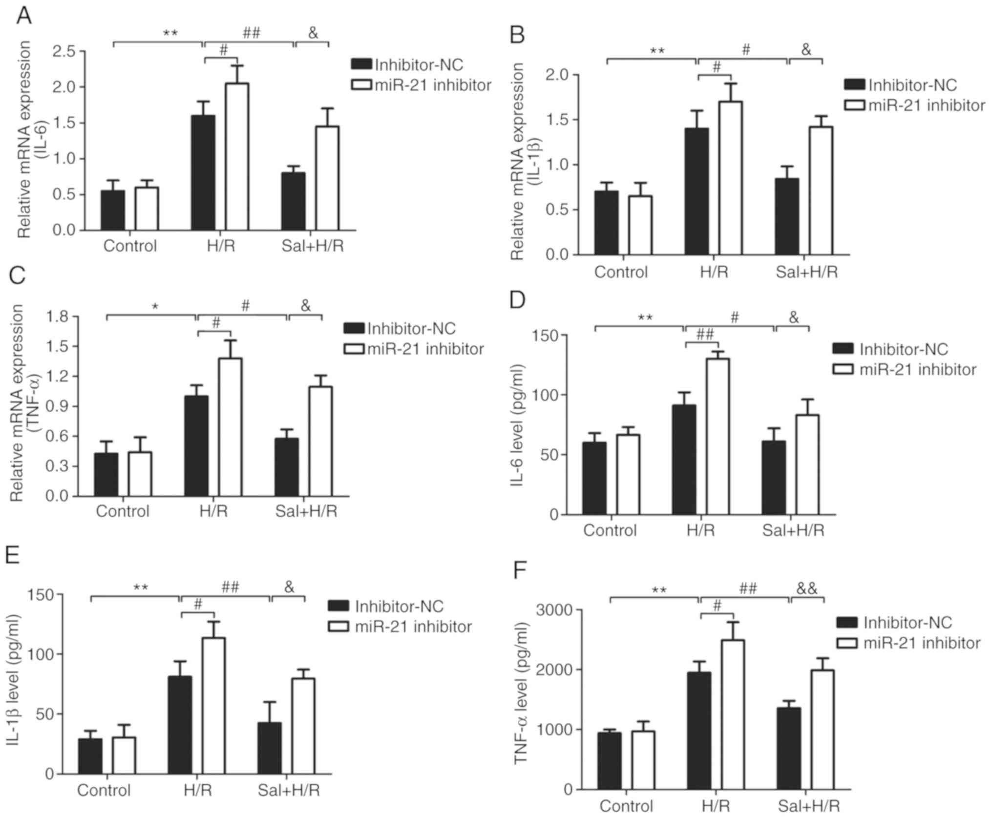 | Figure 5.Effect of the miR-21 inhibitor on
inflammatory factors in H9c2 cells subjected to H/R injury in the
presence or absence of Sal. H9c2 cells were transfected with either
a miR-21 inhibitor or inhibitor-NC. Following transfection, cells
were pretreated with Sal, and were subsequently maintained under
hypoxia for 6 h, followed by 12 h of reoxygenation. mRNA expression
levels of (A) IL-6, (B) IL-1β and (C) TNF-α were determined using
RT-qPCR and normalized to GAPDH. The levels of (D) IL-6, (E) IL-1β
and (F) TNF-α in the culture supernatant were detected by western
blot analysis. Data are presented as the mean ± standard deviation
of three experiments. *P<0.05 and **P<0.01 vs. the control
group; #P<0.05 and ##P<0.01 vs. the H/R
group; &P<0.05 and &&P<0.01
vs. the Sal + H/R control group. H/R, hypoxia/reoxygenation;
miR-21, microRNA-21; IL, interleukin; TNF-α, tumor necrosis
factor-α; NC, negative control; Sal, salidroside. |
Discussion
In the present study, the effects of salidroside on
H/R injury in H9c2 cells and the role of miR-21 in this process was
assessed. The results demonstrated that salidroside attenuated
H/R-induced cytotoxicity and apoptosis, and decreased oxidative
stress and the inflammatory response. Additionally, the results
suggested that miR-21 mediated the protective effects of
salidroside in H/R-induced injury in H9c2 cells. These findings
suggest that salidroside may be an effective means of reducing
myocardial I/R injury and enhancing miR-21 levels may additionally
reduce myocardial I/R injury, highlighting a potentially novel
therapeutic approach.
Salidroside is a major active ingredient obtained
from the medicinal plant Rhodiola rosea and has various
pharmacological properties, including antioxidant (28), anti-inflammatory (29) and cardioprotective effects (26). To date, numerous studies have
demonstrated the protective effects of salidroside on myocardial
injury (30), myocardial hypoxia
(31) and myocardial I/R injury
(13). However, the underlying
molecular mechanisms of salidroside action remained unclear.
Consistent with these studies, the data of the present study also
confirmed that salidroside pretreatment attenuated the H/R-induced
cytotoxicity and apoptosis, inhibiting myocardial I/R injury.
Notably, emerging evidence has shown that myocardial I/R injury
leads to decreased levels of miR-21 and overexpression of miR-21 is
able to effectively inhibit myocardial apoptosis and the
inflammatory response, protecting the myocardium from I/R injury
(21,22,32).
However, the role of miR-21 in the cardioprotective effects of
salidroside has not been reported previously, to the best of our
knowledge. In present study, it was demonstrated, for the first
time, that salidroside reversed the H/R-induced downregulation of
miR-21, and inhibition of miR-21 abrogated the effects of
salidroside treatment in the H/R model of injury in H9c2 cells.
These results suggest that miR-21 mediates the protective effects
of salidroside in myocardial I/R injury.
Myocardial oxidative stress is a major initiator of
the pathological process of cardiac remodeling following I/R
(4). Accumulating evidence has shown
that ROS are the major initiators of myocardial damage in
myocardial I/R injury and the attenuation of oxidative stress in
myocardial cell has been demonstrated to improve myocardial
function following ischemia (4,33). It
has been demonstrated that salidroside may suppress oxidative
stress-induced endothelial dysfunction, cardiomyocyte injury and
necrosis, and cerebral ischemia/reperfusion injury, through
decreasing excessive ROS generation and improving mitochondrial
function (34–37). However, the effect of salidroside in
myocardial I/R injury-induced oxidative stress has not been
studied. In the present study, salidroside pretreatment reduced the
H/R-induced increase in production of ROS and the levels of MDA,
suggesting that salidroside reduced oxidative stress during H/R in
H9c2 cells. Oxidative stress occurs when there is an imbalance
between ROS production and the antioxidant defense systems in
cells, such that the latter becomes overwhelmed (38). SOD and GSH are the primary cellular
defense mechanisms against oxidative stress injury and are the
major intracellular redox buffers in several cell types (39,40). In
the present study, salidroside improved the antioxidant defense
system in H/R-treated H9c2 cells as shown by the increase in the
activities of SOD and GSH-Px. Numerous studies have demonstrated
the ability of miR-21s to preserve an efficient antioxidant
response induced by multiple injuries (41,42). In
the present study, it was demonstrated that inhibition of miR-21
abolished the protective effects of salidroside on oxidative stress
in H/R-treated H9c2 cells. These results suggest that miR-21 may
contribute to the protective effect of salidroside against
H/R-induced oxidative stress in H9c2 cells.
The inflammatory response is another crucial
component in the initiation and exacerbation of myocardial I/R
injury (43,44). I/R increases the local or systemic
release of proinflammatory cytokines, including TNF-α, IL-1β and
IL-6, and further increases cardiac injury, and inhibition of the
I/R-induced inflammatory response alleviates myocardial I/R injury
(45,46). It has been previously hypothesized
that salidroside may exhibit a protective effect on myocardial
injury by inhibiting the inflammatory response (11,14,47).
In vitro and in vivo experiments have demonstrated
that miR-21 regulated the production of various important
pro-inflammatory cytokines secreted during I/R injury (48,49).
However, the role of miR-21 in the protective effects mediated by
salidroside during the inflammatory response induced by myocardial
I/R injury have not been extensively studied. In the present study,
salidroside reversed the H/R-induced increases in expression and
activity of IL-6, IL-1β and TNF-α in H9c2 cells, thus inhibiting
the inflammatory response during H/R, and these effects were
abolished by the miR-21 inhibitor. These results suggest that
miR-21 mediates the salidroside-induced inhibition of the
inflammatory response in H/R-treated H9c2 cells.
In conclusion, the present study demonstrated that
salidroside pretreatment mitigated damage of cardiomyocytes after
H/R-stimulation of H9c2 cells. The cardioprotective effects of
salidroside may be attributed to its ability of suppressing
myocardial oxidative stress and inflammation in vitro,
through enhancing miR-21 expression. Therefore, the results of the
present study suggest that salidroside may be a potential
therapeutic agent for the treatment of myocardial I/R injury and
modulation of miR-21 levels may provide a new strategic option for
limiting damage following a cardiovascular event. However, the
molecular mechanism by which miR-21 protects cultured H9c2 cells
against oxidative stress and inflammation induced by H/R requires
further elucidation.
Acknowledgements
Not applicable.
Funding
The present study was funded by the China Aerospace
Science and Technology Group Corporation Medical and Health
Research Project (grant no. 2017-LCYL-003).
Availability of data and materials
The datasets used and/or analyzed during the present
study are available from the author on reasonable request.
Authors' contribution
BL and HW designed the study and carried out the
experiment. ML, NJ, and JL performed the analysis. MZ participated
in the design of the study. All authors read and approved the final
version of the manuscript.
Ethics approval and consent to
participate
Not applicable.
Patient consent for publication
Not applicable.
Competing interests
The authors declare that they have no competing
interests.
References
|
1
|
Yellon DM and Hausenloy DJ: Myocardial
reperfusion injury. N Engl J Med. 357:1121–1135. 2007. View Article : Google Scholar : PubMed/NCBI
|
|
2
|
Humphries KH, Izadnegahdar M, Sedlak T,
Saw J, Johnston N, Schenck-Gustafsson K, Shah RU, Regitz-Zagrosek
V, Grewal J, Vaccarino V, et al: Sex differences in cardiovascular
disease-impact on care and outcomes. Front Neuroendocrinol.
46:46–70. 2017. View Article : Google Scholar : PubMed/NCBI
|
|
3
|
Gu C, Li T, Jiang S, Yang Z, Lv J, Yi W,
Yang Y and Fang M: AMP-activated protein kinase sparks the fire of
cardioprotection against myocardial ischemia and cardiac ageing.
Ageing Res Rev. 47:168–175. 2018. View Article : Google Scholar : PubMed/NCBI
|
|
4
|
Shvedova M, Anfinogenova Y,
Atochina-Vasserman EN, Schepetkin IA and Atochin DN: c-Jun
N-terminal kinases (JNKs) in myocardial and cerebral
ischemia/reperfusion injury. Front Pharmacol. 9:7152018. View Article : Google Scholar : PubMed/NCBI
|
|
5
|
Yu LM, Di WC, Dong X, Li Z, Zhang Y, Xue
XD, Xu YL, Zhang J, Xiao X, Han JS, et al: Melatonin protects
diabetic heart against ischemia-reperfusion injury, role of
membrane receptor-dependent cGMP-PKG activation. Biochim Biophys
Acta Mol Basis Dis. 1864:563–578. 2018. View Article : Google Scholar : PubMed/NCBI
|
|
6
|
Sun MS, Jin H, Sun X, Huang S, Zhang FL,
Guo ZN and Yang Y: Free radical damage in ischemia-reperfusion
injury: An obstacle in acute ischemic stroke after
revascularization therapy. Oxid Med Cell Longev. 2018:38049792018.
View Article : Google Scholar : PubMed/NCBI
|
|
7
|
González-Montero J, Brito R, Gajardo AI
and Rodrigo R: Myocardial reperfusion injury and oxidative stress:
Therapeutic opportunities. World J Cardiol. 10:74–86. 2018.
View Article : Google Scholar : PubMed/NCBI
|
|
8
|
Yang CF: Clinical manifestations and basic
mechanisms of myocardial ischemia/reperfusion injury. Ci Ji Yi Xue
Za Zhi. 30:209–215. 2018.PubMed/NCBI
|
|
9
|
Kucinskaite A, Briedis V and Savickas A:
Experimental analysis of therapeutic properties of Rhodiola rosea
L. and its possible application in medicine. Medicina (Kaunas).
40:614–619. 2004.PubMed/NCBI
|
|
10
|
Chang X, Luo F, Jiang W, Zhu L, Gao J, He
H, Wei T, Gong S and Yan T: Protective activity of salidroside
against ethanol-induced gastric ulcer via the MAPK/NF-κB pathway
in vivo and in vitro. Int Immunopharmacol.
28:604–615. 2015. View Article : Google Scholar : PubMed/NCBI
|
|
11
|
Zhu L, Wei T, Chang X, He H, Gao J, Wen Z
and Yan T: Effects of salidroside on myocardial injury in vivo
in vitro via regulation of Nox/NF-κB/AP1 Pathway. Inflammation.
38:1589–1598. 2015. View Article : Google Scholar : PubMed/NCBI
|
|
12
|
Chen L, Liu P, Feng X and Ma C:
Salidroside suppressing LPS-induced myocardial injury by inhibiting
ROS-mediated PI3K/Akt/mTOR pathway in vitro and in
vivo. J Cell Mol Med. 21:3178–3189. 2017. View Article : Google Scholar : PubMed/NCBI
|
|
13
|
Sun MY, Ma DS, Zhao S, Wang L, Ma CY and
Bai Y: Salidroside mitigates hypoxia/reoxygenation injury by
alleviating endoplasmic reticulum stress induced apoptosis in H9c2
cardiomyocytes. Mol Med Rep. 18:3760–3768. 2018.PubMed/NCBI
|
|
14
|
Chang X, Zhang K, Zhou R, Luo F, Zhu L,
Gao J, He H, Wei T, Yan T and Ma C: Cardioprotective effects of
salidroside on myocardial ischemia-reperfusion injury in coronary
artery occlusion-induced rats and Langendorff-perfused rat hearts.
Int J Cardiol. 215:532–544. 2016. View Article : Google Scholar : PubMed/NCBI
|
|
15
|
Zhu L, Wei T, Gao J, Chang X, He H, Luo F,
Zhou R, Ma C, Liu Y and Yan T: The cardioprotective effect of
salidroside against myocardial ischemia reperfusion injury in rats
by inhibiting apoptosis and inflammation. Apoptosis. 20:1433–1443.
2015. View Article : Google Scholar : PubMed/NCBI
|
|
16
|
Chen J and Wang DZ: microRNAs in
cardiovascular development. J Mol Cell Cardiol. 52:949–957. 2012.
View Article : Google Scholar : PubMed/NCBI
|
|
17
|
Cheng Y and Zhang C: MicroRNA-21 in
cardiovascular disease. J Cardiovasc Transl Res. 3:251–255. 2010.
View Article : Google Scholar : PubMed/NCBI
|
|
18
|
Oyama Y, Bartman CM, Gile J and Eckle T:
Circadian MicroRNAs in Cardioprotection. Curr Pharm Des.
23:3723–3730. 2017. View Article : Google Scholar : PubMed/NCBI
|
|
19
|
Panagal M, Biruntha M, Vidhyavathi RM,
Sivagurunathan P, Senthilkumar SR and Sekar D: Dissecting the role
of miR-21 in different types of stroke. Gene. 681:69–72. 2019.
View Article : Google Scholar : PubMed/NCBI
|
|
20
|
Pordzik J, Pisarz K, De Rosa S, Jones AD,
Eyileten C, Indolfi C, Malek L and Postula M: The potential role of
platelet-related micrornas in the development of cardiovascular
events in high-risk populations, including diabetic patients: A
review. Front Endocrinol (Lausanne). 9:742018. View Article : Google Scholar : PubMed/NCBI
|
|
21
|
Pan YQ, Li J, Li XW, Li YC, Li J and Lin
JF: Effect of miR-21/TLR4/NF-κB pathway on myocardial apoptosis in
rats with myocardial ischemia-reperfusion. Eur Rev Med Pharmacol
Sci. 22:7928–7937. 2018.PubMed/NCBI
|
|
22
|
Tong Z, Tang Y, Jiang B, Wu Y, Liu Y, Li Y
and Xiao X: Phosphorylation of nucleolin is indispensable to
upregulate miR-21 and inhibit apoptosis in cardiomyocytes. J Cell
Physiol. 234:4044–4053. 2019. View Article : Google Scholar : PubMed/NCBI
|
|
23
|
Xu X, Kriegel AJ, Jiao X, Liu H, Bai X,
Olson J, Liang M and Ding X: miR-21 in ischemia/reperfusion injury:
A double-edged sword? Physiol Genomics. 46:789–797. 2014.
View Article : Google Scholar : PubMed/NCBI
|
|
24
|
Liu K, Ma L, Zhou F, Yang Y, Hu HB, Wang L
and Zhong L: Identification of microRNAs related to myocardial
ischemic reperfusion injury. J Cell Physiol. 234:11380–11390. 2018.
View Article : Google Scholar : PubMed/NCBI
|
|
25
|
Ye Y, Perez-Polo JR, Qian J and Birnbaum
Y: The role of microRNA in modulating myocardial
ischemia-reperfusion injury. Physiol Genomics. 43:534–542. 2011.
View Article : Google Scholar : PubMed/NCBI
|
|
26
|
Cheng J, Wu Q, Lv R, Huang L, Xu B, Wang
X, Chen A and He F: MicroRNA-449a inhibition protects H9C2 cells
against hypoxia/reoxygenation-induced injury by targeting the
notch-1 signaling pathway. Cell Physiol Biochem. 46:2587–2600.
2018. View Article : Google Scholar : PubMed/NCBI
|
|
27
|
Livak KJ and Schmittgen TD: Analysis of
relative gene expression data using real-time quantitative PCR and
the 2(-Delta Delta C(T)) method. Methods. 25:402–408. 2001.
View Article : Google Scholar : PubMed/NCBI
|
|
28
|
Gao J, He H, Jiang W, Chang X, Zhu L, Luo
F, Zhou R, Ma C and Yan T: Salidroside ameliorates cognitive
impairment in a d-galactose-induced rat model of Alzheimer's
disease. Behav Brain Res. 293:27–33. 2015. View Article : Google Scholar : PubMed/NCBI
|
|
29
|
Wang J, Xiao L, Zhu L, Hu M, Wang Q and
Yan T: The effect of synthetic salidroside on cytokines and airway
inflammation of asthma induced by diisocyanate (TDI) in mice by
regulating GATA3/T-bet. Inflammation. 38:697–704. 2015. View Article : Google Scholar : PubMed/NCBI
|
|
30
|
Wang Y, Xu P, Wang Y, Liu H, Zhou Y and
Cao X: The protection of salidroside of the heart against acute
exhaustive injury and molecular mechanism in rat. Oxid Med Cell
Longev. 2013:5078322013. View Article : Google Scholar : PubMed/NCBI
|
|
31
|
Xu ZW, Chen X, Jin XH, Meng XY, Zhou X,
Fan FX, Mao SY, Wang Y, Zhang WC, Shan NN, et al: SILAC-based
proteomic analysis reveals that salidroside antagonizes cobalt
chloride-induced hypoxic effects by restoring the tricarboxylic
acid cycle in cardiomyocytes. J Proteomics. 130:211–220. 2016.
View Article : Google Scholar : PubMed/NCBI
|
|
32
|
Han Q, Zhang HY, Zhong BL, Zhang B and
Chen H: Antiapoptotic effect of recombinant HMGB1 A-box protein via
regulation of microRNA-21 in myocardial ischemia-reperfusion injury
model in rats. DNA Cell Biol. 35:192–202. 2016. View Article : Google Scholar : PubMed/NCBI
|
|
33
|
Francis A and Baynosa R:
Ischaemia-reperfusion injury and hyperbaric oxygen pathways: A
review of cellular mechanisms. Diving Hyperb Med. 47:110–117.
2017.PubMed/NCBI
|
|
34
|
Han J, Xiao Q, Lin YH, Zheng ZZ, He ZD, Hu
J and Chen LD: Neuroprotective effects of salidroside on focal
cerebral ischemia/reperfusion injury involve the nuclear erythroid
2-related factor 2 pathway. Neural Regen Res. 10:1989–96. 2015.
View Article : Google Scholar : PubMed/NCBI
|
|
35
|
Xing SS, Li J, Chen L, Yang YF, He PL, Li
J and Yang J: Salidroside attenuates endothelial cellular
senescence via decreasing the expression of inflammatory cytokines
and increasing the expression of SIRT3. Mech Ageing Dev. 175:1–6.
2018. View Article : Google Scholar : PubMed/NCBI
|
|
36
|
Zhong Z, Han J, Zhang J, Xiao Q, Hu J and
Chen L: Pharmacological activities, mechanisms of action, and
safety of salidroside in the central nervous system. Drug Des Devel
Ther. 12:1479–1489. 2018. View Article : Google Scholar : PubMed/NCBI
|
|
37
|
Zhu Y, Zhang YJ, Liu WW, Shi AW and Gu N:
Salidroside Suppresses HUVECs cell injury induced by oxidative
stress through activating the Nrf2 signaling pathway. Molecules.
21(pii): E10332016. View Article : Google Scholar : PubMed/NCBI
|
|
38
|
Juránek I and Bezek S: Controversy of free
radical hypothesis: Reactive oxygen species-cause or consequence of
tissue injury? Gen Physiol Biophys. 24:263–278. 2005.PubMed/NCBI
|
|
39
|
Sena CM, Leandro A, Azul L, Seiça R and
Perry G: Vascular oxidative stress: Impact and therapeutic
approaches. Front Physiol. 9:16682018. View Article : Google Scholar : PubMed/NCBI
|
|
40
|
Sies H: Oxidative stress: A concept in
redox biology and medicine. Redox Biol. 4:180–183. 2015. View Article : Google Scholar : PubMed/NCBI
|
|
41
|
Peng J, Huang N, Huang S, Li L, Ling Z,
Jin S, Huang A, Lin K and Zou X: Effect of miR-21 down-regulated by
H2O2 on osteogenic differentiation of
MC3T3-E1 cells. Zhongguo Xiu Fu Chong Jian Wai Ke Za Zhi.
32:276–284. 2018.(In Chinese). PubMed/NCBI
|
|
42
|
Shi B, Wang Y, Zhao R, Long X, Deng W and
Wang Z: Bone marrow mesenchymal stem cell-derived exosomal miR-21
protects C-kit+ cardiac stem cells from oxidative injury through
the PTEN/PI3K/Akt axis. PLoS One. 13:e01916162018. View Article : Google Scholar : PubMed/NCBI
|
|
43
|
Ong SB, Hernández-Reséndiz S,
Crespo-Avilan GE, Mukhametshina RT, Kwek XY, Cabrera-Fuentes HA and
Hausenloy DJ: Inflammation following acute myocardial infarction:
Multiple players, dynamic roles, and novel therapeutic
opportunities. Pharmacol Ther. 186:73–87. 2018. View Article : Google Scholar : PubMed/NCBI
|
|
44
|
Wang Z, Hu W, Lu C, Ma Z, Jiang S, Gu C,
Acuña-Castroviejo D and Yang Y: Targeting NLRP3 (nucleotide-binding
domain, leucine-rich-containing family, pyrin domain-containing-3)
inflammasome in cardiovascular disorders. Arterioscler Thromb Vasc
Biol. 38:2765–2779. 2018. View Article : Google Scholar : PubMed/NCBI
|
|
45
|
Kalogeris T, Baines CP, Krenz M and
Korthuis RJ: Ischemia/Reperfusion. Compr Physiol. 7:113–170. 2016.
View Article : Google Scholar : PubMed/NCBI
|
|
46
|
Xu T, Qin G, Jiang W, Zhao Y, Xu Y and Lv
X: 6-gingerol protects heart by suppressing myocardial
ischemia/reperfusion induced inflammation via the
PI3K/Akt-dependent mechanism in rats. Evid Based Complement
Alternat Med. 2018:62096792018. View Article : Google Scholar : PubMed/NCBI
|
|
47
|
He H, Chang X, Gao J, Zhu L, Miao M and
Yan T: Salidroside mitigates sepsis-induced myocarditis in rats by
regulating IGF-1/PI3K/Akt/GSK-3β Signaling. Inflammation.
38:2178–2184. 2015. View Article : Google Scholar : PubMed/NCBI
|
|
48
|
Song N, Zhang T, Xu X, Lu Z, Yu X, Fang Y,
Hu J, Jia P, Teng J and Ding X: miR-21 protects against
ischemia/reperfusion-induced acute kidney injury by preventing
epithelial cell apoptosis and inhibiting dendritic cell maturation.
Front Physiol. 9:7902018. View Article : Google Scholar : PubMed/NCBI
|
|
49
|
Zhang W and Shu L: Upregulation of miR-21
by ghrelin ameliorates ischemia/reperfusion-induced acute kidney
injury by inhibiting inflammation and cell apoptosis. DNA Cell
Biol. 35:417–25. 2016. View Article : Google Scholar : PubMed/NCBI
|