Open Access

Anatomical features of the superficial temporal artery in hemorrhagic moyamoya disease based on CT angiography

  • Authors:
    • Kun Hou
    • Qinglian Li
    • Kan Xu
    • Baofeng Xu
    • Jinlu Yu
  • View Affiliations

  • Published online on: January 27, 2020     https://doi.org/10.3892/etm.2020.8473
  • Pages: 2143-2148
  • Copyright: © Hou et al. This is an open access article distributed under the terms of Creative Commons Attribution License.

Metrics: Total Views: 0 (Spandidos Publications: | PMC Statistics: )
Total PDF Downloads: 0 (Spandidos Publications: | PMC Statistics: )


Abstract

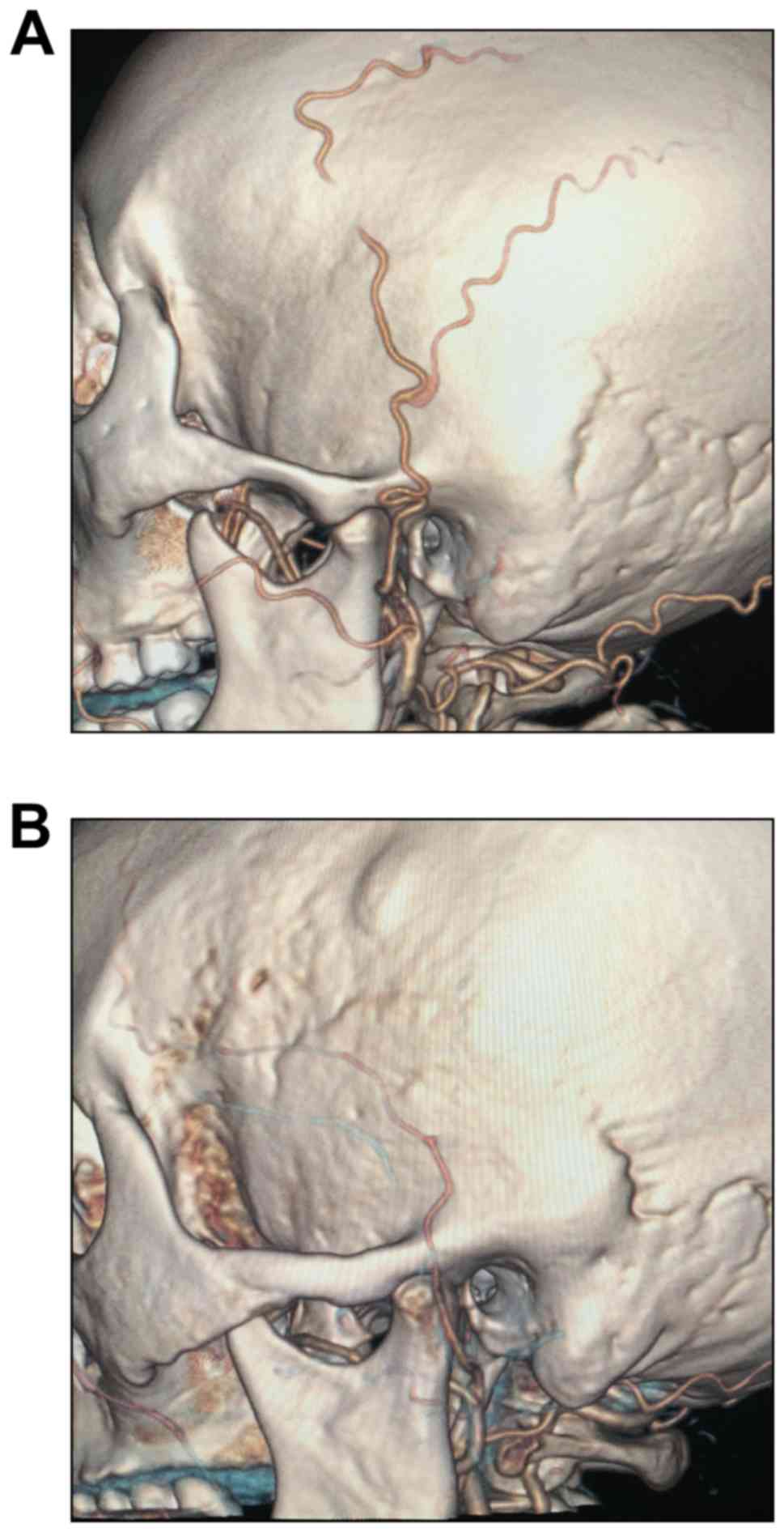
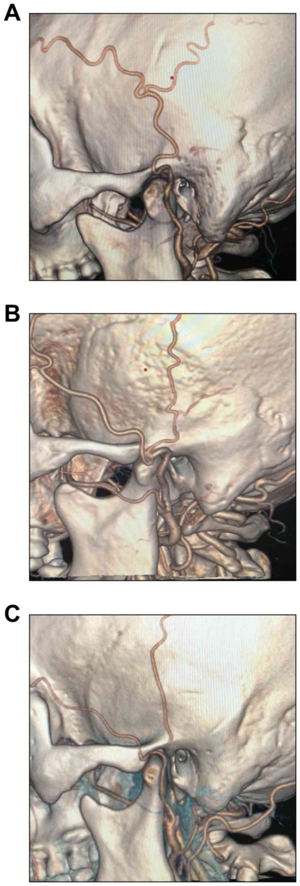
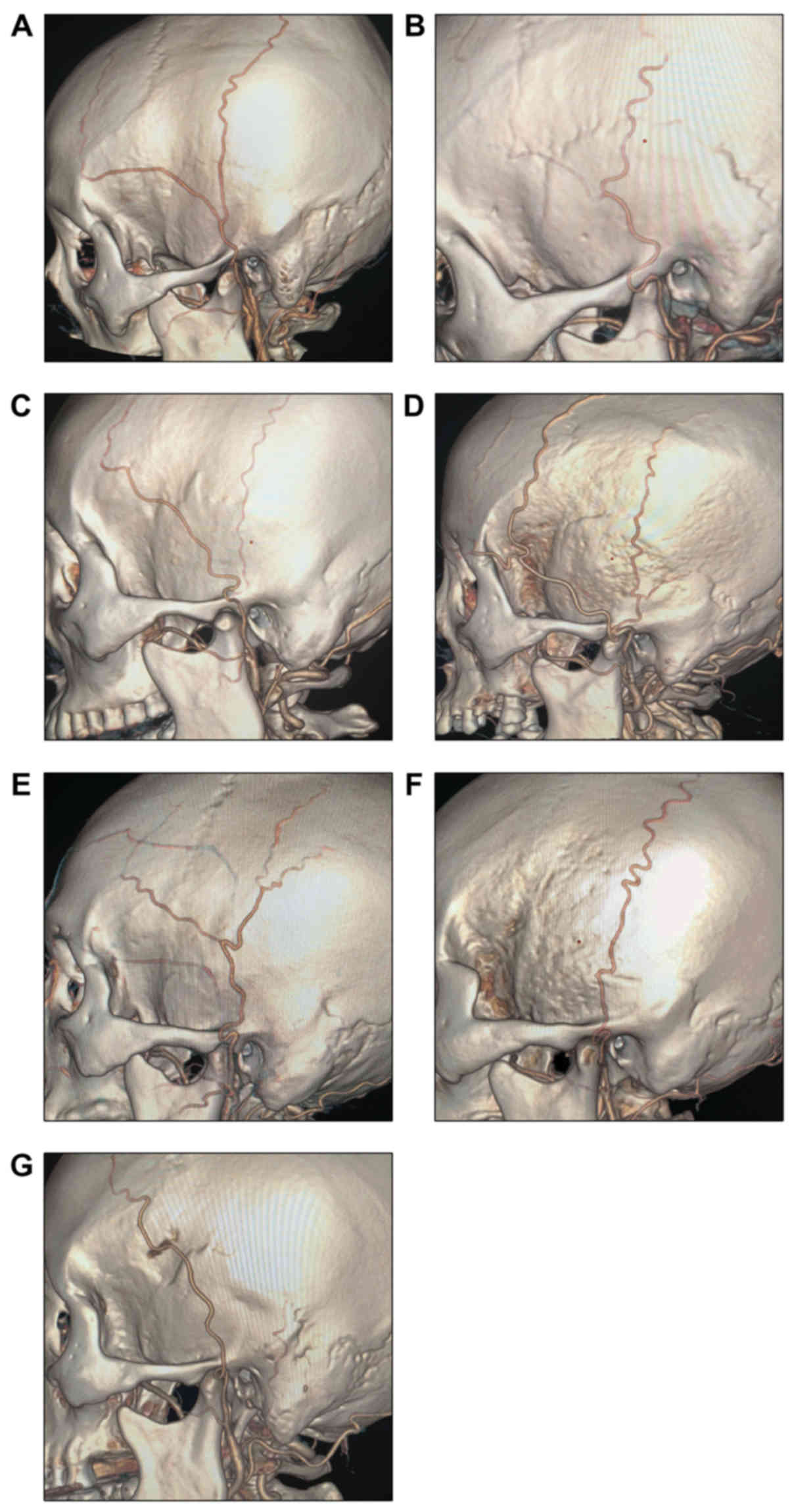
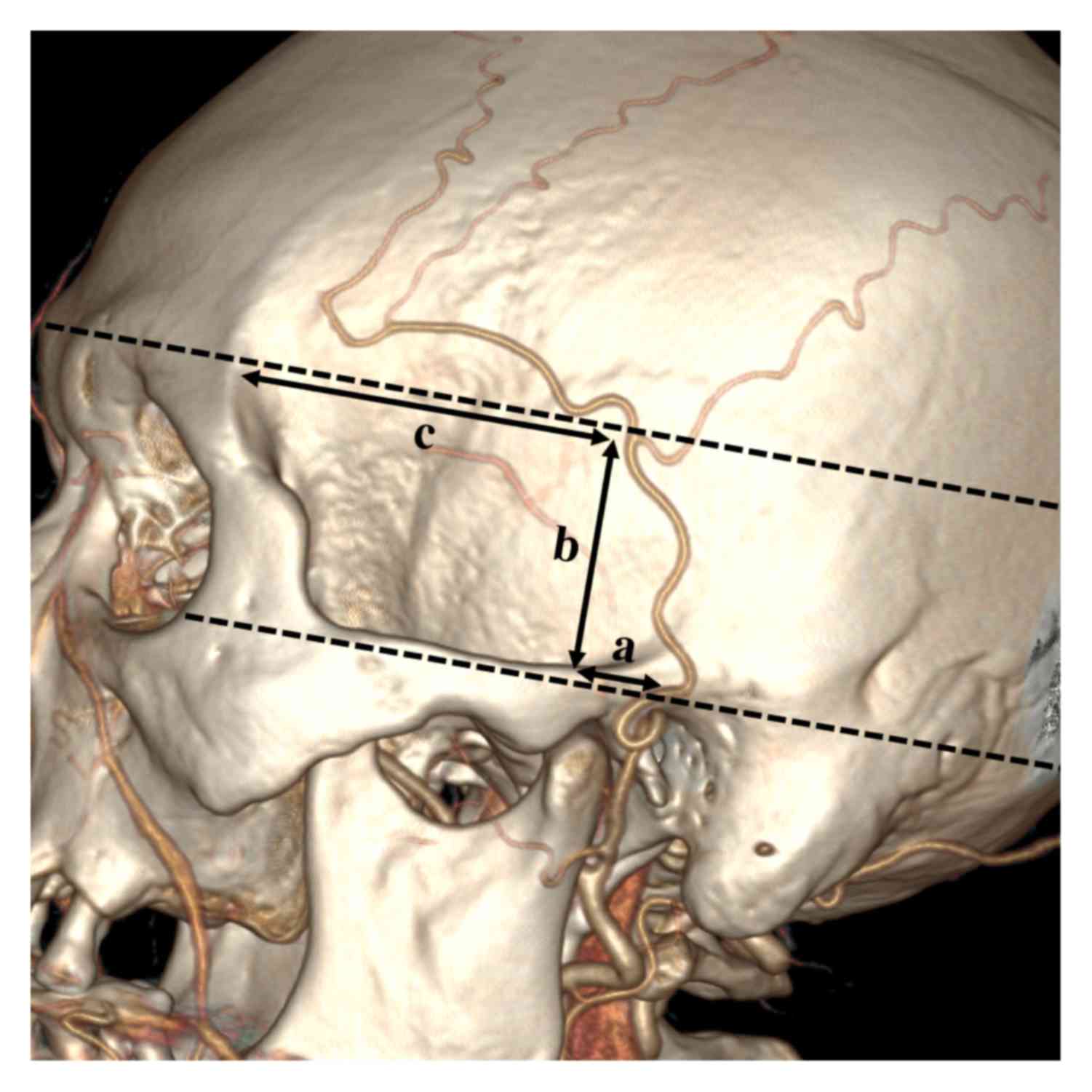
The superficial temporal artery (STA) is an important continuation of the external carotid artery. It is divided into the frontal branch and the parietal branch at or above the zygomatic arch. In the present study, a comparative analysis of the STA in patients with and without moyamoya disease (MMD) was performed using CT angiography. Patients admitted to our institution for spontaneous intracranial hemorrhage were potential candidates. In general, 25 cases (50 sides) in the MMD group and 25 cases (50 sides) in the non‑MMD group were selected for evaluation. The morphology of the STA when crossing the zygomatic arch, the association between the STA bifurcation and the zygomatic arch, the branching characteristics of the STA, the parameters of the STA bifurcation point, the diameter of the STA and the distance from the origin of the STA to the bifurcation point were selected for analysis. There were no significant differences between the two groups with regard to the association between the STA bifurcation and the zygomatic arch, the diameter of the STA or the distance from the origin of the STA to the bifurcation point. However, the bifurcation point of the STA was closer to the posterior edge of the mandibular condyle in the patients with MMD.
View Figures
View References

Related Articles

Journal Cover

March-2020
Volume 19 Issue 3

Print ISSN: 1792-0981
Online ISSN:1792-1015

Sign up for eToc alerts

Recommend to Library

Copy and paste a formatted citation
x
Spandidos Publications style
Hou K, Li Q, Xu K, Xu B and Yu J: Anatomical features of the superficial temporal artery in hemorrhagic moyamoya disease based on CT angiography. Exp Ther Med 19: 2143-2148, 2020
APA
Hou, K., Li, Q., Xu, K., Xu, B., & Yu, J. (2020). Anatomical features of the superficial temporal artery in hemorrhagic moyamoya disease based on CT angiography. Experimental and Therapeutic Medicine, 19, 2143-2148. https://doi.org/10.3892/etm.2020.8473
MLA
Hou, K., Li, Q., Xu, K., Xu, B., Yu, J."Anatomical features of the superficial temporal artery in hemorrhagic moyamoya disease based on CT angiography". Experimental and Therapeutic Medicine 19.3 (2020): 2143-2148.
Chicago
Hou, K., Li, Q., Xu, K., Xu, B., Yu, J."Anatomical features of the superficial temporal artery in hemorrhagic moyamoya disease based on CT angiography". Experimental and Therapeutic Medicine 19, no. 3 (2020): 2143-2148. https://doi.org/10.3892/etm.2020.8473