Introduction
The superficial temporal artery (STA) is an
important continuation of the external carotid artery. It is
divided into the frontal branch and the parietal branch at or above
the zygomatic arch (1). In
neurosurgical practice, the STA is an excellent donor vessel for
extracranial-intracranial bypass (2). In previous studies, the diameter of the
STA, the association between its bifurcation point and the
zygomatic arch, its characteristics when crossing the zygomatic
arch and its classifications were the parameters assessed. Most of
the studies were performed using cadaveric measurements in the
general population and had small sample sizes (3,4).
Certain studies have evaluated the anatomical
characteristics of the STA in patients with moyamoya disease (MMD).
MMD is an idiopathic chronic occlusive cerebrovascular disease
characterized by stenosis of the terminal internal carotid artery
(ICA) and/or the proximal portion of the anterior cerebral artery
and/or the middle cerebral artery, as well as development of basal
moyamoya vessels (5). At present,
extracranial-intracranial bypass is an effective method for the
treatment of MMD and the STA is the major source of donor vessels
(6). However, whether the STA is
also affected in patients with MMD has remained elusive. In the
present study, a comparative analysis of the STA in patients with
and without MMD was performed using CT angiography (CTA).
Materials and methods
Patients
Patients admitted to our institution for spontaneous
intracranial hemorrhage (ICH) between January 2017 and January 2018
were considered as potential candidates. All patients underwent
head CTA. If the CTA indicated steno-occlusive changes in the ICA
terminus and/or the beginning of its two major branches, further
digital subtraction angiography was performed for the definite
diagnosis of MMD. Patients who had an underlying disease that may
cause steno-occlusive changes in the ICA terminus (moyamoya
syndrome) were excluded. According to the CTA and DSA results, ICH
cases were divided into MMD and non-MMD groups. The patients
included were required to have discernible STAs and/or STA branches
on CTA.
Workstation and indexes measured
Raw-image CTA data were reconstructed in the Volume
Rendering program of the GE Workstation (version 4.6; GE
Healthcare) to observe the anatomical characteristics of the
STA.
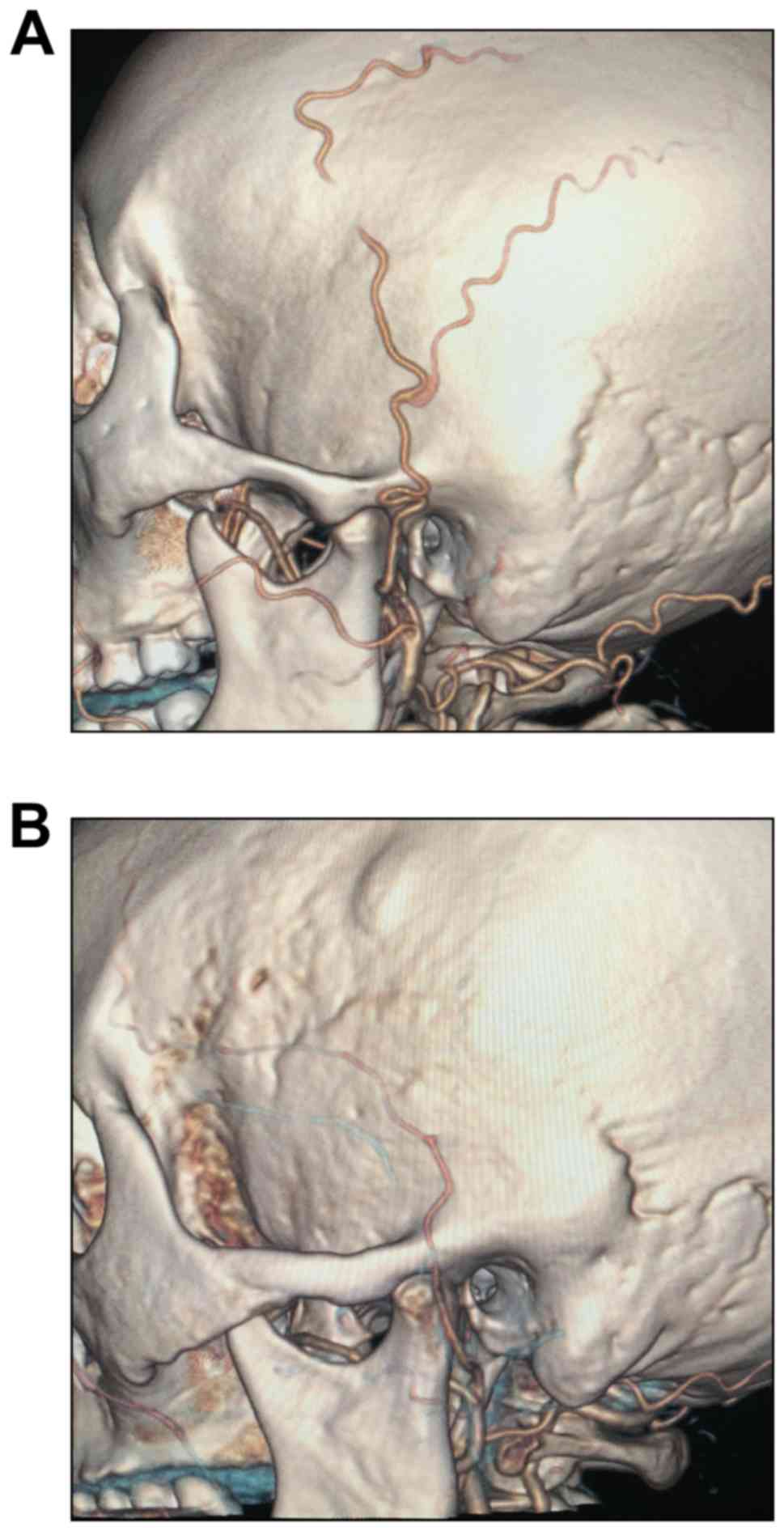
Morphology of the STA when crossing
the zygomatic arch
According to their morphology when crossing the
zygomatic arch, the STAs were divided into the tortuous (T-type;
Fig. 1A) and straight type (S-type;
Fig. 1B).
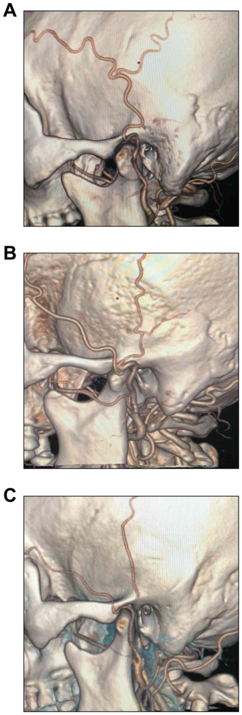
Association between the STA
bifurcation and the zygomatic arch
According to the vertical association between the
STA bifurcation and the zygomatic arch, the STAs were divided into
three types: i) Bifurcation above the upper edge of the zygomatic
arch (Fig. 2A), ii) bifurcation at
the zygomatic arch (Fig. 2B) and
iii) bifurcation below the lower edge of the zygomatic arch
(Fig. 2C).
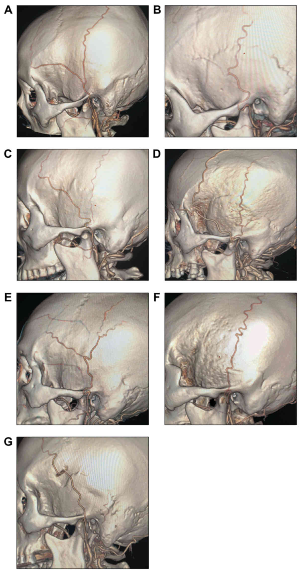
Branching characteristics of the
STA
The STAs were subdivided into the following types
based on their branching characteristics: Type A: STA with equal
bifurcation (Fig. 3A); type B:
Atrophic frontal branch (Fig. 3B);
type C: Atrophic parietal branch (Fig.
3C); type D: Additional frontal branch (Fig. 3D); type E: Additional parietal branch
(Fig. 3E); type F: Parietal branch
only (Fig. 3F); type G: Frontal
branch only (Fig. 3G).
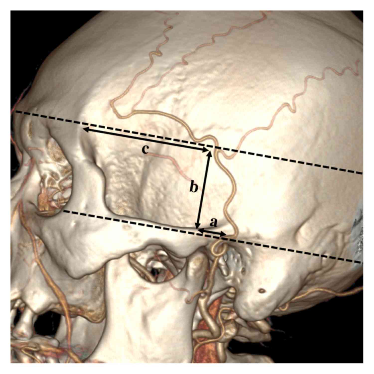
Parameters of the STA bifurcation
point
For patients with an STA bifurcation point above the
zygomatic arch, 3 parameters were used, as illustrated in Fig. 4. Line a was defined as the distance
from the bifurcation point to the posterior edge of the mandibular
condyle. Line a is parallel to the long axis of the zygomatic arch.
Line b was defined as the vertical distance from the STA
bifurcation point to the upper edge of the zygomatic arch. Line c
was defined as the distance from the STA bifurcation point to the
zygomatic process of the frontal bone. Line c is also parallel to
the long axis of the zygomatic arch (Fig. 4).
Diameter of the STA
Cases with bifurcation points below the zygomatic
arch and those whose STA did not bifurcate were excluded from this
measurement. The diameter of the STA was measured at the beginning,
the midpoint of the zygomatic arch and the bifurcation point.
Distance from the origin of the STA to
the bifurcation point
Patients whose STA did not bifurcate were excluded
from this measurement. The Two-Click AVA tool was used to select
two points at the beginning and the bifurcation of the STA, and the
workstation then automatically measured the length.
Statistical analysis
SPSS 18.0 (IBM Corp.) was used for statistical
analysis. The normal distribution for continuous variables was
assessed using a Shapiro-wilk test. Continuous variables are
expressed as the mean ± standard deviation and differences were
assessed with Student's t-test. Categorical variables were
described as the number of cases (percentage) and were assessed
with the χ2 test or Fisher's exact test. P<0.05 was
considered to indicate statistical significance.
Results
General information
A total of 25 consecutive cases (50 sides) were
finally selected as the MMD group and 25 control cases as the
non-MMD group. In the MMD group, the average age of the patients
was 54.6±9.3 years (range, 27–71 years) and the ratio of males to
females was 1.1:1. In the non-MMD group, the average age was
55.8±11.3 years (range, 30–77 years) and the ratio of males to
females was 2.1:1. There was no statistical difference (P=0.248) in
gender ratio between the MMD and non-MMD groups.
Morphology of the STA when crossing
the zygomatic arch
In the MMD group, 44 sides (88%) were type T and 6
sides (12%) were type S. In the non-MMD group, 45 sides (90%) were
type T and 5 sides (10%) were type S. No significant difference was
present between the two groups in terms of the distribution of T-
and S-types (P=0.749; Table I).
 | Table I.Distribution of patients with/without
MMD regarding the morphology of the superficial temporal artery
when crossing the zygomatic arch. |
Table I.
Distribution of patients with/without
MMD regarding the morphology of the superficial temporal artery
when crossing the zygomatic arch.
| Zygomatic arch
crossing typea | MMD (n=50) | Non-MMD (n=50) | P-value |
|---|
| S | 6
(12) | 5
(10) | 0.749 |
| T | 44 (88) | 45 (90) |
|
Association between the STA
bifurcation point and the zygomatic arch
In the MMD group, 8 sides without STA bifurcation
were identified and excluded. In the remaining sides, the
bifurcation points were located above the upper edge of the
zygomatic arch on 37 sides (88.1%) and directly on the zygomatic
arch on 5 sides (11.9%). None of the patients had an STA that
bifurcated below the lower edge of the zygomatic arch. In the
non-MMD group, 8 sides with no bifurcation were excluded. In the
remaining sides, the bifurcation points were located above the
upper edge of the zygomatic arch on 37 sides (88.1%), directly on
the zygomatic arch on 4 sides (9.5%) and below the lower edge of
the zygomatic arch on 1 side (2.4%). No significant difference was
present between the two groups with regard to the distribution of
different associations between the STA bifurcation and the
zygomatic arch (P=1.000; Table
II).
| Table II.Distribution of patients with/without
MMD regarding the location of the STA bifurcation point relative to
the zygomatic arch. |
Table II.
Distribution of patients with/without
MMD regarding the location of the STA bifurcation point relative to
the zygomatic arch.
| Location of STA
bifurcation pointa | Total (n=84) | MMD (n=42) | Non-MMD (n=42) | P-value |
|---|
| Above the upper edge
of the zygomatic arch | 74 (88.1) | 37 (88.1) | 37 (88.1) | 1.000 |
| At the zygomatic
arch | 9
(10.7) | 5
(11.9) | 4 (9.5) |
|
| Below the lower edge
of the zygomatic arch | 1 (1.2) | 0 (0.0) | 1 (2.4) |
|
Branching characteristics of the
STA
In the MMD group, the branching characteristic was
type A, B, C, D, E, F and G on 25 sides (50%), 8 sides (16%), 6
sides (12%), 3 sides (6%), 0 side (0%), 4 sides (8%) and 4 sides
(8%), respectively. In the non-MMD group, the branching
characteristic was type A, B, C, D, E, F and G in 19 sides (38%), 3
sides (6%), 5 sides (10%), 8 sides (16%), 7 sides (14%), 4 sides
(8%) and 4 sides (8%), respectively. Overall, a statistical
difference was indicated in the component ratios of each branching
type between the two groups (P=0.042). Regarding the individual
types, only the incidence of type E was significantly different
between the two groups (P=0.012; Table
III).
| Table III.Branching characteristics of the
superficial temporal artery. |
Table III.
Branching characteristics of the
superficial temporal artery.
| Branching
typea | MMD (n=50) | Non-MMD (n=50) | P-value |
|---|
| Total |
|
| 0.042 |
| A | 25 (50) | 19 (38) | 0.314 |
| B | 8
(16) | 3 (6) | 0.200 |
| C | 6
(12) | 5 (10) | 1.000 |
| D | 3 (6) | 8 (16) | 0.200 |
| E | 0
(0) | 7 (14) | 0.012 |
| F | 4
(8) | 4 (8) | 1.000 |
| G | 4
(8) | 4 (8) | 1.000 |
Comparison of measurements of the STA
bifurcation point
The STA bifurcation point was above the zygomatic
arch on 37 sides in the MMD group and 38 sides in the non-MMD
group. The mean values of lines a, b and c were 13.35±4.07,
27.16±9.83 and 54.71±3.71 mm in the MMD group, respectively. The
mean values of lines a, b and c were 15.43±4.63, 29.60±8.96 and
55.18±6.73 mm in the non-MMD group, respectively. The values of
lines a, b and c were normally distributed and Student's t-test was
used for the intergroup comparison. In the MMD group, line a was
significantly larger than that in the non-MMD group (P=0.045). No
significant difference was noted between the two groups with regard
to lines b and c (Table IV).
| Table IV.Parameters of the superficial temporal
artery bifurcation point (mm). |
Table IV.
Parameters of the superficial temporal
artery bifurcation point (mm).
| Distancea | MMD (n=37) | Non-MMD (n=38) | P-value |
|---|
| a | 13.35±4.07 | 15.43±4.63 | 0.045 |
| b | 27.16±9.83 | 29.60±8.96 | 0.268 |
| c | 54.71±3.71 | 55.18±6.73 | 0.713 |
Diameter of the STA
In the MMD group, the diameters at the beginning (50
sides), the midpoint of the zygomatic arch (50 sides) and the
bifurcation (42 sides) were 2.45±0.46, 1.68±0.44 and 1.95±0.39 mm,
respectively. In the non-MMD group, the diameters at the beginning
(50 sides), the zygomatic arch midpoint (50 sides), and the
bifurcation (42 sides) were 2.41±0.58, 1.71±0.51 and 1.87±0.57 mm,
respectively. The values of the diameters at each point were all
normally distributed. Student's t-test indicated no significant
differences with regard to the diameters of the STA at the
beginning, the midpoint of the zygomatic arch and the bifurcation
between the two groups (Table
V).
| Table V.Diameter of thesuperficial temporal
artery (mm). |
Table V.
Diameter of thesuperficial temporal
artery (mm).
|
| MMD (n=50) | Non-MMD (n=50) |
|
|---|
|
|
|
|
|
|---|
| Location | Cases (n) | Mean ± SD | Cases (n) | Mean ± SD | P-value |
|---|
| Beginning | 50 | 2.45±0.46 | 50 | 2.41±0.58 | 0.690 |
| Midpoint of the
zygomatic arch | 50 | 1.68±0.44 | 49 | 1.71±0.51 | 0.721 |
| Bifurcation | 42 | 1.95±0.39 | 42 | 1.87±0.57 | 0.412 |
Distance from the origin of the STA to
the bifurcation point
The mean distance from the origin of the STA to the
bifurcation point in the MMD group (42 sides) was 73.89±15.47 mm,
and in the non-MMD group (42 sides), it was 78.71±21.68 mm. No
significant difference was present between the two groups (P=0.244;
Table VI).
| Table VI.Distance from the origin of the
superficial temporal artery to the bifurcation point (mm). |
Table VI.
Distance from the origin of the
superficial temporal artery to the bifurcation point (mm).
|
| MMD (n=50) | Non-MMD (n=50) |
|
|---|
|
|
|
|
|
|---|
| Item | Cases (n) | Mean ± SD | Cases (n) | Mean ± SD | P-value |
|---|
| Distance | 42 | 73.89±15.47 | 42 | 78.71±21.68 | 0.244 |
Discussion
The STA has an important role in MMD, as it is the
major donor vessel for the extracranial-intracranial bypass.
However, whether the STA is affected in patients with MMD remained
elusive. As CTA is able to clearly display the external carotid
artery system (7), it was used to
study the anatomical characteristics of the STA in the present
study. After its origination from the external carotid artery, the
STA has two characteristic vessel types (tortuous or straight) when
crossing the zygomatic arch (4). In
the present study, the tortuous type was indicated to be more
common than the straight type in the MMD and the non-MMD groups,
and there was no significant difference between the two groups.
This anatomical trait has a physiological advantage, as higher
tortuosity of the STA allows for more effective extension during
mandibular joint movement and avoids vascular traction when opening
the mouth (6).
In the present study, the diameter and trunk length
of the STA were not different between the MMD and the non-MMD
groups, indicating that MMD did not influence the diameter and
trunk length of the STA; this result is in contrast to what occurs
in the compensatory dilated tortuous middle meningeal artery in
patients with MMD (8). The present
results also indicated that, compared with the diameters above and
below the zygomatic arch, the diameter of the STA at the midpoint
of the zygomatic arch was the smallest. This difference may be a
compensatory result of the movements required for chewing (9).
The diameter of the STA at the zygomatic arch
midpoint was ~1.7 mm, which is generally smaller than that reported
by Chen et al (10) (2.1 mm)
in a Chinese cadaveric study. This difference may be due to the
following factors: First, prior to a cadaveric study, liquid medium
is always infused into the vascular system, which expands the
diameters of vessels. Furthermore, the measured values in cadaveric
studies reflect the external vascular diameter. However, the data
obtained from CTA reflect the internal vascular diameter. Finally,
vascular diameters measured in vivo may also be affected by
systolic and diastolic blood pressure. In a Korean study based on
CTA, the diameter of the STA was ~1.8 mm, which was similar to the
present results (9).
The STA gives rise to the frontal branch and the
parietal branch after crossing the zygomatic arch. The two branches
have an important role in supplying their respective regions of the
scalp. The bifurcation point is undoubtedly the core of the STA
(11). In line with the method
described by Kim et al (9),
the posterior edge of the mandibular condyle, the upper edge of the
zygomatic arch and the zygomatic process of the frontal bone as
facial markers were used in the present study, making the
superficial location of the STA bifurcation more accurate. The
present study also indicated that the bifurcation point of the STA
was closer to the posterior edge of the mandibular condyle in
patients with MMD. Hence, to reduce the possibility of direct
damage to the trunk of the STA during an extended pterional
craniotomy in patients with MMD, it is necessary to make the
incision closer to the anterior edge of the auricle.
Acknowledgements
Not applicable.
Funding
No funding was received.
Availability of data and materials
The datasets used and/or analyzed during the present
study are available from the corresponding author on reasonable
request.
Authors' contributions
JY and KH made substantial contributions to the
conception and design of the work. KH and QL wrote the manuscript.
QL, KX and BX collected and analyzed the data. JY and KH critically
revised the manuscript. All authors read and approved the final
manuscript.
Ethics approval and consent to
participate
The study was approved by the Ethics Committee of
the First Hospital of Jilin University (Changchun, China). Informed
consent for participation in the study or use of their medical data
was obtained from all participants or their legal guardians.
Patient consent for publication
Written informed consent was obtained from the
patients or their guardians for publication of this manuscript and
any accompanying images.
Competing interests
The authors declare that they have no competing
interests.
Glossary
Abbreviations
Abbreviations:
|
CTA
|
CT angiography
|
|
ICH
|
intracranial hemorrhage
|
|
MMD
|
moyamoya disease
|
|
STA
|
superficial temporal artery
|
References
|
1
|
Shin KJ, Shin HJ, Lee SH, Koh KS and Song
WC: Surgical anatomy of the superficial temporal artery to prevent
facial nerve injury during arterial biopsy. Clin Anat. 31:608–613.
2018. View
Article : Google Scholar : PubMed/NCBI
|
|
2
|
Wada K, Otani N, Toyooka T, Takeuchi S,
Tomiyama A and Mori K: Superficial temporal artery to anterior
cerebral artery hemi-bonnet bypass using radial artery graft for
prevention of complications after surgical treatment of partially
thrombosed large/giant anterior cerebral artery aneurysm. J Stroke
Cerebrovasc Dis. 27:3505–3510. 2018. View Article : Google Scholar : PubMed/NCBI
|
|
3
|
Marano SR, Fischer DW, Gaines C and
Sonntag VK: Anatomical study of the superficial temporal artery.
Neurosurgery. 16:786–790. 1985. View Article : Google Scholar : PubMed/NCBI
|
|
4
|
Yonenaga K, Tohnai I, Mitsudo K, Mori Y,
Saijo H, Iwai T, Yonehara Y, Ota Y, Torigoe K and Takato T:
Anatomical study of the external carotid artery and its branches
for administration of superselective intra-arterial chemotherapy
via the superficial temporal artery. Int J Clin Oncol. 16:654–659.
2011. View Article : Google Scholar : PubMed/NCBI
|
|
5
|
Li Q, Qu L, Yuan Y, Xu B, Guo Y, Xu K and
Yu J: Analysis of the clinical characteristics of hemorrhagic
moyamoya disease in the Jilin province of northeastern China: A
single-center study of 212 cases. Biomed Rep. 8:191–197.
2018.PubMed/NCBI
|
|
6
|
Yu J, Shi L, Guo Y, Xu B and Xu K:
Progress on complications of direct bypass for moyamoya disease.
Int J Med Sci. 13:578–587. 2016. View Article : Google Scholar : PubMed/NCBI
|
|
7
|
Korn A, Bender B, Brodoefel H, Hauser TK,
Danz S, Ernemann U and Thomas C: Grading of carotid artery stenosis
in the presence of extensive calcifications: Dual-energy CT
angiography in comparison with contrast-enhanced MR angiography.
Clin Neuroradiol. 25:33–40. 2015. View Article : Google Scholar : PubMed/NCBI
|
|
8
|
Yu J, Guo Y, Xu B and Xu K: Clinical
importance of the middle meningeal artery: A review of the
literature. Int J Med Sci. 13:790–799. 2016. View Article : Google Scholar : PubMed/NCBI
|
|
9
|
Kim BS, Jung YJ, Chang CH and Choi BY: The
anatomy of the superficial temporal artery in adult koreans using
3-dimensional computed tomographic angiogram: Clinical research. J
Cerebrovasc Endovasc Neurosurg. 15:145–151. 2013. View Article : Google Scholar : PubMed/NCBI
|
|
10
|
Chen TH, Chen CH, Shyu JF, Wu CW, Lui WY
and Liu JC: Distribution of the superficial temporal artery in the
Chinese adult. Plast Reconstr Surg. 104:1276–1279. 1999. View Article : Google Scholar : PubMed/NCBI
|
|
11
|
Aveta A, Brunetti B, Tenna S, Segreto F
and Persichetti P: Superficial temporal artery perforator flap:
Anatomic study of number and reliability of distal branches of the
superficial temporal artery and clinical applications in three
cases. Microsurgery. 37:924–929. 2017. View Article : Google Scholar : PubMed/NCBI
|