Introduction
The number of children with diabetes mellitus (DM)
has increased in recent years. DM has become one of the most
serious diseases affecting the physical and mental development of
children (1). The most common type
of diabetes in children is type 1 diabetes (T1D). T1D is an
autoimmune disease caused by an immune-mediated destruction of the
insulin-producing pancreatic β cells. Autoantibodies directed
against islet autoantigens, such as insulin, glutamic acid
decarboxylase 65 (GAD65), islet cell antibody (ICA), islet
antigen-2 (IA-2) and zinc transporter 8 (ZnT8), are markers of
islet autoimmunity, which precedes the clinical onset of T1D
(2). Most patients with T1D have
multiple detectable islet cell autoantibodies in their blood at
diagnosis, with <10% only having one when assessed using a
combination of islet cell autoantibodies and antibodies against
GAD65, IA-2, ICA and insulin. The addition of ZnT8 antibodies may
increase this autoimmunity detection rate to 98% (3).
Insulin deficiency is usually attributed to the
autoimmune destruction of islet β cells in childhood diabetes, but
monogenic diabetes is also a common cause of insulin deficiency
(4). The prevalence of obesity has
increased rapidly and type 2 DM (T2DM) incidence in children has
also been increasing. In previous years, ~12% of young adults
diagnosed with diabetes in the United States have been classified
as T2DM (5,6).
The incidence of monogenic diabetes in children is
low, but the atypical clinical manifestations, limitations of the
detection methods and lack of understanding of such diseases often
renders their diagnosis unclear or wrong (7). In the present study, next generation
sequencing (NGS)-based mutation screening of known causative genes
for glucose metabolism-related genes in antibody-negative childhood
diabetes was conducted and the findings may prove to be helpful for
the improvement of precision treatment.
Materials and methods
Cohort
The medical records of Chinese patients from the
inpatient or outpatient clinics of the Children's Hospital of
Soochow University (Soochow, China) between January 2010 and
December 2017 were analyzed. A total of 64 patients with new-onset
diabetes (>6 m, <16 y) were identified and their initial
clinical characteristics were analyzed. Of which 32 cases with
autoantibody-negative diabetes (male, 16 cases; female, 16 cases)
were screened for ICA, GAD antibody (GADA), IA-2 and ZnT8 at
diagnosis. Having at least one positive antibody and a fasting C
peptide of ≥0.3 ng/ml at initial diagnosis was considered positive
autoimmunity (8-10).
Peripheral blood DNA was extracted from the 32 patients with
autoantibody-negative diabetes and their parents for
high-throughput sequencing of glucose metabolism-related genes.
Pathogenicity analysis of the candidate mutated site has careful
consideration of the patient's clinical presentations and
sequencing result base on Standards and Guidelines for the
Interpretation of Sequence Variants revised by the American College
of Medical Genetics (ACMG). Patients with autoantibody-negative
diabetes were classified into the pathogenic mutation group,
variant of unknown significance (VUS) group and no-mutation group
based on the results, and the three groups of data were
compared.
The group with autoantibody-negative diabetes was
the experimental group. The control group comprised 32 T1D cases
(male, 16 cases; female, 16 cases). The only difference between the
experimental group and the control group is that the experimental
group patients are autoantibody-negative, and the control group
patients tested positive for at least two of the islet
auto-antibodies. T1D was diagnosed according to the criteria of the
American Diabetes Association (11).
All children (experimental and control) present with a very early
form of diabetes (>6 m, <16 y). No significant differences
were observed in age and sex. The control group comprised 32
patients with typical childhood T1D, who had been enrolled during
the same period. After half a year follow-up, the control group
still relied on insulin and the c-peptide was so low that it could
not be detected. The control group patients matched the
experimental group in age and body mass index (BMI). They were
positive for at least two islet auto-antibodies (ICA, GADA, IA-2 or
ZnT8).
The trial exclusion criteria were as follows: i) In
addition to diabetes, other diseases with a definite diagnosis; ii)
chronic inflammatory disease, such as chronic diarrhea and iii)
obesity (95th centile of BMI) and acanthosis nigricans skin.
NGS and validation
A total of 159 glucose metabolism-related genes were
screened, including the known monogenic diabetes and T2DM
susceptibility-related genes, covering the entire coding region and
exon-intron boundaries (±5 bp). The focus was put on screening 13
maturity-onset diabetes of the young (MODY) genes (GCK,
HNF1A, HNF4A, HNF1B, NEUROD1,
INS, CEL, PDX1, PAX4, BLK,
KLF11, KCNJ11 and ABCC8) and 20 neonatal DM
(NDM) genes (GCK, KCNJ11, ABCC8, INS,
PDX1, PTF1A, HNF1B, NEUROD1,
NEUROG3, RFX6, EIF2AK3, FOXP3,
GLIS3, SLC19A2, SLC2A2, IER3IP1,
ZFP57, WFS1, GATA6 and GATA4). DNA
samples from the 32 cases were fragmented using a Scientz08-III
automated sonicator. DNA samples were extracted using a DNA
extraction kit (CWE2100 Blood DNA kit V2; Beijing Kangwei Century
Biotechnology Co., Ltd.) on an 96-channel automatic nucleic acid
extraction machine (Beijing Kangwei Century Biotechnology Co.,
Ltd.). DNA samples (750 ng) were fragmented into 150-200 bp via
ultrasound treatment for 35 min (running for 3 sec with 1 sec
intervals) with 50% ultrasonic intensity at 4˚C, High-throughput
sequencing was performed on an Illumina HiSeq 2500 system and the
data were sequenced using an Illumina sequencer. Data reading and
bioinformatics analysis were performed following Control Software
assessment (The Genome Analysis Toolkit; http://www.broadinstitute.org/gsa/wiki/index.php/Home_Page).
The obtained sequences were separately Basic Local Alignment Search
Tool-matched with GenBank (http://www.ncbi.nlm.nih.gov/genbank) sequences to
confirm the variant sites; suspected variant sites were searched
with MITOMAP (www.mitomap.org/MITOMAP). For polymorphism screening,
gene variants were detected and verified by Sanger sequencing.
For the validation of this panel, parents and family
members of positive patients were analyzed. The detected
pathogenicity of rare variants (minor allele frequency ≤0.01) was
evaluated according to the recommendations of the ACMG for variant
classification and reporting (12).
These guidelines classify variants into 5 categories: Pathogenic,
likely pathogenic, VUS, likely benign and benign. The ACMG criteria
for variant classification are based on a set of different
evaluation fields. Population data was determined from public
genomic databases (1000 Genomes Project, GnomAD and dbSNP)
(https://www.internationalgenome.org,
http://gnomad.broadinstitute.org/about and https://www.ncbi.nlm.nih.gov/SNP/). Other
criteria to consider were the type of variant (e.g. frameshift,
nonsense or essential splice variants) and clinical, functional and
genotype-phenotype data from the literature and disease databases
(Human Gene Mutation Database Professional and PubMed) (http://www.hgmd.cf.ac.uk/ac/index.php
and https://www.ncbi.nlm.nih.gov/pubmed). If such variants
had not been previously reported, they were evaluated to predict
their possible functional significance using in silico
prediction tools, such as SIFT, PolyPhen2 and Mutation Taster
(https://sift.jcvi.org, http://genetics.bwh.harvard.edu/pph2/ and http://www.mutationtaster.org). Rare variants were
considered to be a VUS if the available information had limited or
contradictory evidence for pathogenicity.
Statistical analysis
Statistical analysis was carried out using SPSS
software 21 (IBM Corp.). Data were expressed as numbers with
percentages, the mean ± standard deviation or the median and
interquartile range as appropriate. Measurement data were used
analysis of variance and nonparametric statistics. The results were
subsequently adjusted using the Bonferroni method. Qualitative data
were used chi-square test. The areas under the receiver operator
characteristic (ROC) curves were used to analyze the diagnostic
precision of the optimal fasting C-peptide for the pathogenic
variant and no-variant groups. P<0.05 was considered to indicate
a statistically significant difference.
The Regional Ethics Committee approved the study
protocol, which was carried out in accordance with the World
Medical Association Code of Ethics of the Declaration of Helsinki
for experiments involving humans. Each participant or responsible
adult signed an informed consent form.
Results
Patients
Out of the 32 cases of antibody-negative diabetes,
21 had possible related variants (Tables
I and II), 11 cases presented
with no mutation related to diabetes. A total of 11 genes were
associated with diabetes, out of which 4 were pathogenic, 3 likely
benign and 4 of VUS. A total of 12 genes were associated with
susceptibility to diabetes (Table
II). Results of family pedigree analysis are presented in
Table III.
 | Table IGenes associated with monogenic
diabetes. |
Table I
Genes associated with monogenic
diabetes.
| Proband | Gene | Mutation(s) by
nucleotide | Mutation(s) by amino
acid | ACMG | Disease |
|---|
| N2 | HNF4Α | c.427-4G>A | - | - | MODY1 |
| N4 | CEL | c.115G>A | V39I | Likely benign | MODY8 |
| N6 | BLK | c.659G>A | C220Y | Uncertain
significance | MODY11 |
| N6 | INS | c.217G>T | G73C | Uncertain
significance | MODY10 |
| N8 | KLF11 | c.709A>G | K220E | Uncertain
significance | MODY7 |
| N11 | ABCC8 | c.1730_1741dup
TTGCCTCCCTCT | - | Likely
pathogenic | MODY12 |
| N14 | HNF1B | c.1395c>G | S465R | Pathogenic | MODY5 |
| N15 | CEL | c.115G>A | V39I | Likely benign | MODY8 |
| N17 | ALMS1 | c.3252 G>T | Q1084H | Likely benign |
Alstromsyndrome |
| N17 | ALMS1 | c.3601 G>T | Y1201H | - | - |
| N18 | HNF1B | c.494G>A | R165H | Pathogenic | MODY5 |
| N19 | KCNJ11 | c.602G>A | R201H | Pathogenic | NDM |
| N21 | GCK | c.593A>T | D198V | Pathogenic | MODY2 |
| Table IIGenes associated with diabetes
susceptibility. |
Table II
Genes associated with diabetes
susceptibility.
| Proband | Gene | Mutation(s) by
nucleotide | Mutation(s) by
amino acid | Disease |
|---|
| N1 | HNF1A | c.79A>C | I27L | Type 2
diabetes |
| N2 | GCGR | c.533C>T | A178V | Type 2
diabetes |
| N3 | ELN | c.1831G>T | G611a | Diabetes |
| N5 | PPARG | c.1345C>T | H449Y | Type 2
diabetes |
| N7 | FOXP3 | c.969G>T | E323D | Type 1
diabetes |
| N9 | TBC1D4 | c.3745C>T | R1249W | Type 2
diabetes |
| N10 | GPD2 | c.1825T>A | S609T | Diabetes |
| N10 | LIPC |
c.1271_1275delCAGTG | N422fs | Type 2
diabetes |
| N12 | MTNR1B | c.247G>T | A83S | Type 2
diabetes |
| N13 | CTLA4 | c.563A>G | K188R | Type 1
diabetes |
| N16 | GCGR | c.118G>A | G40S | Type 2
diabetes |
| N20 | HNF1A | 3'UTR
197G>C | - | Type 1
diabetes |
| Table IIIThe results of family pedigree
analysis. |
Table III
The results of family pedigree
analysis.
| Proband | Patient | Father | Mother |
|---|
| N1 | HNF1A
c.79A>C | Normal | HNF1A
c.79A>C |
| N2 | HNF4Α
c.427-4G>A | HNF4Α
c.427-4G>A | Normal |
| N2 | GCGR
c.533C>T | GCGR
c.533C>T | Normal |
| N3 | ELN
c.1831G>T | Normal | ELN
c.1831G>T |
| N4 | CEL
c.115G>A | Normal | CEL
c.115G>A |
| N5 | PPARG
c.1345C>T | Normal | PPARG
c.1345C>T |
| N6 | BLK
c.659G>A | BLK
c.659G>A | Normal |
| N6 | INS
c.217G>T | INS
c.217G>T | Normal |
| N7 | FOXP3
c.969G>T | FOXP3
c.969G>T | Normal |
| N8 | KLF11
c.709A>G | Normal | Normal |
| N9 | TBC1D4
c.3745C>T | Normal | TBC1D4
c.3745C>T |
| N10 | GPD2
c.1825T>A | GPD2
c.1825T>A | Normal |
| N10 | LIPC
c.1271_1275delCAGTG | LIPC
c.1271_1275delCAGTG | Normal |
| N11 | ABCC8
c.1730_1741dupTTGCCTCCCTCT | Normal | Normal |
| N12 | MTNR1B
c.247G>T | MTNR1B
c.247G>T | Normal |
| N13 | CTLA4
c.563A>G | CTLA4
c.563A>G | Normal |
| N14 | HNF1B
c.1395c>G | HNF1B
c.1395c>G | Normal |
| N15 | CEL
c.115G>A | Normal | CEL
c.115G>A |
| N16 | GCGR
c.118G>A | Normal | GCGR
c.118G>A |
| N17 | ALMS1 c.3252
G>T | Normal | ALMS1 c.3252
G>T |
| | ALMS1 c.3601
G>T | Normal | ALMS1 c.3601
G>T |
| N18 | HNF1B
c.494G>A | Normal | Normal |
| N19 | KCNJ11
c.602G>A | Normal | Normal |
| N20 | HNF1A 3'UTR
197G>C | Normal | Normal |
| N21 | GCK
c.593A>T | Normal | GCK
c.593A>T |
A total of 4 antibody-negative patients (12.5%) were
screened for monogenic diabetes, out of which 2 had HNF1B missense
mutations [(c.1395c>G (S465R) and c.494G>A (R165H)], and 1
had a GCK missense mutation [(c.593A>T (D198V)]. A de
novo mutation of KCNJ11, c.602G>A (R201H), was
detected in a 5-day onset patient. These variations have been
reported previously (13-15).
Comparison of the clinical data
Table IV shows the
genetic screening results. No significant differences were observed
in the onset time, age and family history between the group with
pathogenic variants (MODY, 3 cases; NDM, 1 case; T2DM, 3 cases)
(male, 4 cases; female, 3 cases) and the control group. The fasting
C-peptide, diabetic ketoacidosis (DKA) incidence and insulin dosage
were significantly different between the pathogenic variant and
control groups (P<0.05). Fasting C-peptide and glycated
hemoglobin (HbA1c) at first visit, as well as insulin dosage, were
significantly different between the no-variant and control groups
(P<0.05). Fasting C-peptide, DKA incidence and HbA1c at first
visit, as well as insulin dosage, were significantly different
between the pathogenic variant and no-variant groups
(P<0.05).
| Table IVComparison of the clinical data of
the pathogenic mutation group, no-mutation group and control
group. |
Table IV
Comparison of the clinical data of
the pathogenic mutation group, no-mutation group and control
group.
| Data | Pathogenic mutation
group (n=7)a | No-mutation group
(n=11)b | Control group
(n=32)c | X²/F/Z value | P-value |
|---|
| Onset time
(week)d,e | 4.00 (4.00,
52.00) | 4 (0.88,
13.25) | 2.00 (2.00,
4.00) | 2.50 | 0.29 |
| Birth weight
(g)f,g |
2,978.57±522.70i |
3,195.00±404.45 |
3,403.23±379.46 | 4.20 | 0.02 |
| Onset age
(year)f | 9.86±5.00 | 8.80±4.13 | 7.29±4.26 | 1.01 | 0.37 |
| BMI
(kg/m2)f | 18.01±3.50 | 16.60±2.39 | 16.39±1.44 | 4.70 | 0.11 |
| Fasting C-peptide
(ng/ml)e | 0.89 (0.61,
2.90)i | 0.48 (0.38,
0.88)i,j | 0.17 (0.05,
0.37) | 24.74 | <0.01 |
| Daily insulin dose
(IU/kg)f,g |
0.14±0.24i |
0.51±0.30i,j | 0.68±0.21 | 13.77 | <0.01 |
| DKA, n
(%)e,g,h | | | | 10.90 | <0.01 |
| YES | 1
(14.29)i | 8
(72.73)j | 25 (78.13) | | |
| NO | 6
(85.71)i | 3
(27.27)j | 7 (21.83) | | |
| Family history, n
(%)e | | | | 1.41 | 0.49 |
| YES | 4 (57.14) | 3 (27.28) | 11 (34.38) | | |
| NO | 3 (42.86) | 8 (72.73) | 21 (65.62) | | |
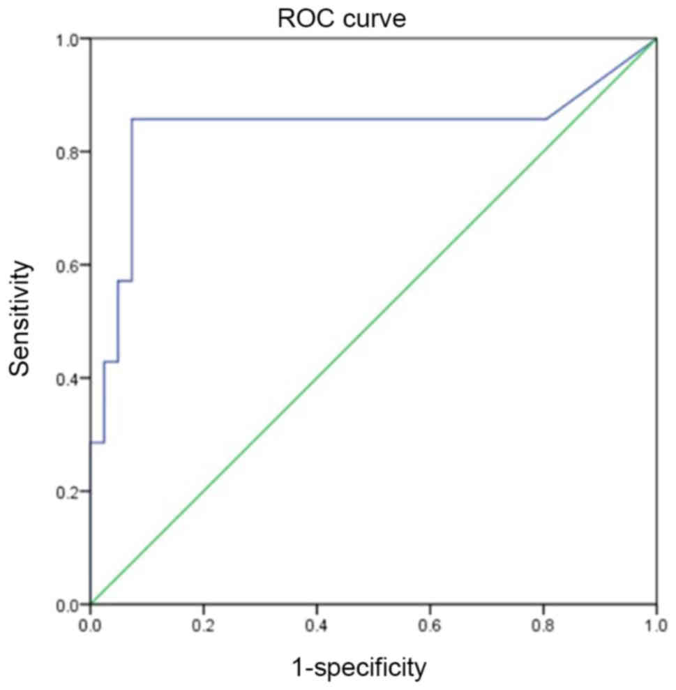
ROC analysis
ROC analysis of the pathogenic variant and
no-variant groups revealed that the optimal fasting C-peptide
cut-off value for predicting diabetic which need to detect gene was
0.64 ng/ml, with a specificity of 85.7%, sensitivity of 92.7% and
AUC of 0.735 (Fig. 1).
Discussion
In the present study, fasting C-peptide levels were
detected in three groups of patients with childhood diabetes: A
pathogenic variant group, a group with no pathogenic variant and an
islet autoantibody-positive control group (Table IV). Fasting C-peptide levels were
increased in the pathogenic variant (0.89 ng/ml) and no-variant
groups (0.48 ng/ml) compared with the control (0.17 ng/ml),
indicating that the autoantibody-negative groups had a better islet
function than the control, which is consistent with the results of
Michels et al (16). The ROC
curve indicated that C-peptide was sensitive and specific in the
autoantibody-negative group, and can be used as a prognostic
indicator for gene detection in such patients.
DKA is considered a typical manifestation of T1D.
With A previous report of DKA in MODY and T2DM, it is now believed
that DKA can occur in all forms of diabetes (17). In the present study, an NDM case
exhibited DKA and hyperglycemic hyperosmolar status, but the
pathogenic variant group had a lower DKA incidence than the other
groups (14 vs. 75 vs. 83.33%), suggesting that DKA incidence is
lower in non-T1D cases. In the present study, there were 2 cases of
MODY5 in the pathogenic variant group. The combined number of cases
may have affected the results. Some monogenic types of diabetes,
such as HNF4A- and HNF1A-MODYs, are associated with macrosomia, and
others, such as HNF1B-, INS- or GCK- (when inherited from the
father) MODYs are associated with a low birth weight. Birth weight
is therefore not a useful marker to distinguish monogenic from
other forms of diabetes (18).
Among the three groups, patients in the pathogenic
variant group used less insulin (0.14±0.24 U/kg vs. 0.51±0.30 U/kg
vs. 0.68±0.21 U/kg). Pörksen et al (19) also believed that patients with
autoantibody-positive diabetes presented with a more severe islet β
cell injury and a higher insulin dose than those with
autoantibody-negative diabetes. A lower HbA1c was observed in the
pathogenic variant group, as compared with the other groups,
indicating that these patients had better blood glucose levels than
those in the other two groups. This may be linked to poor islet
function in T1D and high blood glucose at initial diagnosis.
In recent years, an increasing number of
ketoacidosis cases without precipitating cause have been reported
in children and adults with T2DM (17). It is difficult to determine the type
of diabetes in the initial diagnosis of these people. They were
overweight and even had DKA. However, insulin secretion and insulin
action were significantly impaired in patients with DKA. After a
period of treatment, β cell function and insulin sensitivity were
improved, and insulin treatment was stopped within a few months of
follow-up (20-22).
The present study records 3 cases of this atypical T2D. In the
initial diagnosis of these people, it was difficult to determine
the type of diabetes they presented with. Through the analysis of
gene results and family history, as well as the monitoring of islet
function, 3 children were treated with diet and medicine and they
were not treated with insulin in the follow-up for several
months.
GCGR is a candidate gene for T2DM (23). The Gly40Ser variant of GCGR results
in the substitution of serine for the 40th amino acid glycine in
the encoded protein, reducing the affinity of the receptor agonist
and cyclic adenosine monophosphate synthesis by the target cell.
Gly40Ser variants lead to a decline in receptor function, which, in
theory may be associated with insufficient insulin secretion in
T2DM, ultimately leading to increased blood glucose. Hansen et
al (24) found that
glucagon-mediated insulin secretion was decreased in murine islet
cell tumors with GCGR variants, suggesting that the Gly40Ser
variant may lead to islet β cell dysfunction (25). In the present study, a pathogenic
variant, i.e., Gly40Ser, was found in the GCGR gene of a girl. She
suffered from mental retardation and her mother and grandmother had
diabetes. At initial diagnosis, her fasting C-peptide levels were
2.77 ng/ml and her BMI was 20.8 kg/m2. The mother, who
had T2DM, had the same variant. After the patient had been admitted
to hospital, insulin was used to control her blood glucose and she
was changed to metformin to control her blood glucose levels and
T2DM. This indicated that GCGR is associated with T2DM in the
Chinese population.
Variants in transcription factors expressed in
pancreatic β cells are a major cause of MODY. A de novo
mutation was detected in HNF1B, namely R165H and kidney ultrasound
revealed a renal cyst. This variant has been reported previously
and it is pathogenic (14,26,27). The
patient's daily dose of insulin was 0.62 U/kg and they had no renal
function. Due to the presence of long-term kidney disease in MODY5,
patients had a long-term renal function follow-up to ensure prompt
intervention in case of problems.
Another example of an HNF1B gene variant is the
S465R variant. The S465 site of the HNF1B gene was highly
conserved. HNF1B (S465R) has been detected in 2 Japanese patients
with diabetes. Furuta et al (13) verified that the S465R variant
decreases HNF1B gene activity and proposed that these 2 patients
may have had T2DM instead of MODY5. Several studies have also shown
that dyslipidemia and insulin resistance are features of MODY5,
suggesting that insulin resistance is not an exclusion criterion
for MODY5 (28,29). The patients in this article had the
same loci, but the onset age was 12 years old, there was no insulin
resistance, the patient was not overweight (BMI of 17.2
kg/m2), kidney function was normal at initial diagnosis,
oral medication controlled blood glucose and the fasting C-peptide
levels decreased to 0.61 ng/ml in the second year. Continuing
deterioration in β cell function led to diabetes and the need for
insulin treatment. Patients with MODY5 may therefore be
misdiagnosed as having T2DM.
In addition to screening for common monogenic
diabetes genes, the genes involved in glucose metabolism were also
examined. The LIPC gene that regulates lipid and lipoprotein
metabolism may be a potential candidate gene for T2DM (30). It was reported by Chiu et al
(31) that in mice, LIPC knockout
protected against obesity but did not affect glucose homeostasis.
González-Navarro et al (32)
induced dyslipidemia in LIPC knockout mice receiving a high-fat
diet and demonstrated that LIPC deficiency promoted steatosis and
glucose intolerance. In the present study, a frameshift mutation of
LIPC was detected in a patient with ketoacidosis. The patient had a
random blood glucose of 24 mmol/l, HbA1c of 7.3%, BMI of 17.2
kg/m2 and fasting C-peptide of 0.69 ng/ml at first
visit, as well as normal triglycerides and low- and high-density
lipoprotein cholesterol. Currently (3 years after diagnosis), the
patient's fasting C-peptide levels have been reduced to 0.01 ng/ml.
She requires insulin to control her blood glucose. LIPC markedly
affects protein function and is related to glucose and lipid
metabolism. Through association analysis, the literature supported
the association between LIPC and T2DM, but the specific mechanism
is unknown. Correlation analysis, functional testing and
verification of a large sample is required.
The inclusion criteria for the control were selected
based on a combination of clinical features and 4 auto-antibody
assays. The possibility of misdiagnosing patients with rarer forms
of monogenic diabetes cannot be completely excluded, as patients
may carry variants in other known DM genes not tested in this
study, or in a gene not yet identified as a monogenic cause of
diabetes. The authors' next study will focus on genetic testing for
the control group. The new genetic variant loci discovered also
require further functional verification. VUS verification in the
cells and mouse model is currently being performed by the present
team.
In conclusion, the present study showed that
targeted next-generation sequencing is necessary for identifying
antibody-negative diabetes. Monogenic diabetes is uncommon in
children with antibody-negative diabetes, but attention should be
paid to the presence of monogenic diabetes in antibody-negative
diabetes. Long-term, large-scale studies are also required for the
evaluation of the clinical value of C-peptide levels.
Acknowledgements
Not applicable.
Funding
The current study was supported by the National
Natural Science Foundation of China (grant no. 81700793), the
Science and Technology Program of Suzhou (grant no. SYS201766) and
the Science and Technology Program of Suzhou (grant no.
SYS201635).
Availability of data and materials
All data generated or analyzed during this study are
included in this published article.
Authors' contributions
LC designed the study. XW and FW conducted the data
collection. XW formulated the research question. XW and FW analyzed
the data and wrote the first draft of the manuscript. TC, RX, HW,
XC, DZ and HS contributed to the interpretation and discussion of
the results and commented on the drafts. All authors have read and
approved the final manuscript.
Ethics approval and consent to
participate
Informed consent was obtained from all study
participants and their parents in advance, and all procedures were
performed in accordance with the Declaration of Helsinki and
approved by the ethics committee of Children's Hospital of Soochow
University.
Patient consent for publication
Not applicable.
Competing interests
The authors declare that they have no competing
interests.
References
|
1
|
Rogers MAM, Kim C, Banerjee T and Lee JM:
Fluctuations in the incidence of type 1 diabetes in the United
States from 2001 to 2015: A longitudinal study. BMC Medicine.
15(199)2017.PubMed/NCBI View Article : Google Scholar
|
|
2
|
American Diabetes Association: Diagnosis
and classification of diabetes mellitus. Diabetes Care 37 (Suppl
1): S81-S90, 2014.
|
|
3
|
Krochik AG, Mazza CS, Valdez SN, Stumpo
RR, Papouchado ML, Iacono RF, Cardoso Landaburu AC, Sica MP, Ozuna
B and Poskus E: Immunologic and genetic markers in
insulin-dependent diabetes mellitus (type 1) in an Argentine
population. Medicina (B Aires). 61:279–283. 2001.PubMed/NCBI
|
|
4
|
Sanyoura M, Philipson LH and Naylor R:
Monogenic diabetes in children and adolescents: Recognition and
treatment options. Curr Diab Rep. 18(58)2018.PubMed/NCBI View Article : Google Scholar
|
|
5
|
Carneiro-Sampaio M and Coutinho A:
Early-onset autoimmune disease as a manifestation of primary
immunodeficiency. Front Immunol. 6(185)2015.PubMed/NCBI View Article : Google Scholar
|
|
6
|
Witsch M, Kosteria I, Kordonouri O, Alonso
G, Archinkova M, Besancon S, Birkebæk NH, Bratina N, Cherubini V,
Hanas R, et al: Possibilities and challenges of a large
international benchmarking in pediatric diabetology-The SWEET
experience. Pediatr Diabetes. 17 (Suppl 23):S7–S15. 2016.PubMed/NCBI View Article : Google Scholar
|
|
7
|
Hameed S, Ellard S, Woodhead HJ, Neville
KA, Walker JL, Craig ME, Armstrong T, Yu L, Eisenbarth GS,
Hattersley AT and Verge CF: Persistently autoantibody negative
(PAN) type 1 diabetes mellitus in children. Pediatric Diabetes.
12:142–149. 2011.PubMed/NCBI View Article : Google Scholar
|
|
8
|
Pihoker C, Gilliam LK, Ellard S, Dabelea
D, Davis C, Dolan LM, Greenbaum CJ, Imperatore G, Lawrence JM,
Marcovina SM, et al: Prevalence, characteristics and clinical
diagnosis of maturity onset diabetes of the young due to mutations
in HNF1A, HNF4A, and glucokinase: Results from the SEARCH for
Diabetes in Youth. J Clin Endocrinol Metab. 98:4055–4062.
2013.PubMed/NCBI View Article : Google Scholar
|
|
9
|
Fu J, Wang T, Liu J, Wang X, Zhang Q, Li M
and Xiao X: Using clinical indices to distinguish MODY2 (GCK
Mutation) and MODY3 (HNF1A Mutation) from type 1 diabetes in a
young chinese population. Diabetes Ther. 10:1381–1390.
2019.PubMed/NCBI View Article : Google Scholar
|
|
10
|
Wędrychowicz A, Tobór E, Wilk M,
Ziółkowska-Ledwith E, Rams A, Wzorek K, Sabal B, Stelmach M and
Starzyk JB: Phenotype heterogeneity in glucokinase-maturity-onset
diabetes of the young (GCK-MODY) patients. J Clin Res Pediatr
Endocrinol. 9:246–252. 2017.PubMed/NCBI View Article : Google Scholar
|
|
11
|
Richards S, Aziz N, Bale S, Bick D, Das S,
Gastier-Foster J, Grody WW, Hegde M, Lyon E, Spector E, et al:
Standards and guidelines for the interpretation of sequence
variants: A joint consensus recommendation of the American College
of Medical Genetics and Genomics and the Association for Molecular
Pathology. Genet Med. 17:405–424. 2015.PubMed/NCBI View Article : Google Scholar
|
|
12
|
American Diabetes Association: Glycemic
targets: Standards of medical care in diabetes-2018. Diabetes Care
41 (Suppl 1): S55-S64, 2018.
|
|
13
|
Furuta H, Furuta M, Sanke T, Ekawa K,
Hanabusa T, Nishi M, Sasaki H and Nanjo K: Nonsense and missense
mutations in the human hepatocyte nuclear factor-1beta gene (TCF2)
and their relation to type 2 diabetes in Japanese. J Clin
Endocrinol Metab. 87:3859–3863. 2002.PubMed/NCBI View Article : Google Scholar
|
|
14
|
Wang C, Zhang R, Lu J, Jiang F, Hu C, Zhou
J, Liu F, Zhang F, Qin W, Li M, et al: Phenotypic heterogeneity in
Chinese patients with hepatocyte nuclear factor-1β mutations.
Diabetes Res Clin Pract. 95:119–124. 2012.PubMed/NCBI View Article : Google Scholar
|
|
15
|
Borowiec M, Antosik K, Fendler W, Deja G,
Jarosz-Chobot P, Mysliwiec M, Zmyslowska A, Malecki M, Szadkowska A
and Mlynarski W: Novel glucokinase mutations in patients with
monogenic diabetes-clinical outline of GCK-MD and potential for
founder effect in Slavic population. Clin Genet. 81:278–283.
2012.PubMed/NCBI View Article : Google Scholar
|
|
16
|
Michels A, Zhang L, Khadra A, Kushner JA,
Redondo MJ and Pietropaolo M: Prediction and prevention of type 1
diabetes: Update on success of prediction and struggles at
prevention. Pediatr Diabetes. 16:465–484. 2015.PubMed/NCBI View Article : Google Scholar
|
|
17
|
Ndebele NFM and Naidoo M: The management
of diabetic ketoacidosis at a rural regional hospital in
KwaZulu-Natal. Afr J Prim Health Care Fam Med. 10:e1–e6.
2018.PubMed/NCBI View Article : Google Scholar
|
|
18
|
Besser RE, Flanagan SE, Mackay DG, Temple
IK, Shepherd MH, Shields BM, Ellard S and Hattersley AT:
Prematurity and genetic testing for neonatal diabetes. Pediatrics.
138(e20153926)2016.PubMed/NCBI View Article : Google Scholar
|
|
19
|
Pörksen S, Laborie LB, Nielsen L, Louise
Max Andersen M, Sandal T, de Wet H, Schwarcz E, Aman J, Swift P,
Kocova M, et al: Disease progression and search for monogenic
diabetes among children with new onset type 1 diabetes negative for
ICA, GAD- and IA-2 Antibodies. BMC Endocr Disord.
10(16)2010.PubMed/NCBI View Article : Google Scholar
|
|
20
|
Type 2 diabetes in children and
adolescents. American Diabetes Association. Pediatrics: 105,
671-680, 2000.
|
|
21
|
Umpierrez GE, Woo W, Hagopian WA, Isaacs
SD, Palmer JP, Gaur LK, Nepom GT, Clark WS, Mixon PS and Kitabchi
AE: Immunogenetic analysis suggests different pathogenesis for
obese and lean African-Americans with diabetic ketoacidosis.
Diabetes Care. 22:1517–1523. 1999.PubMed/NCBI View Article : Google Scholar
|
|
22
|
Smiley D, Chandra P and Umpierrez GE:
Update on diagnosis, pathogenesis and management of ketosis-prone
Type 2 diabetes mellitus. Diabetes Manag (Lond). 1:589–600.
2011.PubMed/NCBI View Article : Google Scholar
|
|
23
|
Hager J, Hansen L, Vaisse C, Vionnet N,
Philippi A, Poller W, Velho G, Carcassi C, Contu L, Julier C, et
al: A missense mutation in the glucagon receptor gene is associated
with noninsulindependent diabetes mellitus. Nat Genet. 9:299–304.
1995.PubMed/NCBI View Article : Google Scholar
|
|
24
|
Hansen LH, Abrahamsen N, Hager J, Jelinek
L, Kindsvogel W, Froguel P and Nishimura E: The Gly40Ser mutation
in the human glucagon receptor gene associated with NIDDM results
in a receptor with reduced sensitivity to glucagon. Diabetes.
45:725–730. 1996.PubMed/NCBI View Article : Google Scholar
|
|
25
|
Deng H, Tang WL and Pan Q: Gly40Ser
mutation of glucagon receptor gene and NIDDM in Han nationality.
Hunan Yi Ke Da Xue Xue Bao. 26:291–293. 2001.PubMed/NCBI(In Chinese).
|
|
26
|
Bellanné-Chantelot C, Chauveau D, Gautier
JF, Dubois-Laforgue D, Clauin S, Beaufils S, Wilhelm JM, Boitard C,
Noël LH, Velho G and Timsit J: Clinical spectrum associated with
hepatocyte nuclear factor-1beta mutations. Ann Intern Med.
140:510–517. 2004.PubMed/NCBI View Article : Google Scholar
|
|
27
|
Murphy R, Ellard S and Hattersley AT:
Clinical implications of a molecular genetic classification of
monogenic beta-cell diabetes. Nat Clin Pract Endocrinol Metab.
4:200–213. 2008.PubMed/NCBI View Article : Google Scholar
|
|
28
|
Welters HJ, Senkel S, Klein-Hitpass L,
Erdmann S, Thomas H, Harries LW, Pearson ER, Bingham C, Hattersley
AT, Ryffel GU and Morgan NG: Conditional expression of hepatocyte
nuclear factor-1β, the maturity-onset diabetes of the young-5 gene
product, influences the viability and functional competence of
pancreatic β-cells. J Endocrinol. 190:171–181. 2006.PubMed/NCBI View Article : Google Scholar
|
|
29
|
Pearson ER, Badman MK, Lockwood CR, Clark
PM, Ellard S, Bingham C and Hattersley AT: Contrasting diabetes
phenotypes associated with hepatocyte nuclear factor-1α and-1β
mutations. Diabetes Care. 27:1102–1107. 2004.PubMed/NCBI View Article : Google Scholar
|
|
30
|
Gelling RW, Vuguin PM, Du XQ, Cui L, Rømer
J, Pederson RA, Leiser M, Sørensen H, Holst JJ, Fledelius C, et al:
Pancreatic β-cell overexpression of the glucagon receptor gene
results in enhanced β-cell function and mass. Am J Physiol
Endocrinol Metab. 297:695–707. 2009.PubMed/NCBI View Article : Google Scholar
|
|
31
|
Chiu HK, Qian K, Ogimoto K, Morton GJ,
Wisse BE, Agrawal N, McDonald TO, Schwartz MW and Dichek HL: Mice
lacking hepatic lipase are lean and protected against diet-induced
obesity and hepatic steatosis. Endocrinology. 151:993–1001.
2010.PubMed/NCBI View Article : Google Scholar
|
|
32
|
González-Navarro H, Nong Z, Amar MJ,
Shamburek RD, Najib-Fruchart J, Paigen BJ, Brewer HB Jr and
Santamarina-Fojo S: The ligand-binding function of hepatic lipase
modulates the development of atherosclerosis in transgenic mice. J
Biol Chem. 279:45312–45321. 2004.PubMed/NCBI View Article : Google Scholar
|