Introduction
Epilepsy is part of a group of neurological
disorders characterized by a long-term risk of recurrent unprovoked
seizures (1). According to
statistics from a meta-analysis, >65 million people suffer from
epilepsy worldwide (2). The majority
of patients with epilepsy response well to anti-epileptic drugs,
which control or reduce epileptic seizure occurrence. However, a
number of patients gradually develop recurrent seizures and a
resistance to anti-epileptic drugs (3). Further investigation is required to
examine the underlying molecular mechanism of epilepsy and provide
novel therapeutic approaches to fulfill clinical needs. Seizures
are initiated by neuronal abnormality (4). Kainic acid (KA) is an agonist of
kainite glutamate receptors and can induce their overstimulation,
eventually leading to neuronal excitotoxicity and neuronal cell
death (5). Therefore, KA is used in
model systems to establish temporal lobe epilepsy, in order to
study the mechanism and efficacy of drugs in epileptic seizures
(6).
Inflammation processes have been clinically and
experimentally reported to serve a pivotal role in the generation
of seizures (7,8). It was observed that inflammatory
mediators are upregulated during the development of seizures in the
mouse model (9). In patients with
intractable epilepsy, the elevation of cytokines, including
interleukin (IL)-1β was detected in the brain tissues (10). IL-1β blockers are therefore
considered as a potential treatment approach for patients with
epilepsy (11). Results from studies
using KA model indicated that KA administration activated microglia
and the release of several important cytokines, including tumor
necrosis factor-α (TNF-α), IL-1β, IL-12 and IL-18 (12-15).
IL-1β serves a key role in the recurrence of seizures by mediating
enhanced calcium influx to induce proconvulsant effects (16).
Quercetin is derived from Quercetum (oaks)
and is categorized as a flavonol (17). Previous studies suggested that
quercetin shows anti-carcinogenic, anti-inflammatory, antiviral,
antioxidant and psychostimulant activities (17,18). For
example, quercetin administration can decrease histological signs
of acute inflammation in animals in a dose-dependent manner by
inhibiting the release of chemokine and the lipid peroxidation
end-product malondialdehyde, and increasing antioxidant enzyme
activity (19). Quercetin also
exhibits a neuro-protective function in several central nervous
system disorders, including seizures and Huntington's disease
(20,21). Moghbelinejad et al (22) has suggested that quercetin regulated
GABAA receptor α5, as well as β1 and β3, in a KA-induced seizure
model of mice. However, the potential molecules regulated by
quercetin in a KA-induced seizure remain to be investigated.
Materials and methods
Mouse model
A total of 30 male BALB/c mice (weight 20-22 g; 8
weeks old) were purchased and housed in laboratory conditions
(relative humidity of 45-55%, 12-h-light/dark cycle, freely
available food and water) at room temperature of 20-23˚C.
Experiments were carried out in accordance with the International
Guidelines for Animal Studies regarding the care and use of animals
for experimental purposes (23). The
study was approved by the Ethics Committee of School of Life
Science at the Jiangsu Normal University. KA and quercetin were
bought from Sigma-Aldrich (Merck KGaA).
KA and quercetin were dissolved in saline (0.9% w/v)
and Tween-80 (0.8% v/v), respectively. The mice were divided into
three groups consisting of 10 mice per group. The control group was
intraperitoneally administered with saline (10 µl, 0.9% w/v, i.p.)
+ Tween-80 (10 µl, 0.8% v/v, i.p.) daily for one week and on the
last day they were injected with saline (10 µl, 0.9% w/v, i.p.) +
Tween-80 (10 µl, 0.8% v/v, i.p.) followed by saline (10 µl, 0.9%
w/v, i.p.) injection 30 min later. The mice in the KA group were
injected daily with saline (10 µl, 0.9% w/v, i.p.) + Tween-80 (10
µl, 0.8% v/v, i.p.) for one week and on the last day, the mice were
injected with saline, and KA (10 µl, 10 mg/kg, i.p.) was
subsequently intraperitoneally administered. In the KA+quercetin
group, the mice were intraperitoneally injected with quercetin (10
µl, 100 mg/kg, i.p.) daily for one week and on the last day, KA (10
µl, 10 mg/kg, i.p.) was administered 30 min following injection
with quercetin (10 µl, 100 mg/kg, i.p.). Following injection of KA,
mice were observed for behavioral changes over a period of 2 h. In
accordance with a previous study, the behavioral tests were scored
from 0-6 according to the following criteria: 0, No response; 1,
immobility; 2, rigid posture; 3, scratching/circling/head bobbing;
4, forelimb clonus/rearing/falling; 5, repetitive pattern of 4; and
6, severe tonic-clonic seizures (24). Following observation, mice were
deeply anesthetized with sodium pentobarbital (65 mg/kg,
intraperitoneally) and sacrificed using cervical dislocation. The
hippocampus of each mouse was collected, cleaned with chilled
saline at 4˚C and frozen for subsequent experimentation.
Primary glial cell culture
Experiments were carried out in accordance with the
International Guidelines for Animal Studies regarding the care and
use of animals for experimental purposes (23). The study was approved by the Ethics
Committee of School of Life Science at the Jiangsu Normal
University. Glial cells were derived from 20 postnatal day 1-3
BALB/c mice purchased from the Branch of National Breeder Center of
Rodents. Briefly, 5 neonatal mice were rinsed in 70% ethanol,
followed by a quick decapitation. Afterwards, cerebral cortices
were isolated, meninges were removed and tissue was minced and
incubated with trypsin (0.025%) for 15 min at 37˚C, followed with a
trituration in the presence of DNAse I (50 µg/ml; Sigma-Aldrich;
Merck KGaA) and 20% fetal bovine serum (FBS) in
Ca2+-/Mg2+-free PBS. Cells were suspended in
DMEM (Gibco; Thermo Fisher Scientific, Inc.) supplemented with 10%
FBS (Gibco; Thermo Fisher Scientific, Inc.), 2 mM L-glutamine, 100
U/ml penicillin and 100 g/ml streptomycin, thereafter, cells
(2x10⁶ cells/well) were seeded onto poly-L-ornithine
coated 6-cm diameter Petri dishes and incubated in 95% humidity and
5% CO2 at 37˚C. After incubation for 14 days, microglia
were collected by shaking the mixed glial cell cultures for 1 h.
Thereafter, microglial cells (5x10⁴ cells/well) were
seeded into 96-well plates and incubated in 95% humidity and 5%
CO2 at 37˚C for 1 h, followed by the removal of
non-adhering cells by washing the plates. The final purity of
microglia cells was ~80%.
Isolated microglia cells were then placed in DMEM
followed by treatment with saline + Tween-80 (control group) or KA
alone (100 µM; KA group) or 100 µM KA preceded by 10 nM quercetin
(KA+quercetin group). Treatment with quercetin was performed 30 min
prior to KA treatment. Cells were treated with KA for 24 h then
subjected to the following experiments.
Western blot analysis
TNF-α (cat. no. 11948; 1:1,000), IL-1β (cat. no.
31202; 1:1,000) and NF-κB p65 (cat. no. 8242; 1:2,000) and
phospho-p65 (p-p65; cat. no. 3031; 1:1,000) antibodies were
purchased from Cell Signaling Technology, Inc. Ionized
calcium-binding adapter molecule 1 (IBA1; cat. no. ab178846;
1:2,000) and GAPDH (cat. no. ab181602; 1:10,000) antibodies were
purchased from Abcam. HRP-conjugated secondary antibodies
anti-rabbit (cat. no. ab7083; 1:10,000) and anti-goat (cat. no.
ab7125; 1:10,000) were purchased from Abcam. Hippocampi were
dissected and snap frozen in liquid N2 for subsequent
protein extraction. Proteins from tissues and cells were isolated
with RIPA lysis buffer (Sigma-Aldrich; Merck KGaA). The
concentration of protein lysates was determined by a bicinchoninic
acid kit (Sigma-Aldrich; Merck KGaA). Equal amount of proteins (20
µg) were loaded and separated on an 8% SDS-PAGE gel. The proteins
were transferred to a PVDF membrane, blocked by 5% non-fat milk at
room temperature for 2 h and washed by TBST (TBS contained 0.1%
Tween-20) for 3 times (10 min/time). The membranes were then
incubated with primary antibodies at 4˚C overnight, washed by TBST
for 3 times (10 min/time) and incubated with secondary antibodies
at room temperature for 1 h, washed by TBST for 3 times (10
min/time), successively. The blots were developed with ECL Western
Blot kit (Pierce; Thermo Fisher Scientific, Inc.), according to the
manufacturer's protocol. ImageJ software version 1.8.0 (National
Institutes of Health) was used for densitometry analysis.
ELISA
The microglia cells were collected by scraping
before the removal of the culture medium. Cells were washed with
cold phosphate buffer saline (PBS), re-suspended in Tris-Cl (10 mM;
pH 7.4) and lysed in triplicate. Protein content in the culture
medium was then analyzed by ELISA using the Quantikine TNF-α (cat.
no. MTA00B) and IL-1β (cat. no. MLB00C) ELISA kit (R&D Systems,
Inc.), according to the manufacturer's protocol.
Statistical analysis
The experiment was repeated three times and all data
were calculated by GraphPad Prism 6.0 (GraphPad Software, Inc.) and
presented as mean ± standard deviation. Two groups were compared
with unpaired Student's t-test. Three groups were firstly compared
with one-way ANOVA followed by Newman Keuls analysis. P<0.05 was
considered to indicate a statistically significant difference.
Results
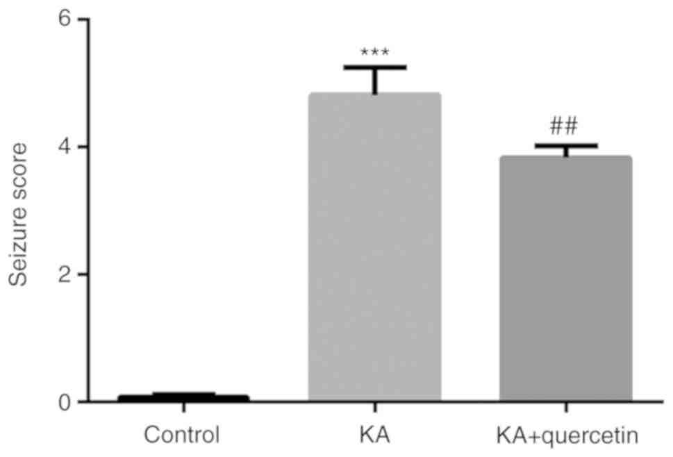
Quercetin attenuates KA-induced
seizure score in mice
Compared with the control group, KA administration
(10 mg/kg) successfully caused seizures with a significantly
increased seizure score (P<0.001, 4.82±0.44 vs. 0.08±0.04),
which in turn was significantly reduced by quercetin administration
(100 mg/kg) in the KA+quercetin group (P<0.01, 3.83±0.18)
(Fig. 1). The results suggested the
potential role of quercetin in relieving KA-induced epilepsy in
mice.
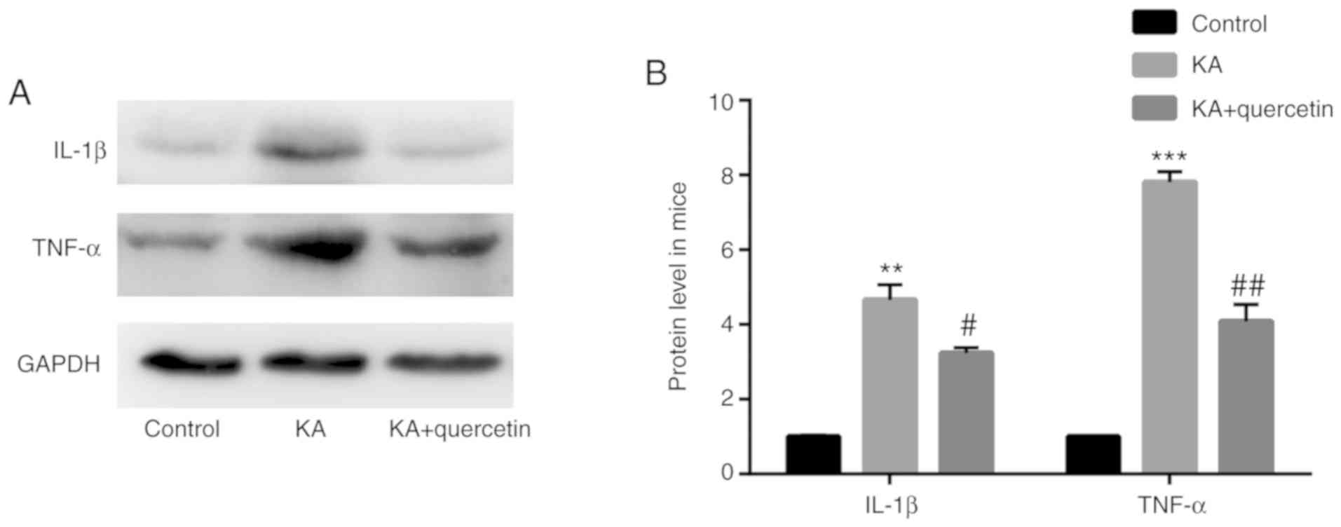
Quercetin attenuates KA-induced
proinflammatory cytokine production in mice
The results of the western blot analysis showed that
mice in the KA group (10 mg/kg) exhibited increased TNF-α
(P<0.001) and IL-1β (P<0.01) protein expression levels, when
compared with those of the control group. In addition, TNF-α
(P<0.01) and IL-1β (P<0.05) protein levels were lower in mice
treated with quercetin (100 mg/kg) in the KA+quercetin group, when
compared with those in the KA group (Fig. 2).
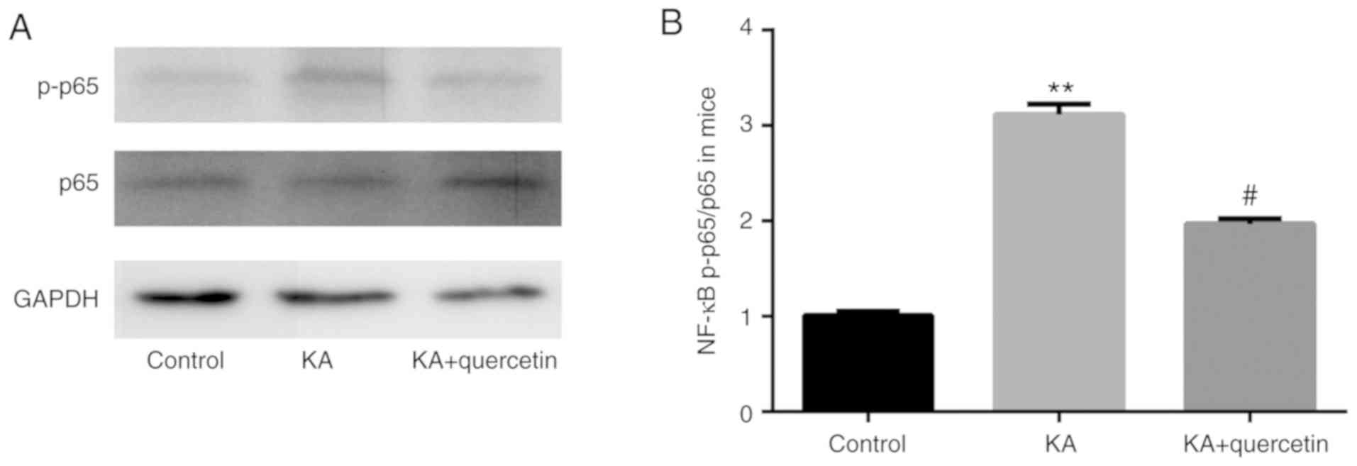
Quercetin attenuates KA-induced
activation of NF-κB in mice
The results of the western blot analysis showed that
there was increased NF-κB phosphorylated (p)-p65 protein expression
level in the KA group (P<0.01, 10 mg/kg), when compared with the
control group, which was subsequently significantly decreased by
quercetin treatment (P<0.05, 100 mg/kg). Meanwhile, there was no
significant difference in NF-κB p65 protein expression level
between the 3 groups (Fig. 3).
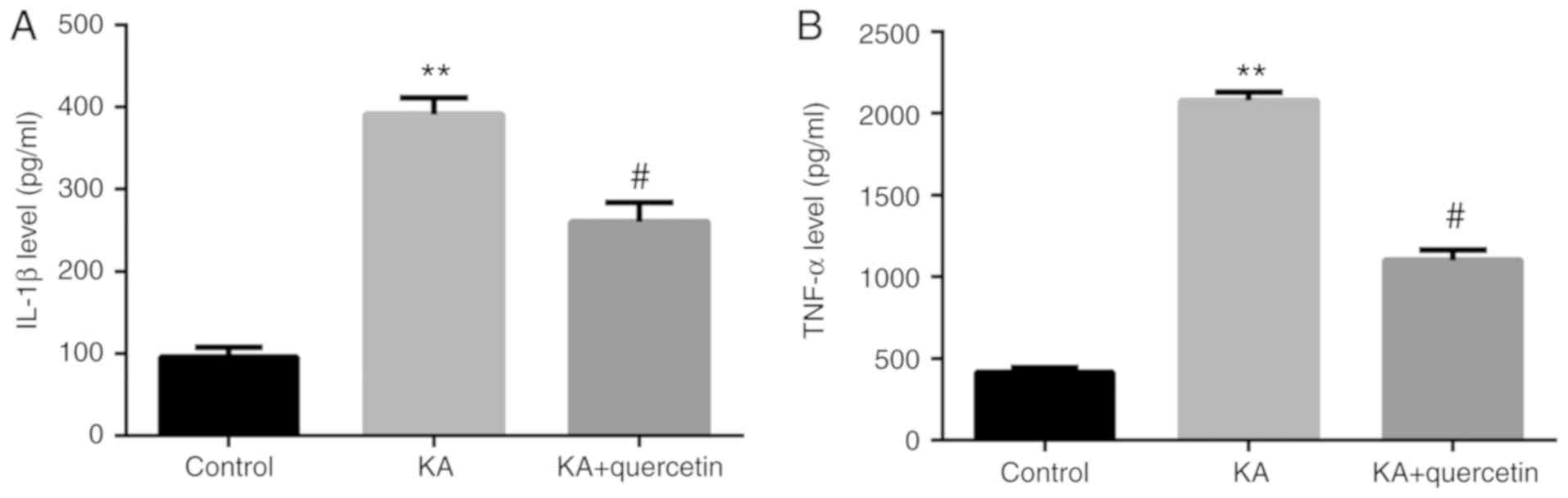
Quercetin attenuates KA-induced
proinflammatory cytokine release from microglia cells
Results from the ELISA demonstrated that the culture
medium of KA (P<0.01, 100 µM)-treated microglia cells expressed
significantly increased protein levels of TNF-α and IL-1β compared
with those of the non-treated group. Furthermore, in the culture
medium of microglia cells, which were pre-treated with quercetin
(P<0.05, 10 nM) prior to KA, TNF-α and IL-1β expression levels
were significantly decreased compared with those of the KA group
(Fig. 4).
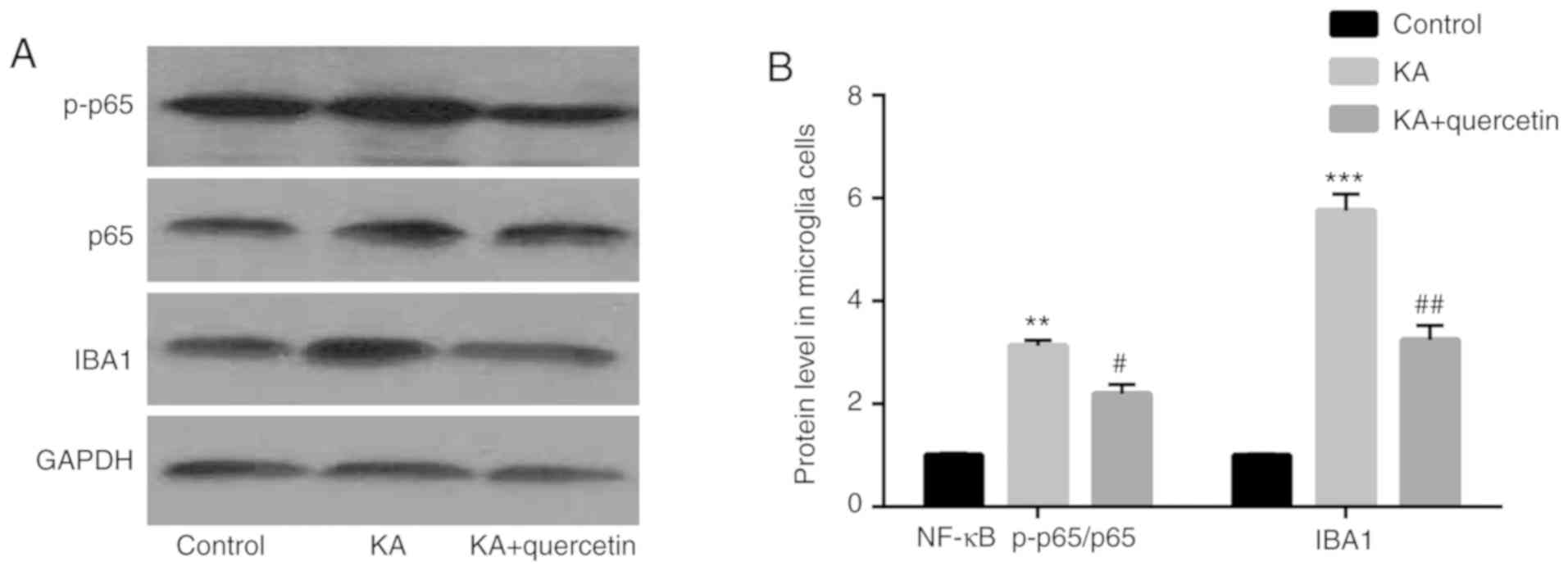
Quercetin attenuates KA-induced
activation of NF-κB and microglia cells
The results of the western blot analysis showed that
compared with the control group, NF-κB p-p65 (P<0.01) expression
levels were increased in the KA (100 µM) group, which were
decreased by quercetin treatment (P<0.05, 10 nM). There was no
significant difference of NF-κB p65 protein expression level among
the 3 groups (Fig. 5). In addition,
compared with the control group, IBA1 (P<0.001) expression
levels were higher in the KA (100 µM) group, which were decreased
by quercetin treatment (P<0.01, 10 nM).
Discussion
The functions of quercetin in animal models of
seizures have been studied and its anticonvulsant properties have
been indicated in rats and mice (25-27).
In addition, clinical and experimental evidence has been reported,
which indicates the association between epilepsy and inflammation
(28,29). In addition, quercetin has been
reported to serve anti-inflammatory roles by inhibiting the
expression of pro-inflammatory cytokines, such as TNF-α and IL-1β,
in neurodegenerative diseases (30),
including Parkinson's disease (31)
and Alzheimer's disease (32). The
aim of the present study was to investigate the potential
therapeutic effects of quercetin and the underlying molecules that
were regulated by quercetin in KA-induced epilepsy. The present
study indicated that KA increased behavioral seizure activities and
pro-inflammatory cytokines in the hippocampus of mice compared with
the control group. However, quercetin decreased both of the
aforementioned effects.
Neuroinflammation is characterized by the activation
of microglia cells (33). Therefore,
once activated, microglial cells take part in the inflammation
process and facilitate the release of cytokines and chemokines,
such as NF-κB, TNF-α and IL-1β (34). Quercetin inhibits the production of
nitric oxide in BV2 microglial cells via NF-κB pathway inactivation
(35). In addition, KA activates the
NF-κB pathway prior to seizure occurrence (36).
Throughout the present study, in comparison with the
control group, it was noted that KA increased TNF-α, IL-1β and
NF-κB expression levels in the hippocampus of mice, and these were
subsequently decreased with quercetin treatment.
KA has been reported to activate microglia cells in
epilepsy (37). The aforementioned
experiments were also repeated in microglia cells in vitro,
in order to examine whether quercetin attenuates KA-induced
epilepsy by inhibiting the activation of microglia cells. It was
indicated that compared with the control group, there were higher
TNF-α and IL-1β expression levels in the culture medium of
microglia cells in the KA group, which were subsequently diminished
by quercetin. Meanwhile, compared with the control group, there
were higher NF-κB and IBA1 expression levels in microglia cells in
the KA group, which were abolished by quercetin, indicating that it
attenuates KA-induced epilepsy by inhibiting the activation of
microglia cells.
In conclusion, the findings of the present study
provide evidence on the role of quercetin in inhibiting KA-induced
epilepsy by microglia cell inactivation and the production of
NF-κB, TNF-α and IL-1β. These findings highlight the potential role
of quercetin in the treatment of epilepsy.
However, there are two limitations of the present
study: One is the lack of analysis of different quercetin doses,
the other is the lack of in vivo and pathology experiments,
these will be the subject of future studies.
Acknowledgements
Not applicable.
Funding
This research was funded by the Priority Academic
Program Development of Jiangsu Higher Education Institutions
(PAPD); the 2013 ‘Qinglan Project’ of the Young and Middle-aged
Academic Leader of Jiangsu College and University, together with
the 2016 ‘333 Project’ Award of Jiangsu Province. The Cultivate
National Science Fund for Distinguished Young Scholars of Jiangsu
Normal University also offered grants. In addition, the present
work also obtained supports by the Major Fundamental Research
Program of the Natural Science Foundation of the Jiangsu Higher
Education Institutions of China (grant no. 13KJA180001), together
with the National Natural Science Foundation of China (grant no.
81571055, 81400902, 81271225, 81171012 and 30950031).
Availability of data and materials
The datasets used and/or analyzed during the current
study are available from the corresponding author on reasonable
request.
Authors' contributions
DW, JL, YZ designed the experiments. DW, ZZhe, SF,
XW, XH, SW collected samples and performed experiments. DW, ZZhe,
YW, ZZha, QS, ML, BH collected and assembled data. DW, ZZhe, JL, YZ
analyzed and interpreted the data. DW, ZZhe, SF, XW, XH, SW, YW,
ZZha, QS, ML, BH were involved in drafting the manuscript and
revising it critically for important intellectual content. All the
authors read and approved the final manuscript.
Ethics approval and consent to
participate
Experiments were carried out in accordance with the
International Guidelines for Animal Studies regarding the care and
use of animals for experimental purposes. The study was approved by
the Ethics Committee of School of Life Science at the Jiangsu
Normal University.
Patient consent for publication
Not applicable.
Competing interests
The authors declare that they have no competing
interests.
References
|
1
|
Sankaraneni R and Lachhwani D:
Antiepileptic drugs - a review. Pediatr Ann. 44:e36–e42.
2015.PubMed/NCBI View Article : Google Scholar
|
|
2
|
Ngugi AK, Bottomley C, Kleinschmidt I,
Sander JW and Newton CR: Estimation of the burden of active and
life-time epilepsy: A meta-analytic approach. Epilepsia.
51:883–890. 2010.PubMed/NCBI View Article : Google Scholar
|
|
3
|
Kwan P, Arzimanoglou A, Berg AT, Brodie
MJ, Allen Hauser W, Mathern G, Moshé SL, Perucca E, Wiebe S and
French J: Definition of drug resistant epilepsy: Consensus proposal
by the ad hoc Task Force of the ILAE Commission on Therapeutic
Strategies. Epilepsia. 51:1069–1077. 2010.PubMed/NCBI View Article : Google Scholar
|
|
4
|
Shapiro LA, Wang L and Ribak CE: Rapid
astrocyte and microglial activation following pilocarpine-induced
seizures in rats. Epilepsia. 49 (Suppl 2):33–41. 2008.PubMed/NCBI View Article : Google Scholar
|
|
5
|
Zhang XM and Zhu J: Kainic acid-induced
neurotoxicity: Targeting glial responses and glia-derived
cytokines. Curr Neuropharmacol. 9:388–398. 2011.PubMed/NCBI View Article : Google Scholar
|
|
6
|
Reddy DS and Kuruba R: Experimental models
of status epilepticus and neuronal injury for evaluation of
therapeutic interventions. Int J Mol Sci. 14:18284–18318.
2013.PubMed/NCBI View Article : Google Scholar
|
|
7
|
Friedman A and Dingledine R: Molecular
cascades that mediate the influence of inflammation on epilepsy.
Epilepsia. 52 (Suppl 3):33–39. 2011.PubMed/NCBI View Article : Google Scholar
|
|
8
|
Pernot F, Heinrich C, Barbier L,
Peinnequin A, Carpentier P, Dhote F, Baille V, Beaup C, Depaulis A
and Dorandeu F: Inflammatory changes during epileptogenesis and
spontaneous seizures in a mouse model of mesiotemporal lobe
epilepsy. Epilepsia. 52:2315–2325. 2011.PubMed/NCBI View Article : Google Scholar
|
|
9
|
Mazarati AM, Lewis ML and Pittman QJ:
Neurobehavioral comorbidities of epilepsy: Role of inflammation.
Epilepsia. 58 (Suppl 3):48–56. 2017.PubMed/NCBI View Article : Google Scholar
|
|
10
|
Choi J, Nordli DR Jr, Alden TD, DiPatri A
Jr, Laux L, Kelley K, Rosenow J, Schuele SU, Rajaram V and Koh S:
Cellular injury and neuroinflammation in children with chronic
intractable epilepsy. J Neuroinflammation. 6(38)2009.PubMed/NCBI View Article : Google Scholar
|
|
11
|
Vezzani A, Friedman A and Dingledine RJ:
The role of inflammation in epileptogenesis. Neuropharmacology.
69:16–24. 2013.PubMed/NCBI View Article : Google Scholar
|
|
12
|
Thompson C, Gary D, Mattson M, Mackenzie A
and Robertson GS: Kainic acid-induced naip expression in the
hippocampus is blocked in mice lacking TNF receptors. Brain Res Mol
Brain Res. 123:126–131. 2004.PubMed/NCBI View Article : Google Scholar
|
|
13
|
Eriksson C, Van Dam AM, Lucassen PJ, Bol
JG, Winblad B and Schultzberg M: Immunohistochemical localization
of interleukin-1beta, interleukin-1 receptor antagonist and
interleukin-1beta converting enzyme/caspase-1 in the rat brain
after peripheral administration of kainic acid. Neuroscience.
93:915–930. 1999.PubMed/NCBI View Article : Google Scholar
|
|
14
|
Chen Z, Duan RS, Concha QH, Wu Q, Mix E,
Winblad B, Ljunggren HG and Zhu J: IL-12p35 deficiency alleviates
kainic acid-induced hippocampal neurodegeneration in C57BL/6 mice.
Neurobiol Dis. 17:171–178. 2004.PubMed/NCBI View Article : Google Scholar
|
|
15
|
Jeon GS, Park SK, Park SW, Kim DW, Chung
CK and Cho SS: Glial expression of interleukin-18 and its receptor
after excitotoxic damage in the mouse hippocampus. Neurochem Res.
33:179–184. 2008.PubMed/NCBI View Article : Google Scholar
|
|
16
|
Vezzani A, Aronica E, Mazarati A and
Pittman QJ: Epilepsy and brain inflammation. Exp Neurol. 244:11–21.
2013.PubMed/NCBI View Article : Google Scholar
|
|
17
|
Li Y, Yao J, Han C, Yang J, Chaudhry MT,
Wang S, Liu H and Yin Y: Quercetin, Inflammation and Immunity.
Nutrients. 8(167)2016.PubMed/NCBI View Article : Google Scholar
|
|
18
|
Zheng J, Wu J, Chen J, Liu J, Lu Y, Huang
C, Hu G, Wang X and Zeng Y: Therapeutic effects of quercetin on
early inflammation in hypertriglyceridemia-related acute
pancreatitis and its mechanism. Pancreatology. 16:200–210.
2016.PubMed/NCBI View Article : Google Scholar
|
|
19
|
Dong YS, Wang JL, Feng DY, Qin HZ, Wen H,
Yin ZM, Gao GD and Li C: Protective effect of quercetin against
oxidative stress and brain edema in an experimental rat model of
subarachnoid hemorrhage. Int J Med Sci. 11:282–290. 2014.PubMed/NCBI View Article : Google Scholar
|
|
20
|
Nassiri-Asl M, Hajiali F, Taghiloo M,
Abbasi E, Mohseni F and Yousefi F: Comparison between the effects
of quercetin on seizure threshold in acute and chronic seizure
models. Toxicol Ind Health. 32:936–944. 2016.PubMed/NCBI View Article : Google Scholar
|
|
21
|
Chakraborty J, Singh R, Dutta D, Naskar A,
Rajamma U and Mohanakumar KP: Quercetin improves behavioral
deficiencies, restores astrocytes and microglia, and reduces
serotonin metabolism in 3-nitropropionic acid-induced rat model of
Huntington's Disease. CNS Neurosci Ther. 20:10–19. 2014.PubMed/NCBI View Article : Google Scholar
|
|
22
|
Moghbelinejad S, Alizadeh S, Mohammadi G,
Khodabandehloo F, Rashvand Z, Najafipour R and Nassiri-Asl M: The
effects of quercetin on the gene expression of the GABAA receptor
α5 subunit gene in a mouse model of kainic acid-induced seizure. J
Physiol Sci. 67:339–343. 2017.PubMed/NCBI View Article : Google Scholar
|
|
23
|
McGrath JC, Drummond GB, McLachlan EM,
Kilkenny C and Wainwright CL: Guidelines for reporting experiments
involving animals: The ARRIVE guidelines. Br J Pharmacol.
160:1573–1576. 2010.PubMed/NCBI View Article : Google Scholar
|
|
24
|
Morrison RS, Wenzel HJ, Kinoshita Y,
Robbins CA, Donehower LA and Schwartzkroin PA: Loss of the p53
tumor suppressor gene protects neurons from kainate-induced cell
death. J Neurosci. 16:1337–1345. 1996.PubMed/NCBI View Article : Google Scholar
|
|
25
|
Nassiri-Asl M, Moghbelinejad S, Abbasi E,
Yonesi F, Haghighi MR, Lotfizadeh M and Bazahang P: Effects of
quercetin on oxidative stress and memory retrieval in kindled rats.
Epilepsy Behav. 28:151–155. 2013.PubMed/NCBI View Article : Google Scholar
|
|
26
|
Nieoczym D, Socała K, Raszewski G and Wlaź
P: Effect of quercetin and rutin in some acute seizure models in
mice. Prog Neuropsychopharmacol Biol Psychiatry. 54:50–58.
2014.PubMed/NCBI View Article : Google Scholar
|
|
27
|
Singh T, Kaur T and Goel RK: Adjuvant
quercetin therapy for combined treatment of epilepsy and comorbid
depression. Neurochem Int. 104:27–33. 2017.PubMed/NCBI View Article : Google Scholar
|
|
28
|
Choi J and Koh S: Role of brain
inflammation in epileptogenesis. Yonsei Med J. 49:1–18.
2008.PubMed/NCBI View Article : Google Scholar
|
|
29
|
Wheless JW, Clarke DF, Arzimanoglou A and
Carpenter D: Treatment of pediatric epilepsy: European expert
opinion, 2007. Epileptic Disord. 9:353–412. 2007.PubMed/NCBI View Article : Google Scholar
|
|
30
|
Spagnuolo C, Moccia S and Russo GL:
Anti-inflammatory effects of flavonoids in neurodegenerative
disorders. Eur J Med Chem. 153:105–115. 2018.PubMed/NCBI View Article : Google Scholar
|
|
31
|
Bournival J, Plouffe M, Renaud J,
Provencher C and Martinoli MG: Quercetin and sesamin protect
dopaminergic cells from MPP+-induced neuroinflammation
in a microglial (N9)-neuronal (PC12) coculture system. Oxid Med
Cell Longev. 2012(921941)2012.PubMed/NCBI View Article : Google Scholar
|
|
32
|
Moreno LCGEI, Puerta E, Suárez-Santiago
JE, Santos-Magalhães NS, Ramirez MJ and Irache JM: Effect of the
oral administration of nanoencapsulated quercetin on a mouse model
of Alzheimer's disease. Int J Pharm. 517:50–57. 2017.PubMed/NCBI View Article : Google Scholar
|
|
33
|
Chen WW, Zhang X and Huang WJ: Role of
neuroinflammation in neurodegenerative diseases (Review). Mol Med
Rep. 13:3391–3396. 2016.PubMed/NCBI View Article : Google Scholar
|
|
34
|
Rahimifard M, Maqbool F, Moeini-Nodeh S,
Niaz K, Abdollahi M, Braidy N, Nabavi SM and Nabavi SF: Targeting
the TLR4 signaling pathway by polyphenols: A novel therapeutic
strategy for neuroinflammation. Ageing Res Rev. 36:11–19.
2017.PubMed/NCBI View Article : Google Scholar
|
|
35
|
Kang CH, Choi YH, Moon SK, Kim WJ and Kim
GY: Quercetin inhibits lipopolysaccharide-induced nitric oxide
production in BV2 microglial cells by suppressing the NF-κB pathway
and activating the Nrf2-dependent HO-1 pathway. Int
Immunopharmacol. 17:808–813. 2013.PubMed/NCBI View Article : Google Scholar
|
|
36
|
Miller JA, Kirkley KA, Padmanabhan R,
Liang LP, Raol YH, Patel M, Bialecki RA and Tjalkens RB: Repeated
exposure to low doses of kainic acid activates nuclear factor kappa
B (NF-κB) prior to seizure in transgenic NF-κB-EGFP reporter mice.
Neurotoxicology. 44:39–47. 2014.PubMed/NCBI View Article : Google Scholar
|
|
37
|
Bosco DB, Zheng J, Xu Z, Peng J, Eyo UB,
Tang K, Yan C, Huang J, Feng L, Wu G, et al: RNAseq analysis of
hippocampal microglia after kainic acid-induced seizures. Mol
Brain. 11(34)2018.PubMed/NCBI View Article : Google Scholar
|