Introduction
Breast cancer is one of the most diverse and complex
types of human malignancy, which is a leading cause of mortality
among women worldwide (1). In the
USA, breast cancer diagnoses are particularly common, accounting
for ~30% of all newly diagnosed malignancies; it was reported that
breast cancer led to various malgnancies, which seriously affected
patients' quality of life (1-3).
Gemcitabine (2',2'-difluorodeoxycytidine, dFdC) is a
pyrimidine nucleoside analog that inhibits DNA replication and
transcription, leading to cell apoptosis (4). It is frequently applied as a single
agent as salvage chemotherapy following several lines of treatment
(5). Considering prior treatment,
dose and schedule, response rates to gemcitabine were typically
14-42% (5). The combined use of
gemcitabine with capecitabine, vinorelbine, platinum and other
chemotherapeutic agents can enhance the anticancer effects of the
overall chemotherapy (6). Since
Gemcitabine has been recommended by Chinese breast cancer diagnosis
and treatment guide (2017) as an effective method in treating
advanced breast cancer (7).
Enhancing the therapeutic effects gemcitabine would be of great
significance for the treatment of this disease.
MicroRNAs (miRNAs or miRs) are a class of
evolutionarily conserved non-coding RNAs that typically consist of
~22 nucleic acids (8). They normally
serve as post-transcriptional regulators of gene expression by
binding to the 3'untranslated regions (3'UTRs) of target mRNAs to
either inhibit translation or promote degradation (8). miRNAs have been reported to participate
in a number of physiological processes, including proliferation,
apoptosis, cell movement and stem cell renewal (8). Accumulating evidence has revealed that
a wide variety of miRNAs are involved in oncogenesis (9). Of note, miR-187-3p has been revealed to
be downregulated in colorectal cancer (10), renal cell carcinoma (11), prostate cancer (12), lung cancer (13) and breast cancer (14). In addition, miR-187-3p expression has
also been previously associated with patient prognosis in breast
cancer (14). However, the precise
function of miR-187-3p in the pathophysiology of cancer remain
controversial, as a previous study has demonstrated that miR-187
overexpression promotes lymphoma progression and enhances
resistance to chemotherapy in peripheral T-cell lymphoma (15).
Fibroblast growth factor 9 (FGF9) is a member of the
FGF family, which includes 23 family members each serving key
functions including cellular differentiation, proliferation and
tumorigenesis (16). FGFs bind to
and activate FGF receptors, leading to the further activation of
developmental signaling pathways that are responsible for a variety
of biological functions (17). The
overexpression of FGF9 has been previously identified as a novel
unfavorable prognostic indicator in lung cancer (18). Intracellular signaling pathways
activated by FGF serve important roles in a wide range of
malignancies, including breast cancer (17,19).
Fillmore et al (20) revealed
that estrogen could activate FGF9/FGFR3/T box transcription factor
3 signaling to increase the numbers of breast cancer stem-like
cells, whilst Yin et al (21)
have previously reported that the miRNA-FGF9 pathway is important
for pleuropulmonary blastoma development.
In the present study, it was revealed that the
overexpression of miR-187-3p inhibited MDA-MB-231 cell
proliferation, promoted apoptosis and reduced resistance to
gemcitabine. Mechanistically, miR-187-3p overexpression resulted in
the downregulation of FGF9 expression to regulate gemcitabine
sensitivity in breast cancer cells, implicating miR-187-3p as a
promising therapeutic target in the treatment of breast cancer.
Materials and methods
Clinical patient tissue samples
A total of 30 breast cancer tumor tissue samples and
matched adjacent non-tumor tissue samples, 5 cm away from the
tumors, were collected at Chifeng Municipal Hospital (Chifeng,
China) from June 2015 to July 2017. All samples were collected from
women aged between 27 and 65 years with an average age of 48±11
years. Patients who have received any chemo- or radio- therapies
were excluded from the study. Written informed consent was provided
by all participants prior to enrollment. The present study was
approved by the Ethics Committee of Chifeng Municipal Hospital
(approval no. 20150602CFMH; Chifeng, China). All tissue samples
were immediately frozen in liquid nitrogen following surgery and
stored in a -80˚C refrigerator prior to use.
Cell culture and reagents
MDA-MB-231 human breast cancer cell line was
purchased from the American Type Culture Collection and was
subsequently cultured in DMEM (Life Technologies; Thermo Fisher
Scientific, Inc.) supplemented with 10% FBS (HyClone; GE Healthcare
Life Sciences) and 1% penicillin-streptomycin solution (Life
Technologies; Thermo Fisher Scientific, Inc.) in a humidified
atmosphere at 37˚C and 5% CO2. Gemcitabine was purchased
from Sigma-Aldrich (Merck KGaA).
Transient transfection
miR-187-3p mimic (50 nM,
5'-GGCCGACGUUGUGUUCUGUGCU-3') and miR-NC mimic (50 nM,
5'-UCGCUUGGUGCAGGUCGGGAA-3') were purchased from Shanghai
GenePharma Co., Ltd., pcDNA3.1 (2 µg) and pcDNA-FGF9 (2 µg) were
purchased from Addgene, Inc. All transfections were performed into
MDA-MB-231 using Lipofectamine® 2000 transfection
reagent (Invitrogen; Thermo Fisher Scientific, Inc.). After
incubation for 48 h, cells were collected for the subsequent
studies.
RNA extraction and reverse
transcription-quantitative PCR (RT-qPCR
Total RNA was extracted from cultured MDA-MB-231
cells and tissues using TRIzol® (Invitrogen; Thermo
Fisher Scientific, Inc.) and cDNA synthesis was performed at 37˚C
for 15 min and 85˚C for 5 sec using a PrimeScript™ RT
reagent kit (Takara Bio, Inc.) according to the manufacturer's
protocols. RT-qPCR was performed in triplicate using
SYBR® Premix Ex Taq™ (Takara Bio, Inc.) in a
Bio-Rad CFX96 Real-Time PCR System (Bio-rad Laboratories Inc.). The
thermocycling conditions were as follows: 95˚C for 30 sec, followed
by 35 cycles of 95˚C for 5 sec and 60˚C for 30 sec. Relative levels
of miR-187-3p were normalized to that of U6 small nucleolar RNA,
whereas those of FGF9 were normalized to GAPDH. The
2-ΔΔCq method was used to quantify relative gene
expression (22). The primer
sequences used were listed as follows: Stem loop primer,
5'-CTCAACTGGTGTCGTGGAGTCGGCAATTCAGTTGAGCCGGCT-3'; miR-187-3p
forward, 5'-GCCGAGTCGTGTCTTGTGTT-3' and reverse,
5'-CTCAACTGGTGTCGTGGA-3'; U6 forward, 5'-CTCAACTGGTGTCGTGGA-3' and
reverse, 5'-CTCAACTGGTGTCGTGGA-3'; FGF9 forward,
5'-ATGGCTCCCTTAGGTGAAGTT-3' and reverse,
5'-CCCAGGTGGTCACTTAACAAAAC-3'; GAPDH forward,
5'-CAATGACCCCTTCATTGACC-3' and reverse,
5'-GACAAGCTTCCCGTTCTCAG-3'.
Cell viability
Cell viability was assessed by performed a cell
counting kit-8 assay (CCK-8; Dojindo Molecular Technologies, Inc.)
according to the manufacturer's protocol. Cells
(~5x10³/well) were seeded into 96-well plates. Following
treatment with ascending concentrations of Gemcitabine (0.25, 0.5,
1, 2 and 4 nM) for 24 h at 37˚C and co-transfection with miR-187-3p
or miR-NC mimic and pcDNA3.1-FGF9 or pcDNA3.1 plasmid for 48 h, 10
µl CCK-8 solution was added into each well and incubated at 37˚C
for 2 h. Absorbance at 450 nm was subsequently measured in each
well using a spectrophometer to determine cell viability.
Apoptosis assay
An Annexin-V/Dead Cell Apoptosis kit (Invitrogen;
Thermo Fisher Scientific, Inc.) was used to perform cell apoptosis
assay according to manufacturer's protocol. Cells were harvested
and washed in cold PBS, after which they were then diluted to
~1x10⁶ cells/ml using 1X Annexin-binding buffer in 100
µl per assay. Cells were subsequently treated with 5 µl Alexa
Fluor® 488 annexin V and 1 µl 100 µg/ml PI per assay
suspension. A total of 400 µl annexin-binding buffer was added and
samples were incubated at room temperature for 15 min. Stained
cells were analyzed using BD FACSCaliburTM flow
cytometer (BD Biosciences) coupled with FlowJo software (version
10.2; FlowJo LLC).
Western blot analysis
Antibodies against FGF9 (cat. no. PA5-23719;
1:1,000) and β-catenin (cat. no. 71-2700; 1:1,000) were obtained
from Thermo Fisher Scientific, Inc. Anti-c-Myc (cat. no. 5605;
1:1,000) and Cyclin D1 (cat. no. 2978; 1:1,000) antibodies were
purchased from Cell Signaling Technology, Inc. GAPDH mouse
monoclonal antibodies (cat. no. ab8245; 1:10,000) was obtained from
Abcam. Anti-mouse (cat. no. CW0221S; 1:10,000) and anti-rabbit
(cat. no. CW0234S; 1:10,000) secondary antibodies were purchased
from Beijing ComWin Biotech Co., Ltd.
Following collection, cells were washed twice with
cold PBS and lysed in cold RIPA buffer (Beyotime Institute of
Biotechnology) supplemented with protease inhibitor cocktail
(Sigma-Aldrich, Merck KGaA). Samples were then incubated on ice for
30 mins. Lysates were subsequently centrifuged at 12,000 x g at 4˚C
for 15 min and protein concentration was measured using
Bicinchoninic Acid Protein Assay kit (Thermo Fisher Scientific,
Inc.). Equal quantities (20 µg/well) of protein were separated by
8% SDS-PAGE, transferred to polyvinylidene fluoride membranes (EMD
millipore) and incubated with the respective aforementioned
antibodies. Membranes were developed using SuperSignal™
West Femto Maximum Sensitivity Substrate (Thermo Fisher Scientific,
Inc.) and image analysis was performed using the
ImageQuant™ LAS 4000 software (GE Healthcare Life
Sciences).
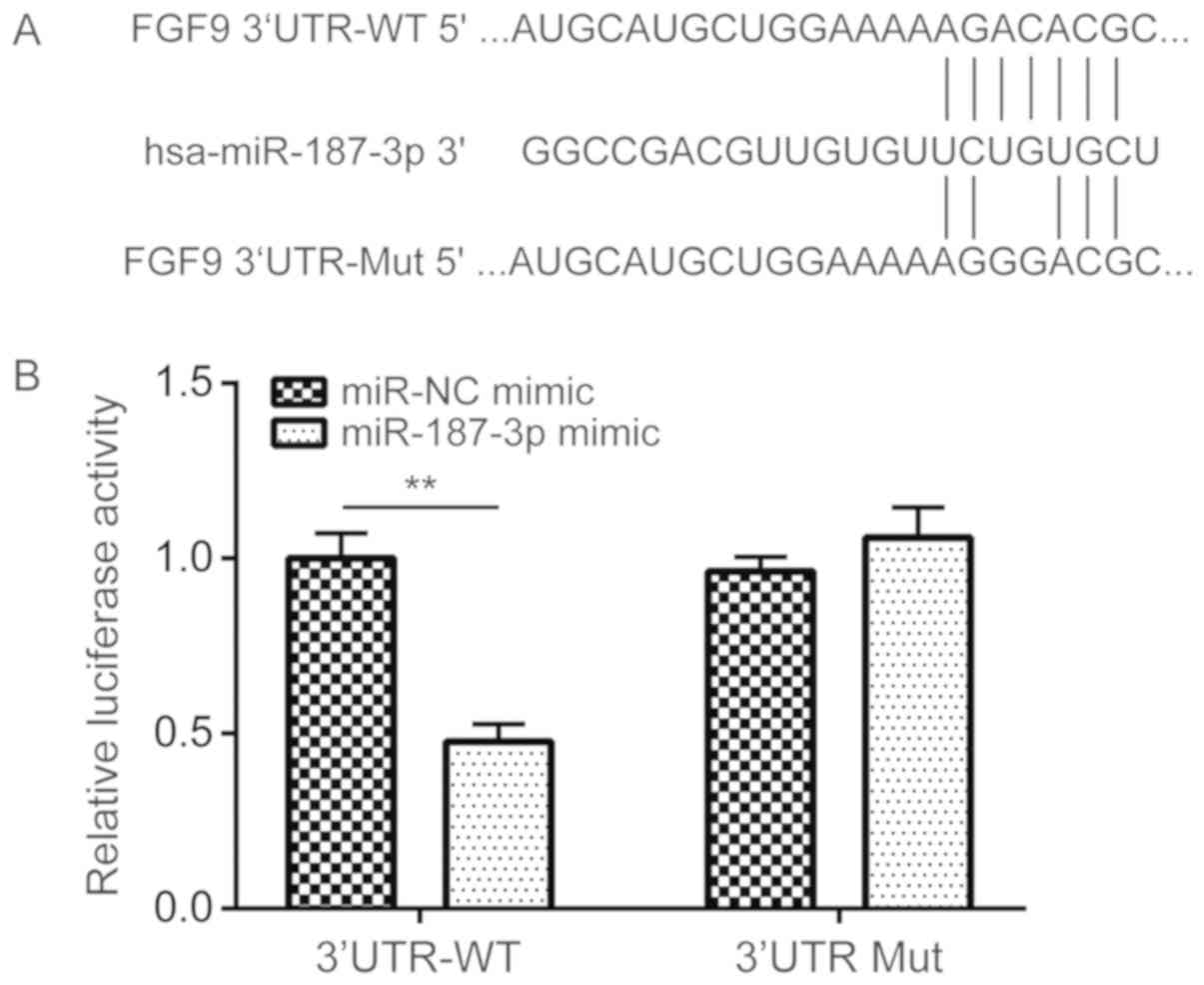
Dual-luciferase reporter gene
assay
The TargetScan miRNA target database (http://www.targetscan.org/) predicted that a putative
miR-187-3p binding site was present on the 3'-UTR of FGF9.
Therefore, the FGF9 3'-UTR region was amplified from cDNA isolated
from MDA-MB-231 cells and inserted into the XbaI restriction site
of pGL3 Luciferase Reporter vector (Promega Corporation) with the
primer pairs as listed: WT FGF9, forward,
5'-GCTCTAGACAAAGACAGTTTCTTCAC-3', reverse,
5'-GCTCTAGATTTTCAAAACTCTGTAAT-3'. In addition, two site mutations
were introduced into the wild-type (WT) pGL3-FGF9 3'UTR WT to
construct the mutant (Mut) FGF9 3'UTR plasmid using the Quicksite
mutation kit (Agilent Technologies, Inc.) with the listed primer
pairs: MT FGF9, forward, 5'-CGGAAAAAGACGGGCCACGACAGG-3', reverse,
5'-CCTGTCGTGGCCCGTCTTTTTCCG-3'. MDA-MB-231 cells were
co-transfected with 100 nM FGF9 3'UTR-WT or FGF9 3'UTR-Mut plasmids
and 100 nM miR-187-3p mimic or negative mimic control using
Lipofectamine® 2000 transfection reagent (Invitrogen;
Thermo Fisher Scientific, Inc.) according to manufacturer's
protocol for 48 h. Luciferase activity was evaluated using the
Dual-Glo® Luciferase assay system according to
manufacturer's protocol (Promega Corporation). The luciferase
activity was normalized to Renilla luciferase activity.
Statistical analysis
All experiments in the present study were performed
three times independently. GraphPad Prism 5.0 software (GraphPad
Software, Inc.) was used for statistical analysis and all data were
presented as mean ± standard deviation. Student's t-test was used
for all comparisons between two groups whereas one-way ANOVA
followed by Student-Newman-Keuls test was used for comparison of
differences between ≥3 groups. Pearson's correlation analysis was
performed to analyze the correlation between FGF mRNA and
miR-187-3p expression in the 30 pairs of breast cancer tumor and
corresponding matched adjacent non-tumor tissue samples collected
from patients with breast cancer. P<0.05 was considered to
indicate a statistically significant difference.
Results
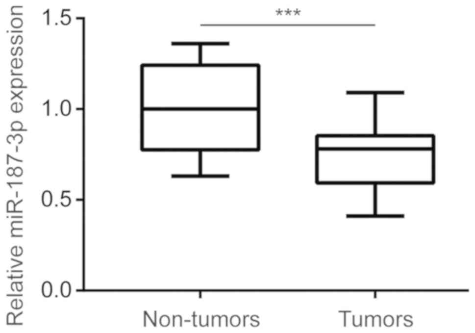
miR-187-3p expression is lower in
breast cancer tumor tissues
To investigate the potential role of miR-187-3p in
breast cancer, miR-187-3p expression was measured in 30 pairs of
breast cancer tumor and corresponding matched adjacent non-tumor
tissue samples collected from patients with breast cancer. RT-qPCR
analysis revealed that miR-187-3p expression was significantly
reduced in breast cancer tumor tissues compared with non-tumor
tissues (Fig. 1), indicating that
miR-187-3p may serve a role in the development of breast
cancer.
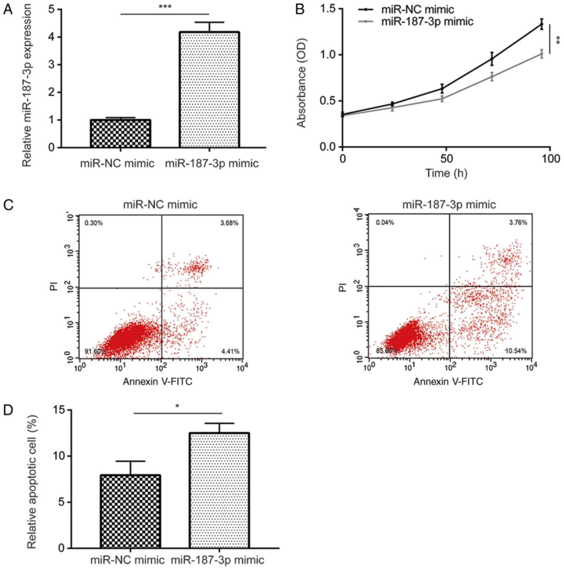
Overexpression of miR-187-3p inhibits
cell viability and promotes apoptosis in MDA-MB-231 cells
The physiological function and underlying mechanism
of miR-187-3p in breast cancer remain poorly understood. Therefore,
MDA-MB-231 cells were successfully transfected with the miR-187-3p
mimic to overexpress miR-187-3p compared with the miR-NC mimic
(Fig. 2A). Compared with the miR-NC
mimic, miR-187-3p overexpression was subsequently revealed to
significantly inhibit MDA-MB-231 cell viability (Fig. 2B). Additionally, compared with the
miR-NC mimic, overexpression of miR-187-3p significantly promoted
MDA-MB-231 cell apoptosis (Fig. 2C
and D). The results indicated that
miR-187-3p overexpression reduced MDA-MB-231 cell viability and
promoted apoptosis.
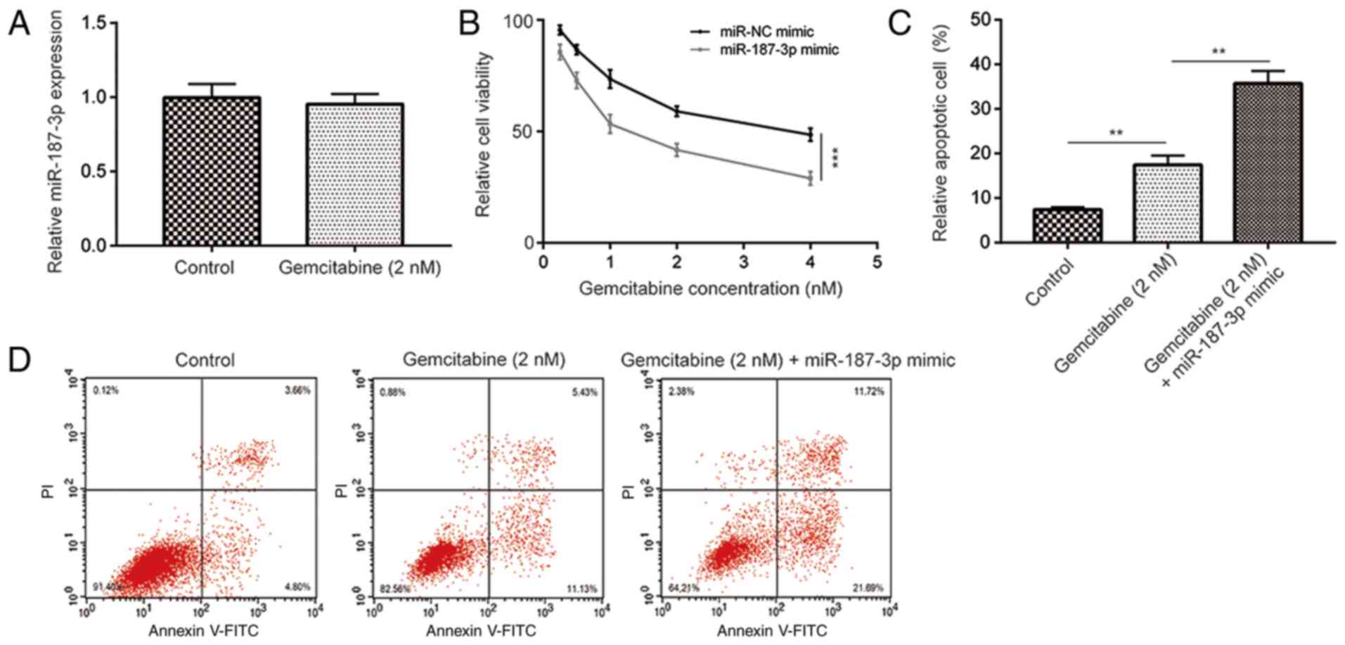
Overexpression of miR-187-3p enhances
gemcitabine sensitivity in MDA-MB-231 cells
Liang et al (23) discovered that miR-187-3p was one of
the most significantly downregulated miRNAs in multidrug-resistant
MCF7 cells compared to parental MCF7 cells. Although gemcitabine is
frequently applied as a therapeutic agent for advanced breast
cancer, it remains associated with limited efficacy (7,24). In
this present study, it was determined that, compared to the
control, gemcitabine (2 nM) treatment did not significantly alter
miR-187-3p expression in MDA-MB-231 cells (Fig. 3A). However, compared with the miR-NC
mimic, overexpression of miR-187-3p significantly enhanced the
gemcitabine-mediated (1, 2, 3 and 4 nM) reduction of cell viability
in MDA-MB-231 cells (Fig. 3B) whilst
significantly potentiating gemcitabine-induced MDA-MB-231 cell
apoptosis (Fig 3C and D). These results indicated that miR-187-3p
overexpression removed gemcitabine resistance in breast cancer
cells.
miR-187-3p regulates FGF9 expression
by binding to its 3'-UTR
Following the indication that miR-187-3p enhanced
the efficacy of gemcitabine, the underlying mechanism was
elucidated. According to the TargetScan database, FGF9 was
predicted to be a potential target gene of miR-187-3p, as a
putative miR-187-3p binding site was observed on the 3'-UTR of FGF9
(Fig. 4A). WT FGF9 3'-UTR and Mut
FGF9 3'-UTR luciferase reporter gene plasmids were constructed to
verify this potential association. Dual-luciferase reporter gene
assay data demonstrated that, compared with the miR-NC mimic,
transfection with the miR-187-3p mimic significantly reduced
relative luciferase activity in MDA-MB-231 cells co-transfected
with the WT FGF9 3'-UTR plasmid (Fig.
4B).
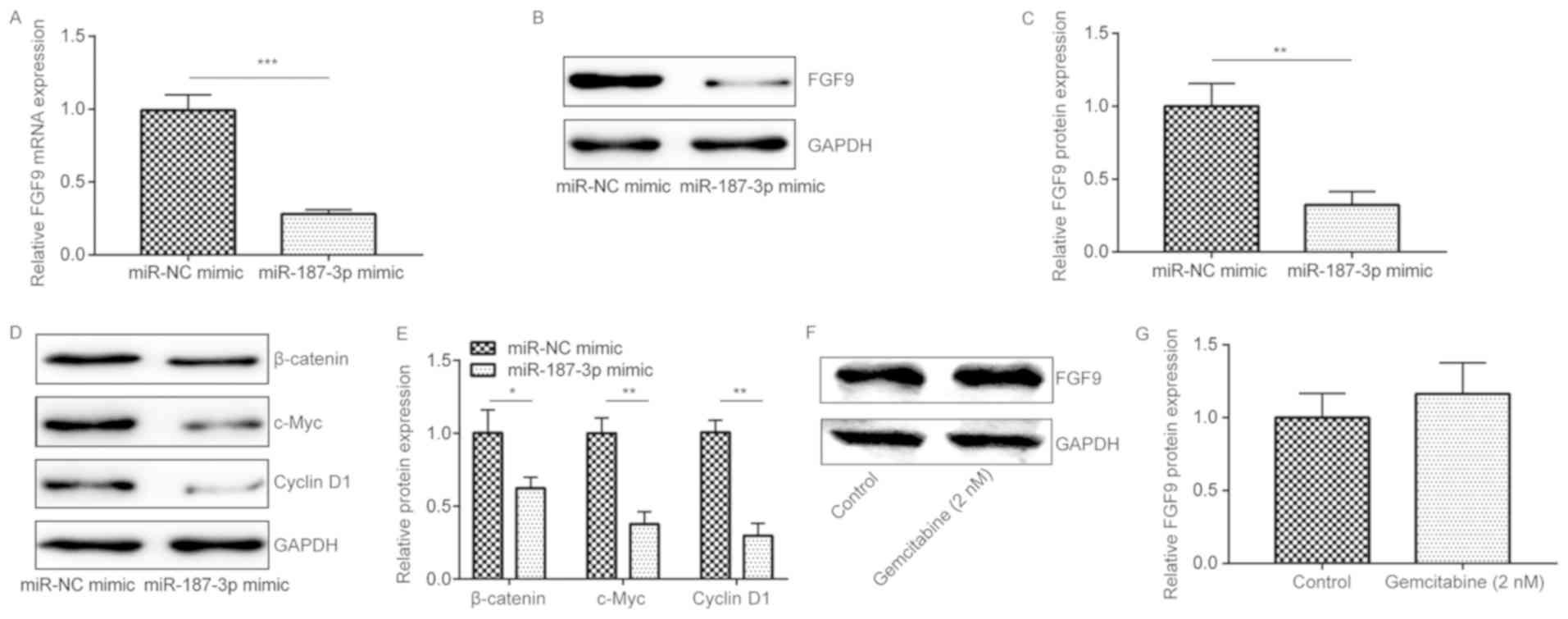
miR-187-3p overexpression negatively
regulates FGF9 expression and the associated downstream Wnt
pathway
To investigate the regulatory effects of miR-187-3p
on FGF9, RT-qPCR and western blot analysis were performed to
measure FGF9 mRNA and protein expression in MDA-MB-231 cells
following miR-187-3p overexpression. It was revealed that, compared
with the miR-NC mimic, miR-187-3p overexpression significantly
reduced FGF9 mRNA and protein expression (Fig. 5A-C). A previous study demonstrated
that FGF9 can regulate Wnt/β-catenin signaling and that
deficiencies in this pathway have resulted in reduced mesenchymal
proliferation (25). In addition,
FGF9 has been reported to be a target for the Wnt/β-catenin pathway
in ovarian endometrioid adenocarcinomas (26). Following miR-187-3p overexpression,
β-catenin, c-Myc and Cyclin D1 protein expression were revealed to
be significantly reduced compared with the miR-NC mimic (Fig. 5D and E). In addition, compared with the control,
gemcitabine treatment did not significantly alter FGF9 expression
in MDA-MB-231 cells (Fig. 5F and
G). The results indicated that
miR-187-3p regulated breast cancer progression by negatively
regulating FGF9 expression, which in turn inactivated the Wnt
signaling pathway.
Overexpression of FGF9 reverses
gemcitabine- and miR-187-3p-mediated cell viability inhibition and
promotion of MDA-MB-231 cell apoptosis
To explore the functional association of miR-187-3p
and FGF9 on gemcitabine sensitivity in breast cancer cells, a
pcDNA3.1-FGF9 recombinant plasmid was constructed, which was
co-transfected with the miR-187-3p mimic into MDA-MB-231 cells.
Compared with the pcDNA3.1, transfection with the pcDNA3.1-FGF9
plasmid increased FGF9 expression in MDA-MB-231 cells (Fig. 6A and B). Compared with the miR-NC mimic and
pcDNA3.1, overexpression of miR-187-3p significantly reduced the
FGF9 protein expression, which was significantly reversed by
co-transfection with the FGF9 plasmid (Fig. 6C and D). Cell viability and apoptosis assays were
performed to measure the effect of miR-187-3p and FGF9
overexpression on gemcitabine sensitivity in breast cancer cells.
In the presence of gemcitabine, co-transfection with the FGF9
plasmid significantly reversed miR-187-3p overexpression-mediated
reduction in cell viability (Fig.
6E) and promotion of apoptosis (Fig.
6F and G). Taken together, the
results indicated that overexpression of miR-187-3p increased
gemcitabine sensitivity in MDA-MB-231 cells by suppressing FGF9
expression.
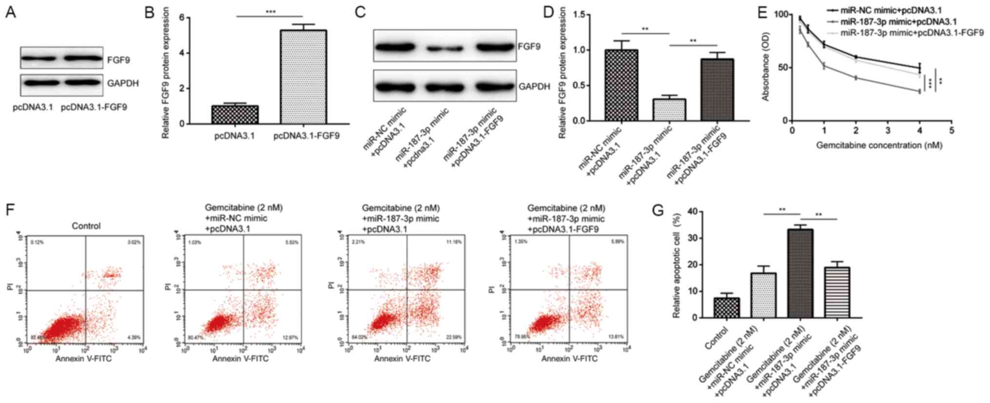 | Figure 6Overexpression of FGF9 reversed the
inhibitory effects of gemcitabine and miR-187-3p on MDA-MB-231
cells. (A) Representative western blotting images revealed that
compared with pcDNA3.1, transfection with pcDNA3.1-FGF9 increased
FGF9 expression in MDA-MB-231 cells. (B) Quantified data from (A)
demonstrating that compared with pcDNA3.1, pcDNA3.1-FGF9
transfection significantly increased FGF9 protein expression. (C)
Compared with cells co-transfected with miR-NC mimic and pcDNA3.1
plasmid, cells co-transfected with miR-187-3p mimic and pcDNA3.1
plasmid exhibited reduced FGF9 protein expression, which was
reversed by co-transfection with miR-187-3p mimic and pcDNA3.1-FGF9
plasmid. (D) Quantified data from (C) demonstrating that the
differences observed in (C) were statistically significant. (E)
Compared with the miR-NC mimic and pcDNA3.1 group, co-transfection
with the miR-187-3p mimic and pcDNA3.1 plasmid significantly
reduced MDA-MB-231 cell viability at each concentration of
gemcitabine (0.25, 0.5, 1, 2 and 4 nM), which was reversed by
co-transfection with the recombinant FGF9 plasmid. (F) Compared
with the control group, co-transfection with the miR-187-3p mimic
and pcDNA3.1 plasmid significantly enhanced gemcitabine (2
nM)-induced MDA-MB-231 cell apoptosis, which was reversed by
co-transfection with the recombinant FGF9 plasmid. (G) Quantified
data from (F) demonstrating that the differences observed in (F)
were statistically significant. Data are presented as the mean ±
standard deviation. **P<0.01,
***P<0.001 as indicated. FGF9. Fibroblast growth
factor 9; miR, microRNA; NC, negative control. |
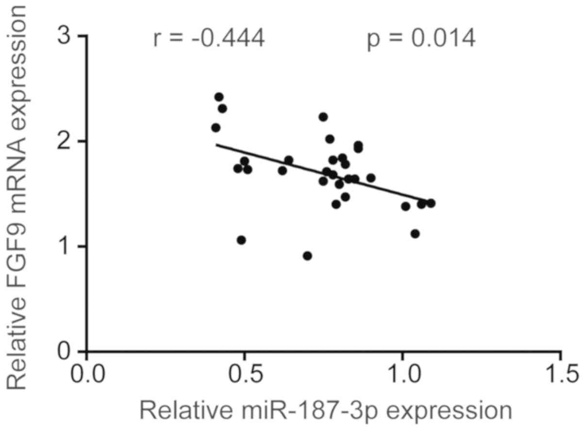
miR-187-3p expression negatively
correlates with that of FGF9 mRNA in breast tumor tissues
Following observations that miR-187-3p negatively
regulates FGF9 in breast cancer cells, the potential correlation
between miR-187-3p and FGF9 mRNA expression was determined in 30
breast cancer tumor and their matched adjacent non-tumor tissue
samples. The results revealed that FGF9 mRNA expression was
negatively correlated with miR-187-3p expression (Fig. 7), indicating that miR-187-3p may
regulate FGF9 during breast cancer pathogenesis.
Discussion
Breast cancer is the most commonly diagnosed type of
cancer in women, with its incidence ranking the highest among all
malignancies (27). Accumulating
evidence has demonstrated that targeted therapies offer superior
therapeutic efficacy compared with conventional approaches
(28). Recently, miRNAs have been
widely demonstrated to be associated with a variety of diseases
including breast cancer (29).
Gasparini et al (30)
reported that the downregulation of miR-27a and miR-30e in patients
with breast cancer correlated with poor patient outcomes, whereas
the overexpression of miR-155 and miR-493 correlated with good
patient outcomes. In addition, Kumaraswamy et al (31) revealed that miR-146a expression
correlated positively with that of breast cancer gene 1, which in
turn negatively regulated epidermal growth factor receptor
expression in breast cancer. The miR-200 family has previously been
demonstrated to serve important roles in the cell migration,
metastasis, proliferation and invasion of breast cancer (32,33).
miRNA expression profiles may influence responses to cancer
chemotherapy (34). miRNAs regulate
cancer drug resistance via a number of methods, including the
expression of specific drug targets, survival and/or apoptosis
signaling, cell proliferation and cell cycle progression (35). Furthermore, miR-200c was revealed to
be reduced in the doxorubicin-resistant MCF7 breast cancer cell
line (MCF7/ADR), where the overexpression of miR-200c increased
MCF7/ADR sensitivity to epirubicin (36). In addition, the expression of miR-7,
miR-326 and miR-345 were revealed to be markedly lower in the MCF7
the cisplatin-resistant cell line in comparison with the MCF7 cell
line (37). The function of
miR-187-3p in cancer remains controversial, as it has been
previously reported to be downregulated in many diseases, including
prostate cancer (38), retinal
ganglion (39), type 2 diabetes
(40), non-small cell lung cancer
(13) and breast cancer (14). miR-187-3p has been previously
associated with a number of different malignancies (13,14).
Using a miRNA microarray, Liang et al (23) determined that miR-187-3p was one of
the most significantly downregulated miRNAs in the
chemotherapy-resistant MCF7 cells compared with parental MCF7
cells. However, the function of miR-187-3p on breast cancer drug
resistance has yet to be clearly elucidated. In the present study,
it was revealed that miR-187-3p expression was significantly lower
in breast cancer tumor tissue compared with matched non-tumor
tissues collected from patients with breast cancer. miR-187-3p
overexpression significantly reduced cell viability and promoted
cell apoptosis, indicating that miR-187-3p is a tumor suppressor in
breast cancer. Although gemcitabine has been used for the treatment
of metastatic breast cancer since 2004(41), its therapeutic use in cancer
chemotherapy has been impeded at least in part by drug resistance
(42). Chaudhari et al
(43) performed miRNA microarray
analysis, which determined that doxorubicin treatment increased
miR-187-3p expression in human cardiomyocytes. In the present
study, cell viability and cell apoptosis assays revealed the effect
of miR-187-3p on breast cancer gemcitabine sensitivity. Data from
these analyses, it was indicated that miR-187-3p overexpression
increased gemcitabine sensitivity in breast cancer cells.
Although Fillmore et al (20) have previously explored the role of
FGF9 signaling in breast cancer cells, the association between
miRNA expression and FGF9 in breast cancer remain unclear.
According to the TargetScan database, FGF9 was predicted to be a
potential target gene of miR-187-3p, which was subsequently
confirmed by luciferase assays and observations that miR-187-3p
negatively regulated FGF9 expression.
Target genes of miR-187-3p were involved in the
regulation of signaling pathways, including phosphatidylinositol
3-kinase-AKT and hypoxia-inducible factor 1 signaling pathways in
epileptic rats (44). Hendrix et
al (45) revealed that FGF9 was
a downstream target of the Wnt signaling pathway, which may be an
indirect transcription target of β-catenin in ovarian endometrioid
adenocarcinomas. Abdel-Rahma et al (46) demonstrated that 80% FGF9 mutations
were associated with membranous β-catenin expression in colorectal
and endometrial carcinomas. In addition, FGF9 has been reported as
a target for the Wnt/β-catenin pathway in ovarian endometrioid
adenocarcinomas (26).
Interestingly, FGF9 interacted with Wnt/β-catenin signaling in a
feed-forward loop in lung development, as determined by Yin et
al (25). Wnt signaling is
associated with drug resistance in cancer (47). In addition, Shi et al
(48) elucidated that LGR5
suppressed docetaxel resistance in breast cancer through the
inactivation of the Wnt/β-catenin signaling. However, information
on the association between miR-187-3p and Wnt/β-catenin in breast
cancer pathophysiology remains unclear. In the current study,
overexpression of miR-187-3p in MDA-MB-231 cells significantly
reduced the protein expression of β-catenin, c-Myc and Cyclin D1.
In conclusion, miR-187-3p negatively regulated FGF9 expression,
which inactivated Wnt/β-catenin signaling and increased breast
cancer sensitivity to gemcitabine-mediated toxicity.
Taken together, the results of the present study
suggested that miR-187-3p increased gemcitabine sensitivity by
targeting FGF9 expression in breast cancer cells, indicating that
miR-187-3p may be a therapeutic target for patients with breast
cancer.
Acknowledgements
Not applicable.
Funding
No funding was received.
Availability of data and materials
The datasets used and/or analyzed during the present
study are available from the corresponding author on reasonable
request.
Authors' contributions
YW, YQ, WL, HY and XY carried out experiments and
analyzed the data. LT and JL collected the clinical samples and
analyzed the data. LL designed the study, analyzed the data and
prepared the manuscript. All author read and approved the final
version of the manuscript.
Ethics approval and consent to
participate
The present study was approved by the Ethic
Committee of Chifeng Municipal Hospital. Written consent was
provided by all participants prior to the study.
Patient consent for publication
Not applicable.
Competing interests
The authors declare that they have no competing
interests.
References
|
1
|
Siegel RL, Miller KD and Jemal A: Cancer
statistics, 2018. CA Cancer J Clin. 68:7–30. 2018.PubMed/NCBI View Article : Google Scholar
|
|
2
|
Feng Y, Spezia M, Huang S, Yuan C, Zeng Z,
Zhang L, Ji X, Liu W, Huang B, Luo W, et al: Breast cancer
development and progression: Risk factors, cancer stem cells,
signaling pathways, genomics, and molecular pathogenesis. Genes
Dis. 5:77–106. 2018.PubMed/NCBI View Article : Google Scholar
|
|
3
|
Strollo SE, Fallon EA, Gapstur SM and
Smith TG: Cancer-related problems, sleep quality, and sleep
disturbance among long-term cancer survivors at 9-years post
diagnosis. Sleep Med. 65:177–185. 2020.PubMed/NCBI View Article : Google Scholar
|
|
4
|
Tripathy D: Overview: Gemcitabine as
single agent therapy for advanced breast cancer. Clin Breast
Cancer. 3 (Suppl 1):8–11. 2002.PubMed/NCBI View Article : Google Scholar
|
|
5
|
Heinemann V: Role of gemcitabine in the
treatment of advanced and metastatic breast cancer. Oncology.
64:191–206. 2003.PubMed/NCBI View Article : Google Scholar
|
|
6
|
Hernandez-Aya LF and Ma CX: Chemotherapy
principles of managing stage IV breast cancer in the United States.
Chin Clin Oncol. 5:1–15. 2016.PubMed/NCBI View Article : Google Scholar
|
|
7
|
Blackstein M, Vogel CL, Ambinder R, Cowan
J, Iglesias J and Melemed A: Gemcitabin as first line therapy in
patient with metastatic breast cancer: A phase II trail. Oncology.
62:2–8. 2002.PubMed/NCBI View Article : Google Scholar
|
|
8
|
Bartel DP: MicroRNAs: Genomics,
biogenesis, mechanism, and function. Cell. 116:281–297.
2004.PubMed/NCBI View Article : Google Scholar
|
|
9
|
Lorio MV and Croce CM: MicroRNAs in
cancer: Small molecules with a huge impact. J Clin Oncol.
27:5848–5856. 2009.PubMed/NCBI View Article : Google Scholar
|
|
10
|
Zhang F, Luo Y, Shao Z, Xu L, Liu X, Niu
Y, Shi J, Sun X, Liu Y, Ding Y and Zhao L: MicroRNA-187, a
downstream effector of TGF beta pathway, suppresses Smad-mediated
epithelial-mesenchymal transition in colorectal cancer. Cancer
Lett. 373:203–213. 2016.PubMed/NCBI View Article : Google Scholar
|
|
11
|
Zhao J, Lei T, Xu C, Li H, Ma W, Yang Y,
Fan S and Liu Y: MicroRNA-187, down-regulated in clear cell renal
cell carcinoma and associated with lower survival, inhibits cell
growth and migration though targeting B7-H3. Biochem Biophys Res
Commun. 438:439–444. 2013.PubMed/NCBI View Article : Google Scholar
|
|
12
|
Casanova-Salas I, Masiá E, Armiñán A,
Calatrava A, Mancarella C, Rubio-Briones J, Scotlandi K, Vicent MJ
and López-Guerrero JA: MiR-187 targets the androgen-regulated gene
ALDH1A3 in prostate cancer. PLoS One. 10(e0125576)2015.PubMed/NCBI View Article : Google Scholar
|
|
13
|
Sun C, Li S, Yang C, Xi Y, Wang L, Zhang F
and Li D: MicroRNA-187-3p mitigates non-small cell lung cancer
(NSCLC) development through down-regulation of BCL6. Biochem
Biophys Res Commun. 471:82–88. 2016.PubMed/NCBI View Article : Google Scholar
|
|
14
|
Mulrane L, Madden SF, Brennan DJ, Gremel
G, McGee SF, McNally S, Martin F, Crown JP, Jirström K, Higgins DG,
et al: Mir-187 is an independent prognostic factor in breast cancer
and confers increased invasive potential in vitro. Clin Cancer Res.
18:6702–6713. 2012.PubMed/NCBI View Article : Google Scholar
|
|
15
|
Yan ZX, Wu LL, Xue K, Zhang QL, Guo Y,
Romero M, Leboeuf C, Janin A, Chen SJ, Wang L and Zhao WL:
MicroRNA187 overexpression is related to tumor progression and
determines sensitivity to bortezomib in peripheral T-cell lymphoma.
Leukemia. 28:880–887. 2014.PubMed/NCBI View Article : Google Scholar
|
|
16
|
Turner N and Grose R: Fibroblast growth
factor signaling: From development to cancer. Nat Rev Cancer.
10:116–129. 2010.PubMed/NCBI View
Article : Google Scholar
|
|
17
|
Grose R and Dickson C: Fibroblast growth
factor signaling in tumorigenesis. Cytokine Growth Factor Rev.
16:179–186. 2005.PubMed/NCBI View Article : Google Scholar
|
|
18
|
Ueda T, Volinia S, Okumura H, Shimizu M,
Taccioli C, Rossi S, Alder H, Liu CG, Oue N, Yasui W, et al:
Relation between microRNA expression and progression and prognosis
of gastric cancer: A microRNA expression analysis. Lancet Oncol.
11:136–146. 2010.PubMed/NCBI View Article : Google Scholar
|
|
19
|
Ornitz DM and Itoh N: Fibroblast growth
factors. Genome Biol. 2(REVIEWS 3005)2001.PubMed/NCBI View Article : Google Scholar
|
|
20
|
Fillmore CM, Gupta PB, Rudnick JA,
Caballero S, Keller PJ, Lander ES and Kuperwasser C: Estrogen
expands breast cancer stem-like cells through paracrine FGF/Tbx3
signaling. Proc Natl Acad Sci USA. 107:21737–21742. 2010.PubMed/NCBI View Article : Google Scholar
|
|
21
|
Yin Y, Castro AM, Hoekstra M, Yan TJ,
Kanakamedala AC, Dehner LP, Hill DA and Ornitz DM: Fibroblast
growth factor 9 regulation by MicroRNAs controls lung development
and links DICER1 loss to the pathogenesis of pleuropulmonary
blastoma. PLoS Genet. 11(e1005242)2015.PubMed/NCBI View Article : Google Scholar
|
|
22
|
Livak KJ and Schmittgen TD: Analysis of
relative gene expression data using real-time quantitative PCR and
the 2(-Delta Delta C(T)) method. Methods. 25:402–408.
2001.PubMed/NCBI View Article : Google Scholar
|
|
23
|
Liang Z, Wu H, Xia J, Li Y, Zhang Y, Huang
K, Wagar N, Yoon Y, Cho HT, Scala S and Shim H: Involvement of
miR-326 in chemotherapy resistance of breast cancer through
modulating expression of multidrug resistance-associated protein 1.
Biochem Pharmacol. 79:817–824. 2010.PubMed/NCBI View Article : Google Scholar
|
|
24
|
Modi S and Seidman AD: Single-agent
gemcitabine in the treatment of advanced breast cancer. Clin Breast
Cancer. 4 (Suppl 3):S101–S106. 2004.PubMed/NCBI View Article : Google Scholar
|
|
25
|
Yin Y, Wang F and Ornitz DM: Mesothelial-
and epithelial-derived FGF9 have distinct functions in the
regulation of lung development. Development. 138:3169–3177.
2011.PubMed/NCBI View Article : Google Scholar
|
|
26
|
Schwartz DR, Wu R, Kardia SL, Levin AM,
Huang CC, Shedden KA, Kuick R, Misek DE, Hanash SM, Taylor JM, et
al: Novel candidate targets of beta-catenin/T-cell factor signaling
identified by gene expression profiling of ovarian endometrioid
adenocarcinomas. Cancer Res. 63:2913–2922. 2003.PubMed/NCBI
|
|
27
|
Parvizpour S, Razmara J and Omidi Y:
Breast cancer vaccination comes to age: Impacts of bioinformatics.
Bioimpacts. 3:223–235. 2018.PubMed/NCBI View Article : Google Scholar
|
|
28
|
Shabnam B, Padmavathi G, Banik K, Girisa
S, Monisha J, Sethi G, Fan L, Wang L, Mao X and Kunnumakkara AB:
Sorcin a potential molecular target for cancer therapy. Transl
Oncol. 11:1379–1389. 2018.PubMed/NCBI View Article : Google Scholar
|
|
29
|
Temian DC, Pop LA, Irimie AI and
Berindan-Neagoe I: The epigenetics of triple-negative and
basal-like breast cancer: Current knowledge. J Breast Cancer.
21:233–243. 2018.PubMed/NCBI View Article : Google Scholar
|
|
30
|
Gasparini P, Cascione L, Fassan M, Lovat
F, Guler G, Balci S, Irkkan C, Morrison C, Croce CM, Shapiro CL and
Huebner K: MicroRNA expression profiling identifies a four microRNA
signature as a novel diagnostic and prognostic biomarker in triple
negative breast cancers. Oncotarget. 5:1174–1184. 2014.PubMed/NCBI View Article : Google Scholar
|
|
31
|
Kumaraswamy E, Wendt KL, Augustine LA,
Stecklein SR, Sibala EC, Li D, Gunewardena S and Jensen RA: BRCA1
regulation of epidermal growth factor receptor (EGFR) expression in
human breast cancer cells involves microRNA-146a and is critical
for its tumor suppressor function. Oncogene. 34:4333–4346.
2015.PubMed/NCBI View Article : Google Scholar
|
|
32
|
Humphries B, Wang Z, Oom AL, Fisher T, Tan
D, Cui Y, Jiang Y and Yang C: MicroRNA-200b targets protein kinase
Cɑ and suppresses triple-negative breast cancer metastasis.
Carcinogenesis. 35:2254–2263. 2014.PubMed/NCBI View Article : Google Scholar
|
|
33
|
Tsouko E, Wang J, Frigo DE, Aydoğdu E and
Williams C: MiR-200a inhibits migration of triple-negative breast
cancer cells through direct repression of the EPHA2 oncogene.
Carcinogenesis. 36:1051–1060. 2015.PubMed/NCBI View Article : Google Scholar
|
|
34
|
Blower PE, Chung JH, Verducci JS, Lin S,
Park JK, Dai Z, Liu CG, Schmittgen TD, Reinhold WC, Croce CM, et
al: MicroRNAs modulate the chemosensitivity of tumor cells. Mol
Cancer Ther. 7:1–9. 2008.PubMed/NCBI View Article : Google Scholar
|
|
35
|
Gottesman MM, Lav O, Hall MD and Gillet
JP: Toward a better understanding of the complexity of cancer drug
resistance. Annu Rev Pharmacol Toxicol. 56:85–102. 2016.PubMed/NCBI View Article : Google Scholar
|
|
36
|
Chen J, Tian W, Cai H, He H and Deng Y:
Down-regulation of microRNA-200c is associated with drug resistance
in human breast cancer. Med Oncol. 29:2527–2534. 2012.PubMed/NCBI View Article : Google Scholar
|
|
37
|
Pogribny IP, Filkowski JN, Tryndyak VP,
Golubov A, Shpyleva SI and Kovalchuk O: Alterations of microRNAs
and their targets are associated with acquired resistance of MCF-7
breast cancer cells to cisplatin. Int J Cancer. 127:1785–1794.
2010.PubMed/NCBI View Article : Google Scholar
|
|
38
|
Casanova-Salas I, Rubio-Briones J,
Calatrava A, Mancarella C, Masiá E, Casanova J, Fernández-Serra A,
Rubio L, Ramírez-Backhaus M, Armiñán A, et al: Identification of
miR-187 and miR-182 as biomarkers of early diagnosis and prognosis
in patients with prostate cancer treated with radical
prostatectomy. J Urol. 192:252–259. 2014.PubMed/NCBI View Article : Google Scholar
|
|
39
|
Zhang QL, Wang W, Li J, Tian SY and Zhang
TZ: Decreased miR-187 induces retinal ganglion cell apoptosis
through upregulating SMAD7 in glaucoma. Biomed Pharm Ther.
75:19–25. 2015.PubMed/NCBI View Article : Google Scholar
|
|
40
|
Locke JM, da Silva Xavier G, Dawe HR,
Rutter GA and Harries LW: Increased expression of miR-187 in human
islets from individuals with type 2 diabetes is associated with
reduced glucose-stimulated insulin secretion. Diabetologia.
57:122–128. 2014.PubMed/NCBI View Article : Google Scholar
|
|
41
|
Barton-Burke M: Gemcitabine: A
pharmacologic and clinical overview. Cancer Nurs. 22:176–183.
1999.PubMed/NCBI View Article : Google Scholar
|
|
42
|
Dyawanapelly S, Kumar A and Chourasia MK:
Lessons learned from gemcitabine: Impact of therapeutic carrier
systems and gemcitabine's drug conjugates on cancer therapy. Crit
Rev Ther Drug Carrier Syst. 1:63–96. 2017.PubMed/NCBI View Article : Google Scholar
|
|
43
|
Chaudhari U, Nemade H, Gaspar JA,
Hescheler J, Hengstler JG and Sachinidis A: MicroRNAs as early
toxicity signatures of doxorubicin in human-induced pluripotent
stem cell-derived cardiomyocytes. Arch Toxicol. 90:3087–3098.
2016.PubMed/NCBI View Article : Google Scholar
|
|
44
|
Zhang S, Kou Y, Hu C and Han Y: MicroRNA
profiling in the dentate gyrus in epileptic rats: The role of
miR-187-3p. Medicine (Baltimore). 96(e6744)2017.PubMed/NCBI View Article : Google Scholar
|
|
45
|
Hendrix ND, Wu R, Kuick R, Schwartz DR,
Fearon ER and Cho KR: Fibroblast growth factor 9 has oncogenic
activity and is a downstream target of wnt signaling in ovarian
endometrioid adenocarcinomas. Cancer Res. 66:1354–1362.
2006.PubMed/NCBI View Article : Google Scholar
|
|
46
|
Abdel-Rahman WM, Kalinina J, Shoman S,
Eissa S, Ollikainen M, Elomaa O, Eliseenkova AV, Bützow R,
Mohammadi M and Peltomäki P: Somatic FGF9 mutations in colorectal
and endometrial carcinomas associated with membranous β-catenin.
Hum Mutat. 29:390–397. 2008.PubMed/NCBI View Article : Google Scholar
|
|
47
|
Galluzzi L, Spranger S, Fuchs E and
López-Soto A: WNT signaling in cancer immunosurveillance. Trends
Cell Biol. 18:30143–30150. 2018.PubMed/NCBI View Article : Google Scholar
|
|
48
|
Shi S, Chen X, Liu H, Yu K, Bao Y, Chai J,
Gao H and Zou L: LGR5 acts as a target of miR-340-5p in the
suppression of cell progression and drug resistance in breast
cancer via Wnt/β-catenin pathway. Gene. 18:31039–31044.
2018.PubMed/NCBI View Article : Google Scholar
|