Introduction
The calcaneus epiphysitis, also known as Sever's
disease, was first reported in detail by James Warren Sever in
1912. The diseased population is child and adolescent who have
emerged and have not closed the calcaneal epiphysis. The clinical
manifestations are localized pain and swelling of the heel. The
positive medial-lateral compression test is helpful for the
diagnosis of the disease. Many factors such as chronic injury,
acute strain and cold stimulation can cause pain in the calcaneal
nodules, and high-intensity physical activity can aggravate the
pain (1). Its pathogenesis is
diverse, and the basic pathology is that the calcaneal callus is
subjected to repetitive microfracture damage, causing aseptic
inflammation of the calcaneal epiphysis, i.e. surrounding soft
tissue and ligament (2,3). There is currently no single explanation
for all cases of the disease. The X-ray shows an increase in the
density of the calcaneal epiphysis, and a radiolucent line of the
epiphysis - that is, a fragmentation of the epiphysis (4,5), but
this phenomenon can also occur in healthy children and adolescents
(6). Because the mechanical
structure of the calcaneus is relatively weak, it is prone to
injury (7). This study compared the
X-ray image of 98 cases of Sever's disease patients and 120 cases
of healthy children and adolescents. Among the 98 patients, 6
underwent MRI, the images showed abnormal signal. Statistical
analysis and images processing was performed to provide a basis for
the diagnosis of Sever's disease.
Patients and methods
General information
Children aged 8-15 years (8-10)
who underwent X-ray examination of the lateral ankle or lateral
calcaneus in People's Hospital of Rizhao (Rizhao, China) from May
2014 to April 2018 were retrospectively collected. There were 98
patients diagnosed as Sever's disease, including 72 males and 26
females. A total of 120 healthy children and adolescents were also
collected, 73 males and 47 females. Of the 98 patients, 6 underwent
MRI. Inclusion criteria: Complete and accurate personal
information, clear images, no metabolic and endocrine disorders, no
history of heel trauma, no dislocation of the ankle joint, and no
heel pain in the healthy population.
The study was approved by the Ethics Committee of
People's Hospital of Rizhao. Patients who participated in this
research had complete clinical data. The signed informed consents
were obtained from the patients or the guardians.
Inspection method
DR used the DR-VR/S 2.0 digital X-ray camera
produced by Philips, the Netherlands. The lateral view of the ankle
joint or the lateral view of the heel was X-ray plain film, the
focal length was 110 cm, and the center line was at the
talocalcaneal joint. Under the control of the automatic exposure
system, the tube voltage varied from 45 to 60 KV, and the tube
current varied from 1.5 to 4 mAs.
MRI adopted GE 1.5T magnetic resonance scanner and
barrel coil, sagittal FSE T1WI (TR/TE=540 ms/9.9 ms), sagittal,
coronal FSE T2WI (TR/TE=2,180 ms/27.8 ms), sagittal FSE STIR T2WI =
1,960 ms/31 ms, layer thickness was 4 mm, FOV was 16 cm.
Observation indicators
The manifestation of the Sever's disease and normal
calcaneal epiphysis on the lateral X-ray film was observed: The
density of the epiphysis (compact and cancellous bone type), and
the radiolucent line of the epiphysis (Figs.
1-3) was observed. The characteristics of changes in MRI signal
of Sever's disease were observed.
Statistical analysis
The statistical software package of SPSS 19.00 (IBM
Corp., Armonk, NY, USA) was used. The χ2 test was used
for the analysis between groups. P<0.05 for the difference was
statistically significant.
Results
Distribution of sex in Sever's disease
group and control group at different ages (Table I)
Among Sever's patients, the male incidence rate was
79.59%, the female incidence rate was 20.41%, the average age of
onset was 11.35, the average age of onset for male was 11.49, and
the average age of onset for female was 10.80.
The distribution and number of
calcaneus epiphysis signs in Sever's disease group and control
group at different ages (Table II).
Comparison of the density of calcaneus
In the Sever's group, the calcaneus epiphyses
compact type: 77 males (78.57%) and 19 females (19.39%); cancellous
bone type: 1 male (1.02%) and 1 female (1.02%).
In the control group, the calcaneus epiphyses
compact type: 50 males (41.67%) and 26 females (21.67%); cancellous
bone type: 23 males (19.17%) and 21 females (17.50%) (Table II).
There was a statistically significant difference in
the density of calcaneal epiphysis between the Sever's disease
group and the control group (χ2=38.85, P<0.05).
Comparison of the radiolucent line of
the calcaneus
In the Sever's group, the translucent line of the
calcaneus epiphysis was observed in: 78 males (79.59%) and 20
females (20.41%).
In the control group, the translucent line of the
calcaneus epiphysis was observed in: 15 males (12.50%) and 10
females (8.33%).
There was a statistically significant difference
between the Sever's disease group and the control group in terms of
the radiolucent line (χ2=137.51, P<0.05).
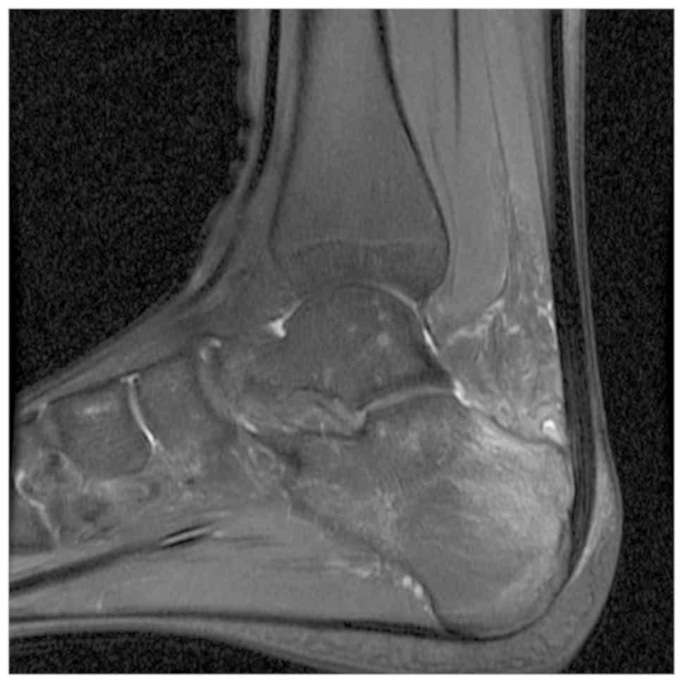
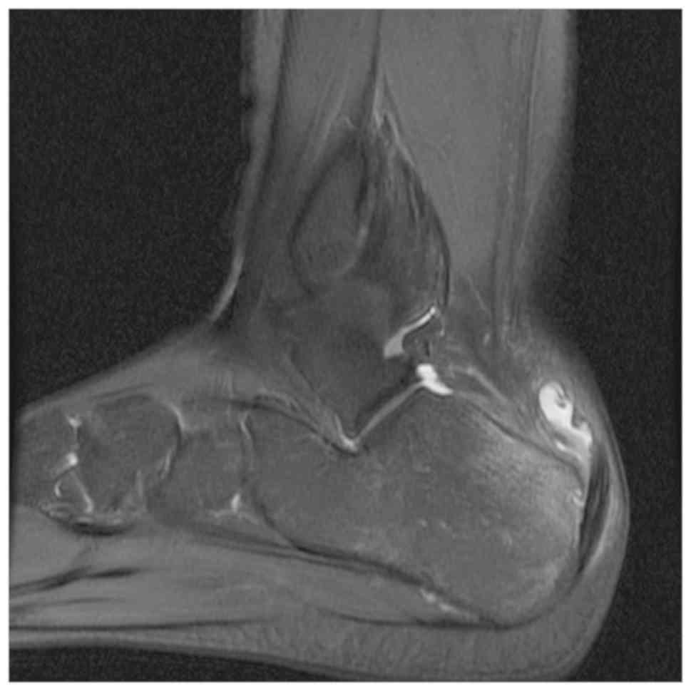
Sever's disease signal changes on MRI
images
The 6 patients who underwent MRI had different
degrees of bone marrow edema, joint effusion and surrounding soft
tissue swelling in MRI. Among them, 2 cases had high signal T2
weighed fat suppressed in the Achilles tendon attachment, and 1
case showed posterior Achilles tendon bursa effusion (Figs. 4 and 5).
Discussion
The results of this study suggested that the X-ray
of Sever's disease showed an increase in the density of the
epiphysis, a radiolucent line and a statistically significant
comparison with the control group, which is consistent with
previous studies (9,11-13).
However, in the calcaneus epiphysis development of healthy children
and adolescents, increased density and radiolucent line is also
observed (4,7-9). It
has been suggested in the literature that some patients with severe
Sever's disease have reduced density and atrophy of the calcaneus
epiphysis (4,14). In the present study, patients with
thinning and atrophy of the epiphysis have not been observed, which
may be related to the small number of samples and the increased
attention of parents. Radiolucent lines can also be seen in the
developmental period of normal developing children's calcaneus,
accounting for about 1/3, usually 1 to 2 lines, and radiolucent
lines were observed in all Sever's patients (11), with higher incidence in male than
female, higher incidence in those with larger amount of exercise,
obesity and in the preferential foot. Sever's disease is mostly
bilateral, and the incidence of boys is higher than that of girls
(15). The average age of onset of
girls in this study was at the age of 10.08 years, and the average
age of onset of boys was 11.49 years, which is consistent with the
pattern of growth and development of girls earlier than boys.
Due to the high cost of MRI examination and the long
waiting time required, there are few patients who choose this
examination, and it is impossible to do statistical analysis. The
signs and symptoms can only be observed according to a few cases.
The nature of Sever's disease is the process of microfracture of
bone cells and repairing after ischemic death. MRI has a good soft
tissue resolution and is sensitive to bone marrow edema, synovial
thickening, joint effusion and soft tissue edema (16). Sever's disease is a self-limiting
disease, mainly characterized by pain, swelling, and fewer other
symptoms such as fever. It is usually treated conservatively by
appropriately limiting physical activity and suggesting rest to
relieve the stimulation to the heel from pulling the Achilles
tendon, or by physical methods such as scaffold support, cold
compress, traction to alleviate the heel pain to achieve
self-healing, most patients only need the clinician physical
examination and the addition of lateral calcaneal view films, CT or
MRI may be required for very few serious cases with poor prognosis
from conservative treatment. Given the radioactivity of X-rays, it
is still controversial whether Sever's patients need to take a
film, but taking X-rays can rule out other lesions of the calcaneus
such as fractures, osteomyelitis, calcaneal tumors or tumor-like
lesions.
In conclusion, the main clinical manifestation of
Sever's disease is local pain of the calcaneus. The X-ray is mainly
characterized by increased density of the calcaneus epiphysis, and
the appearance of radiolucent line. It is sometimes difficult to
diagnose by X-ray film alone, the combination with the medical
history or MRI examination is required. MRI is the most accurate
imaging examination method for the damage of the epiphysis
(17), which can provide more
reliable image information for the observation and prognosis of
Sever's disease osteophyte soft tissue injury, and provide
direction for guiding clinical treatment (18,19).
This study was aimed to provide some help for clinical and imaging
diagnosis of Sever's disease.
Acknowledgements
Not applicable.
Funding
No funding was received.
Availability of data and materials
The datasets used and/or analyzed during the present
study are available from the corresponding author on reasonable
request.
Authors' contributions
YG conceived the study and drafted the manuscript.
JL and YL acquired the data. QL and SX analyzed the data and
revised the manuscript. All authors read and approved the final
manuscript.
Ethics approval and consent to
participate
The study was approved by the Ethics Committee of
People's Hospital of Rizhao (Rizhao, China). Patients who
participated in this research had complete clinical data. The
signed informed consents were obtained from the patients or the
guardians.
Patient consent for publication
Not applicable.
Competing interests
The authors declare that they have no competing
interests.
References
|
1
|
Ogden JA, Ganey TM, Hill JD and Jaakkola
JI: Sever's injury: A stress fracture of the immature calcaneal
metaphysis. J Pediatr Orthop. 24:488–492. 2004.PubMed/NCBI View Article : Google Scholar
|
|
2
|
Mustapić M, Borić I, Lepur D, Zadravec D
and Visković K: Sever's disease complicated with osteomyelitis.
Acta Clin Croat. 53:252–255. 2014.PubMed/NCBI
|
|
3
|
James AM, Williams CM and Haines TP: Heel
raises versus prefabricated orthoses in the treatment of posterior
heel pain associated with calcaneal apophysitis (Sever's Disease):
A randomised control trial. J Foot Ankle Res. 3(3)2010.PubMed/NCBI View Article : Google Scholar
|
|
4
|
Cassas KJ and Cassettari-Wayhs A:
Childhood and adolescent sports-related overuse injuries. Am Fam
Physician. 73:1014–1022. 2006.PubMed/NCBI
|
|
5
|
Kose O, Celiktas M, Yigit S and Kisin B:
Can we make a diagnosis with radiographic examination alone in
calcaneal apophysitis (Sever's disease)? J Pediatr Orthop B.
19:396–398. 2010.PubMed/NCBI View Article : Google Scholar
|
|
6
|
Puffinbarger WR, Gruel CR, Herndon WA and
Sullivan JA: Osteomyelitis of the calcaneus in children. J Pediatr
Orthop. 16:224–230. 1996.PubMed/NCBI View Article : Google Scholar
|
|
7
|
Ramponi DR and Baker C: Sever's Disease
(Calcaneal Apophysitis). Adv Emerg Nurs J. 41:10–14.
2019.PubMed/NCBI View Article : Google Scholar
|
|
8
|
Howard R: Diagnosing and treating Sever's
disease in children. Emerg Nurse. 22:28–30. 2014.PubMed/NCBI View Article : Google Scholar
|
|
9
|
Ceylan HH and Caypinar B: Incidence of
calcaneal apophysitis in Northwest Istanbul. BMC Musculoskelet
Disord. 19:267–272. 2018.PubMed/NCBI View Article : Google Scholar
|
|
10
|
Perhamre S, Lazowska D, Papageorgiou S,
Lundin F, Klässbo M and Norlin R: Sever's injury: A clinical
diagnosis. J Am Podiatr Med Assoc. 103:361–368. 2013.PubMed/NCBI View
Article : Google Scholar
|
|
11
|
Liberson A, Lieberson S, Mendes DG,
Shajrawi I, Ben Haim Y and Boss JH: Remodeling of the calcaneus
apophysis in the growing child. J Pediatr Orthop B. 4:74–79.
1995.PubMed/NCBI View Article : Google Scholar
|
|
12
|
Scharfbillig RW, Jones S and Scutter SD:
Sever's disease: What does the literature really tell us? J Am
Podiatr Med Assoc. 98:212–223. 2008.PubMed/NCBI View
Article : Google Scholar
|
|
13
|
Weiner DS, Morscher M and Dicintio MS:
Calcaneal apophysitis: Simple diagnosis, simpler treatment. J Fam
Pract. 56:352–355. 2007.PubMed/NCBI
|
|
14
|
Wiegerinck JI, Yntema C, Brouwer HJ and
Struijs PA: Incidence of calcaneal apophysitis in the general
population. Eur J Pediatr. 173:677–679. 2014.PubMed/NCBI View Article : Google Scholar
|
|
15
|
Davison MJ, David-West SK and Duncan R:
Careful assessment the key to diagnosing adolescent heel pain.
Practitioner. 260:30–33. 2016.PubMed/NCBI
|
|
16
|
Pouletaut P, Claude I, Winzenrieth R, Ho
Ba Tho MC and Sebag G: Automated analysis of MR image of hip:
Geometrical evaluation of the Legg-Calve-Perthes disease. Med Eng
Phys. 27:415–424. 2005.PubMed/NCBI View Article : Google Scholar
|
|
17
|
Park YH, Lim JW, Choi GW and Kim HJ:
Quantitative magnetic resonance imaging analysis of the common site
of Acute Achilles tendon rupture: 5 to 8 cm above the distal end of
the calcaneal insertion. Am J Sports Med. 47:2374–2379.
2019.PubMed/NCBI View Article : Google Scholar
|
|
18
|
Kose O: Do we really need radiographic
assessment for the diagnosis of non-specific heel pain (calcaneal
apophysitis) in children? Skeletal Radiol. 39:359–361.
2010.PubMed/NCBI View Article : Google Scholar
|
|
19
|
Rossi I, Rosenberg Z and Zember J: Normal
skeletal development and imaging pitfalls of the calcaneal
apophysis: MRI features. Skeletal Radiol. 45:483–493.
2016.PubMed/NCBI View Article : Google Scholar
|