Introduction
Trigeminal neuralgia is a severe unilateral facial
pain involving one or more branches of the trigeminal nerve
(1,2). It is usually unilateral, severe,
transient, and recurrent, causing great pain to patients. As the
disease progresses, patients may have difficulty eating, talking
and maintaining facial hygiene in fear of triggering the pain,
which has a profound impact on the quality of life (1,3,4). Neuropathic pain is common in trigeminal
neuralgia, peripheral neuropathy, and multiple sclerosis (5). Primary trigeminal neuralgia is caused
by the compression of the trigeminal nerve by adjacent blood
vessels (6), which is recurrent and
very difficult to treat conservatively. Its unbearable severe pain
has serious effects on the physical and psychological health of
patients (7-9), so
its treatment has become the focus of clinical research.
The current preferred surgical treatment for primary
trigeminal neuralgia is microvascular decompression (MVD) (10). MVD, a minimally invasive
interventional technique which is able to identify the pain nerve,
can effectively isolate the responsible vessels compressing the
roots of the trigeminal nerve and the brainstem so as to relieve
the compression, and repair the pain nerve under the operating
microscope to eradicate the trigeminal pain source. This minimally
invasive technique completely retains the function of blood vessels
and nerves, is an effective treatment for trigeminal neuralgia
(11,12). However, clinical data revealed that
MVD is not effective for some patients, leaving poorly relieved
pain and a large postoperative trauma (13). Percutaneous stereotactic
radiofrequency rhizotomyroot (PSR) is also common in treating
primary trigeminal neuralgia (14),
suitable for cases like recurrent disease after surgery and failed
identification of the responsible blood vessels by MVD. PSR only
partially cuts off the sensory roots, so it can save the function
of the patient's movement roots and relieve the patient's facial
pain in a very effective manner (15). However, clinical records show that
some patients treated with PSR are prone to a variety of
complications and a poor prognosis (16).
To find a better treatment for primary trigeminal
neuralgia, in this study, the clinical efficacy of MVD and MVD
combined with PSR in primary trigeminal neuralgia was compared to
confirm the effect of MVD and PSR on primary trigeminal neuralgia
to provide reference for the treatment of this disease.
Patients and methods
General information
A prospective analysis was performed. A total of 141
patients with primary trigeminal neuralgia admitted into Shandong
Provincial Hospital (Jinan, China) from May 2011 to June 2013 were
collected. Among them, 63 patients (26 male and 37 female, aged
37.6±7.9 years) with MVD were studied as group A, while the other
78 (45 male and 33 female, aged 36.5±8.3 years) with MVD combined
with PSR were studied as group B.
Inclusion and exclusion criteria
Inclusion criteria: Patients diagnosed with primary
trigeminal neuralgia according to the criteria of the International
Headache Society (IHS); patients with no neurological deficits;
patients with poor efficacy from drug treatment; patients with
significant nerve compression; patient with vascular compression
according to imaging examination. The present study was approved by
the Ethics Committee of Shandong Provincial Hospital (SPH201104).
All patients and their families signed the informed consent.
Exclusion criteria: Patients with cardiovascular and
cerebrovascular diseases, malignant tumors, mental disorders,
symptoms of secondary trigeminal neuralgia such as multiple
sclerosis or tumor nerve compression symptoms; patients with
surgery contraindications; pregnant or lactating women.
Treatment methods
All surgery procedures were performed by the same
physician. Patients were generally anesthetized and placed in the
prone position, with head down at approximately 15 degrees to
expose the mastoid of the operating side at the highest level of
the head. In the longitudinal incision in the hairline behind the
ear, the retrosigmoid sinus approach was made to fully expose the
posterior margin of the mastoid. A hole was drilled in the
posterior occipital lacunae of the mastoid and was expanded by a
rongeur to make a bone window of approximately 1.5 to 2.5 cm and
the transverse sinus and sigmoid sinus were exposed. The mastoid
air cell was closed by bone wax. The dura mater was given a ‘⊥’ cut
and suspended. The subarachnoid space was opened under the
microscope, and the cerebellar hemisphere was lightly pulled to the
inner side with the brain plate. The arachnoid was sharply cut with
a micro-scissor to identify the compressing vessels and isolate it
from the initial segment of the trigeminal nerve root. The Teflon
cotton was placed between the responsible vessels and the brain
stem to decompress the blood vessels. 6 hours after the completion
of MVD, PSR was performed to detect the presence of thick veins on
the surgical site to compress the trigeminal nerve. The arachnoid
band between the vein and the nerve or the brainstem was cut, and
the veins were free. Isolation, distance or suspension were
selected to decompress according to the type of venous compression
(electrocoagulation shearing can be performed in the case of
difficult separation), and then PSR is performed on the sensory
nerves 1/3 to 1/5 of the posterior lateral of the trigeminal
sensory root. Under the microscope, the trigeminal nerve root was
carefully and sharply cut off. The operation is performed with
caution. It is strictly required to avoid mechanical damage to
blood vessels and nerves.
Postoperative treatment
The patient was treated with intravenous
dexamethasone to avoid postoperative rejection. Routine
examinations were performed after the surgery to monitor the
recovery and the complications. If no abnormalities were observed,
patients were allowed to discharge one week after the surgery.
Outcome measures
Main outcome measure: The postoperative surgical
outcomes of the two groups were observed. The efficacy criteria are
shown in Table I.
 | Table I.Efficacy evaluation. |
Table I.
Efficacy evaluation.
| Response | Criteria |
|---|
| Cured | The pain completely
disappeared and no drug was needed. |
| Marked response | The pain was relieved
by 90%, the drug was used occasionally or in a small dose. |
| Moderate
response | The pain was relieved
to a certain degree, the dosage of the drug was reduced by 50%, or
the multiple pain turned into single pain. |
| No response | The pain was not
relieved. |
Secondary outcome measures: The incidence of
postoperative complication in the two groups was observed; the risk
factors affecting the efficacy were explored by the multivariate
analysis; the quality of life after the treatment of the two groups
was monitored; the 5-year recurrence rate of the two groups was
observed.
Statistical methods
Statistical analysis was performed using SPSS 19.0
(Chicago SPSS Co., Ltd.), and the collected data were visualized
using GraphPad Prism 7 (San Diego Graphpad Software Co., Ltd.). The
count data were expressed with rate (%) and compared by the
Chi-square test. The measurement data were expressed with the mean
± standard deviation (mean ± SD) and compared between two groups by
the independent sample t-test. Logistic regression test was used
for multivariate analysis. K-M survival curve was used to analyze
the 5-year recurrence of patients. A statistical difference was
recognized at P<0.05.
Results
General clinical data of group A and
group B
Group A (patients treated with MVD) and group B
(patients treated with MVD combined with PSR) were not
statistically different in age, sex, BMI (kg/m2),
painful side, clinical symptoms, compressing vessels, compression
degree, duration of disease, place of residence, smoking or
drinking (P>0.05). Details are shown in Table II.
| Table IIGeneral clinical data of group A and
group B [n (%)]. |
Table II
General clinical data of group A and
group B [n (%)].
| Factors | Group A (n=63) | Group B (n=78) | t/χ2
value | P-value |
|---|
| Sex | | | 3.760 | 0.053 |
|
Male | 26 (41.27) | 45 (57.69) | | |
|
Female | 37 (58.73) | 33 (42.31) | | |
| Age (years) | 37.6±7.9 | 36.5±8.3 | 0.799 | 0.426 |
| BMI
(kg/m2) | 23.57±2.15 | 24.07±2.13 | 1.380 | 0.170 |
| Painful side | | | 0.263 | 0.608 |
|
Left
side | 32 (50.79) | 43 (55.13) | | |
|
Right
side | 31 (49.21) | 35 (44.87) | | |
| Clinical
symptoms | | | 0.564 | 0.453 |
|
Typical | 34 (53.97) | 47 (60.26) | | |
|
Not
typical | 29 (46.03) | 31 (39.74) | | |
| Compressing
vessels | | | 0.983 | 0.612 |
|
Artery
compression | 18 (28.57) | 17 (21.79) | | |
|
Venous
compression | 20 (31.75) | 25 (32.05) | | |
|
Mixed
compression | 25 (39.68) | 36 (46.15) | | |
| Compression
degree | | | 0.017 | 0.897 |
|
Displaced | 30 (44.44) | 38 (42.31) | | |
|
Not
displaced | 33 (47.62) | 40 (46.15) | | |
| Duration of disease
(years) | | | 5.425 | 0.994 |
|
<5 | 25 (39.68) | 31 (39.74) | | |
|
≥5 | 38 (60.32) | 47 (60.26) | | |
| Place of
residence | | | 0.101 | 0.751 |
|
Urban
area | 29 (46.03) | 38 (48.72) | | |
|
Rural
area | 34 (53.97) | 40 (51.28) | | |
| Smoking | | | 0.015 | 0.901 |
|
Yes | 26 (41.27) | 33 (42.31) | | |
|
No | 37 (58.73) | 45 (57.69) | | |
| Drinking | | | 0.726 | 0.394 |
|
Yes | 23 (36.51) | 34 (43.59) | | |
|
No | 40 (63.49) | 44 (56.41) | | |
Evaluation of the efficacy in group A
and group B
The total effective rate was 88.89% in group A,
lower than that in group B (96.15%), but the difference was not
statistically significant (P>0.05) (Table III).
| Table IIIEvaluation of the efficacy in group A
and group B [n (%)]. |
Table III
Evaluation of the efficacy in group A
and group B [n (%)].
| Response | Group A (n=63) | Group B (n=78) | χ2
value | P-value |
|---|
| Cured | 38 (60.32) | 48 (61.54) | 0.022 | 0.883 |
| Marked
response | 10 (15.87) | 15 (19.23) | 0.269 | 0.604 |
| Moderate
response | 8 (12.70) | 12 (15.38) | 0.207 | 0.650 |
| No response | 7 (11.11) | 3 (3.85) | 2.792 | 0.095 |
| Total effective
rate | 56 (88.89) | 75 (96.15) | 2.792 | 0.095 |
The incidence of complications after
treatment in group A and group B
The number of patients experiencing nausea and
vomiting, peripheral facial paralysis, hearing loss, cerebrospinal
fluid leakage, subcutaneous effusion in group A were 13, 5, 3, 1,
and 3, respectively, and the total incidence of adverse reaction in
group A was 39.68%, while the numbers in group B were 5, 3, 1, 0,
and 1, respectively, and the total incidence of adverse reaction in
group B was 12.82%. The difference between the two groups in the
adverse reactions was statistically significant (P<0.05).
Additional details are shown in Table
IV.
| Table IVThe incidence of complications after
treatment in group A and group B [n (%)]. |
Table IV
The incidence of complications after
treatment in group A and group B [n (%)].
| Group | Nausea and
vomiting | Peripheral facial
paralysis | Hearing loss | Cerebrospinal fluid
leakage | Subcutaneous
effusion | Total | χ2
value | P-value |
|---|
| Group A (n=63) | 13 (20.63) | 5 (7.94) | 3 (4.76) | 1 (1.59) | 3 (4.76) | 25 (39.68) | 13.48 | <0.001 |
| Group B (n=78) | 5 (6.41) | 3 (3.85) | 1 (1.28) | 0 (0) | 1 (1.28) | 10 (12.82) | | |
Univariate analysis of the
efficacy
The two groups of patients were divided into the
cured group (n=86) and the not-cured group (n=55) according to
efficacy. The univariate analysis of the clinical data of the two
groups demonstrated that the cured group and the not-cured group
were not statistically different in sex, age, BMI
(kg/m2), painful side, compressing vessels, smoking,
drinking, and place of residence (P>0.05), but statistically
different in the decompression degree, duration of disease,
compression degree, and clinical symptoms (P<0.05). Additional
details are shown in Tables
V-VII.
Comparison of postoperative quality of
life between group A and group B
The quality of life of patients at one year of
treatment in group B was significantly better than that of patients
in group B in terms of physiological role, overall health,
physiological function, vitality, physical pain, mental health,
emotional function, social function (P<0.05). More details are
shown in Table VIII.
| Table VIIIComparison of postoperative quality
of life between group A and group B. |
Table VIII
Comparison of postoperative quality
of life between group A and group B.
| Quality of life
score | Group B (n=78) | Group A (n=63) | t value | P-value |
|---|
| Physiological
role |
36.19±6.61a | 25.53±5.28 | 8.068 | <0.001 |
| Overall health |
39.62±8.34a | 28.39±8.70 | 5.967 | <0.001 |
| Physiological
function |
42.52±11.09a | 33.65±10.11 | 3.785 | <0.001 |
| Vitality |
44.45±6.91a | 35.98±6.67 | 5.647 | <0.001 |
| Physical pain |
46.94±8.81a | 37.05±8.01 | 5.318 | <0.001 |
| Mental health |
48.18±7.28a | 36.72±7.23 | 7.152 | <0.001 |
| Emotional
function |
43.42±8.78a | 32.97±8.19 | 5.573 | <0.001 |
| Social
function |
43.72±7.89a | 36.87±7.16 | 4.117 | <0.001 |
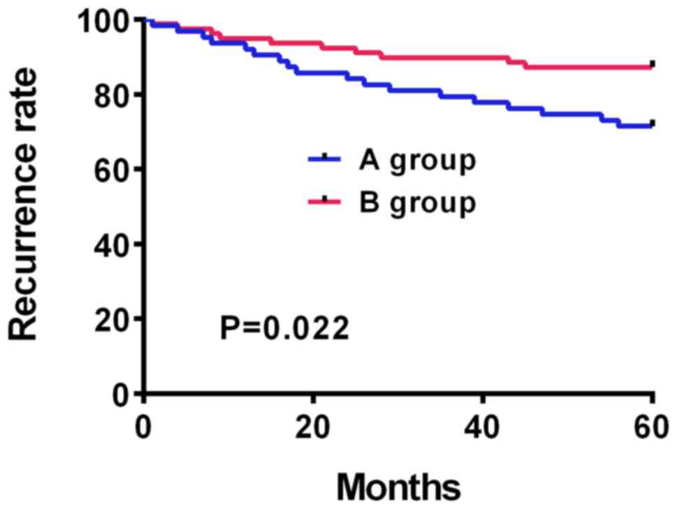
The 5-year recurrence rate in both
groups
According to the statistics, all 141 patients and
their families were successfully followed up. Five years after the
surgery, 18 patients in group A had recurrent disease, with a
recurrence rate of 28.57%, and 10 patients in group B had recurrent
disease, with a recurrence rate of 12.82%. The figure of the 5-year
recurrence of the two groups displayed that the 5-year recurrence
rate of group B was statistically lower than that of group A
(P=0.022) (Fig. 1).
Discussion
Primary trigeminal neuralgia is debilitating,
characterized by unilateral facial pain following the sensory
distribution of cranial nerve V (17). Most patients with long-term use of
drugs can develop drug resistance and experience side effects
(18), and thus need surgery. MVD is
the most common surgery for primary trigeminal neuralgia, but it is
limited by age, pain level, medical comorbidities, past surgery
procedures, and patient preference (19,20).
Minimally invasive techniques such as PSR are designed to damage
nerves for pain control (21,22). To
discover better treatments for primary trigeminal nerves, the
application of MVD combined with PSR was investigated in this
study.
This study found that MVD combined with PSR
treatment had better efficacy than MVD alone. The study by Zeng
et al (23) compared the
efficacy of MVD combined with PSR and MVD for primary trigeminal
neuralgia and found that MVD combined with PSR was better than MVD
alone because the pain was completely eliminated soon after PSR
combined with MVD, which is similar to the results of the present
study. The study by Zeng et al only listed the pain relief
rate of the patient for two years, but in this study, the follow-up
time was extended and it was found that the recurrence rate of
patients receiving MVD combined with PSR was significantly lower
than that of patients receiving MVD alone. It was speculated that
MVD combined with PSR can identify responsible blood vessels more
accurately, treat patients with different symptoms and eliminate
the pain more efficiently. The study by Du et al (24) employed percutaneous balloon
compression of trigeminal ganglion for recurrent trigeminal
neuralgia after MVD because they believed MVD does not achieve a
100% cure rate and it carries high risk and low success rate for
recurrent MVD, suggesting that MVD treatment is not suitable for
all the patients. MVD is a non-destructive treatment. With
identified pain nerve by medical equipment, MVD can relieve the
compression of the roots of the trigeminal nerve and preserve the
functions of blood vessels and nerves. PSR can remove the root
nerve to stop the pain. MVD is not suitable for patients whose
responsible vessels are not determined, besides, it causes large
trauma. Considering different needs of different patients, it was
speculated that MVD combined with PSR treatment can be applied to
more patients to achieve better efficacy. The postoperative
complications of the two treatments were compared and the results
showed that MVD combined with PSR treatment had significantly fewer
complications than MVD, indicating that MVD combined with PSR may
be a better treatment for primary trigeminal neuralgia. A
univariate analysis was performed on the efficacy of the two groups
before the multivariate analysis and a statistical difference was
found between the patients cured and not cured in the degree of
decompression, duration of disease, degree of compression, and
clinical symptoms. Multivariate analysis was then performed by
logistic regression test and revealed that the decompression
degree, duration of the disease, the compression degree, and the
clinical symptoms were risk factors for the patient's poor
efficacy. Such results suggest that more attention should be paid
to select suitable treatment for patients with poor decompression
and compression degree, long disease duration, and severe clinical
symptoms to achieve better efficacy. The following comparison of
quality of life scores between the two groups demonstrated that
patients treated with MVD combined with PSR had a higher quality of
life and a lower 5-year recurrence rate than patients treated with
MVD alone. This further suggests that the efficacy of MVD combined
with PSR is better.
In summary, MVD combined with PSR in the treatment
of primary trigeminal neuralgia has better efficacy, fewer
complications, higher quality of life of patients, and lower 5-year
recurrence rate than MVD alone. The risk factors affecting the
efficacy of patients include the decompression degree, duration of
disease, compression degree, and clinical symptoms.
Acknowledgements
Not applicable.
Funding
No funding was received.
Availability of data and materials
The datasets used and/or analyzed during the present
study are available from the corresponding author on reasonable
request.
Authors' contributions
GZ, XS and BF conceived and designed the study. GZ,
ZZ, HY and XZ were responsible for the collection, analysis and
interpretation of the data. GZ drafted the manuscript. GZ and BF
revised the manuscript critically for important intellectual
content. GZ wrote the manuscript. All authors read and approved the
final manuscript.
Ethics approval and consent to
participate
The study was approved by the Ethics Committee of
Shandong Provincial Hospital (Jinan, China). Signed informed
consents were obtained from the patients and/or the guardians.
Patient consent for publication
Not applicable.
Competing interests
The authors declare that they have no competing
interests.
References
|
1
|
Moore D, Chong MS, Shetty A and Zakrzewska
JM: A systematic review of rescue analgesic strategies in acute
exacerbations of primary trigeminal neuralgia. Br J Anaesth.
123:e385–e396. 2019.PubMed/NCBI View Article : Google Scholar
|
|
2
|
Zhang YP, Wang Y, Xia WG and Song AQ:
Triple Puncture for primary trigeminal neuralgia: A randomized
clinical trial. Curr Med Sci. 39:638–644. 2019.PubMed/NCBI View Article : Google Scholar
|
|
3
|
Hu H, Chen L, Ma R, Gao H and Fang J:
Acupuncture for primary trigeminal neuralgia: A systematic review
and PRISMA-compliant meta-analysis. Complement Ther Clin Pract.
34:254–267. 2019.PubMed/NCBI View Article : Google Scholar
|
|
4
|
Pokhrel D, Sood S, McClinton C, Saleh H,
Badkul R, Jiang H, Stepp T, Camarata P and Wang F: Linac-based
stereotactic radiosurgery (SRS) in the treatment of refractory
trigeminal neuralgia: Detailed description of SRS procedure and
reported clinical outcomes. J Appl Clin Med Phys. 18:136–143.
2017.PubMed/NCBI View Article : Google Scholar
|
|
5
|
Qin ZL, Yang LQ, Li N, Yue JN, Wu BS, Tang
YZ, Guo YN, Lai GH and Ni JX: Clinical study of cerebrospinal fluid
neuropeptides in patients with primary trigeminal neuralgia. Clin
Neurol Neurosurg. 143:111–115. 2016.PubMed/NCBI View Article : Google Scholar
|
|
6
|
Montano N, Conforti G, Di Bonaventura R,
Meglio M, Fernandez E and Papacci F: Advances in diagnosis and
treatment of trigeminal neuralgia. Ther Clin Risk Manag.
11:289–299. 2015.PubMed/NCBI View Article : Google Scholar
|
|
7
|
Wolf A and Kondziolka D: Trigeminal
neuralgia and other facial neuralgias. Prog Neurol Surg.
34:273–278. 2019.PubMed/NCBI View Article : Google Scholar
|
|
8
|
Marchetti M, Pinzi V, De Martin E,
Ghielmetti F and Fariselli L: Radiosurgery for trigeminal
neuralgia: The state of art. Neurol Sci. 40 (Suppl 1):153–157.
2019.PubMed/NCBI View Article : Google Scholar
|
|
9
|
Ding Y, Li H, Hong T, Zhu Y, Yao P and
Zhou G: Combination of pulsed radiofrequency with continuous
radiofrequency thermocoagulation at low temperature improves
efficacy and safety in V2/V3 primary trigeminal neuralgia. Pain
Physician. 21:E545–E553. 2018.PubMed/NCBI
|
|
10
|
Chen ZW, Ma JB, Xie KY, Huang B, Yao M,
Fei Y and Zhang L: A study of the relations of foramen rotundum
structure direction and the approach of percutaneous puncturing of
radiofrequency thermocoagulation for treating V2 of primary
trigeminal neuralgia. Zhonghua Yi Xue Za Zhi. 98:436–439. 2018.(In
Chinese). PubMed/NCBI View Article : Google Scholar
|
|
11
|
Liang C, Wang XY, Li J, Zhang M and Liu
HF: Modified backward avulsion used in the treatment of
third-branch primary trigeminal neuralgia. J Craniofac Surg.
29:e87–e90. 2018.PubMed/NCBI View Article : Google Scholar
|
|
12
|
Mistry AM, Niesner KJ, Lake WB, Forbes JA,
Shannon CN, Kasl RA, Konrad PE and Neimat JS: Neurovascular
compression at the root entry zone correlates with trigeminal
neuralgia and early microvascular decompression outcome. World
Neurosurg. 95:208–213. 2016.PubMed/NCBI View Article : Google Scholar
|
|
13
|
Ma S, Agarwalla PK, van Loveren HR and
Agazzi S: Successful microvascular decompression for trigeminal
neuralgia secondary to a persistent trigeminal artery. Oper
Neurosurg (Hagerstown). 16:18–22. 2019.PubMed/NCBI View Article : Google Scholar
|
|
14
|
Breeze RE and Craig DB: Microvascular
decompression for trigeminal neuralgia: A durable, noncompressive
technique using teflon secured with mini clip. Oper Neurosurg
(Hagerstown). 16:580–582. 2019.PubMed/NCBI View Article : Google Scholar
|
|
15
|
Wang Y, Yang Q, Cao D, Seminowicz D,
Remeniuk B, Gao L and Zhang M: Correlation between nerve atrophy,
brain grey matter volume and pain severity in patients with primary
trigeminal neuralgia. Cephalalgia. 39:515–525. 2019.PubMed/NCBI View Article : Google Scholar
|
|
16
|
Anichini G, Iqbal M, Rafiq NM, Ironside JW
and Kamel M: Sacrificing the superior petrosal vein during
microvascular decompression. Is it safe? Learning the hard way.
Case report and review of literature. Surg Neurol Int. 7 (Suppl
14):S415–S420. 2016.PubMed/NCBI View Article : Google Scholar
|
|
17
|
Hussain MA, Konteas A, Sunderland G,
Franceschini P, Byrne P, Osman-Farah J and Eldridge P:
Re-exploration of microvascular decompression in recurrent
trigeminal neuralgia and intraoperative management options. World
Neurosurg. 117:e67–e74. 2018.PubMed/NCBI View Article : Google Scholar
|
|
18
|
Kang IH, Park BJ, Park CK, Malla HP, Lee
SH and Rhee BA: A clinical analysis of secondary surgery in
trigeminal neuralgia patients who failed prior treatment. J Korean
Neurosurg Soc. 59:637–642. 2016.PubMed/NCBI View Article : Google Scholar
|
|
19
|
Wang DD, Raygor KP, Cage TA, Ward MM,
Westcott S, Barbaro NM and Chang EF: Prospective comparison of
long-term pain relief rates after first-time microvascular
decompression and stereotactic radiosurgery for trigeminal
neuralgia. J Neurosurg. 128:68–77. 2018.PubMed/NCBI View Article : Google Scholar
|
|
20
|
Romanelli P, Conti A, Bianchi L, Bergantin
A, Martinotti A and Beltramo G: Image-guided robotic radiosurgery
for trigeminal neuralgia. Neurosurgery. 83:1023–1030.
2018.PubMed/NCBI View Article : Google Scholar
|
|
21
|
Chen JN, Yu WH, Du HG, Jiang L, Dong XQ
and Cao J: Prospective comparison of redo microvascular
decompression and percutaneous balloon compression as primary
surgery for recurrent trigeminal neuralgia. J Korean Neurosurg Soc.
61:747–752. 2018.PubMed/NCBI View Article : Google Scholar
|
|
22
|
Zhang D, Meng Y, Hai H, Yu XT and Ma YW:
Radial extracorporeal shock wave therapy in an individual with
primary trigeminal neuralgia: A case report and literature review.
Am J Phys Med Rehabil. 97:e42–e45. 2018.PubMed/NCBI View Article : Google Scholar
|
|
23
|
Zeng YJ, Zhang H, Yu S, Zhang W and Sun
XC: Efficacy and safety of microvascular decompression and gamma
knife surgery treatments for patients with primary trigeminal
neuralgia: A prospective study. World Neurosurg. 116:e113–e117.
2018.PubMed/NCBI View Article : Google Scholar
|
|
24
|
Du Y, Yang D, Dong X, Du Q, Wang H and Yu
W: Percutaneous balloon compression (PBC) of trigeminal ganglion
for recurrent trigeminal neuralgia after microvascular
decompression (MVD). Ir J Med Sci. 184:745–751. 2015.PubMed/NCBI View Article : Google Scholar
|