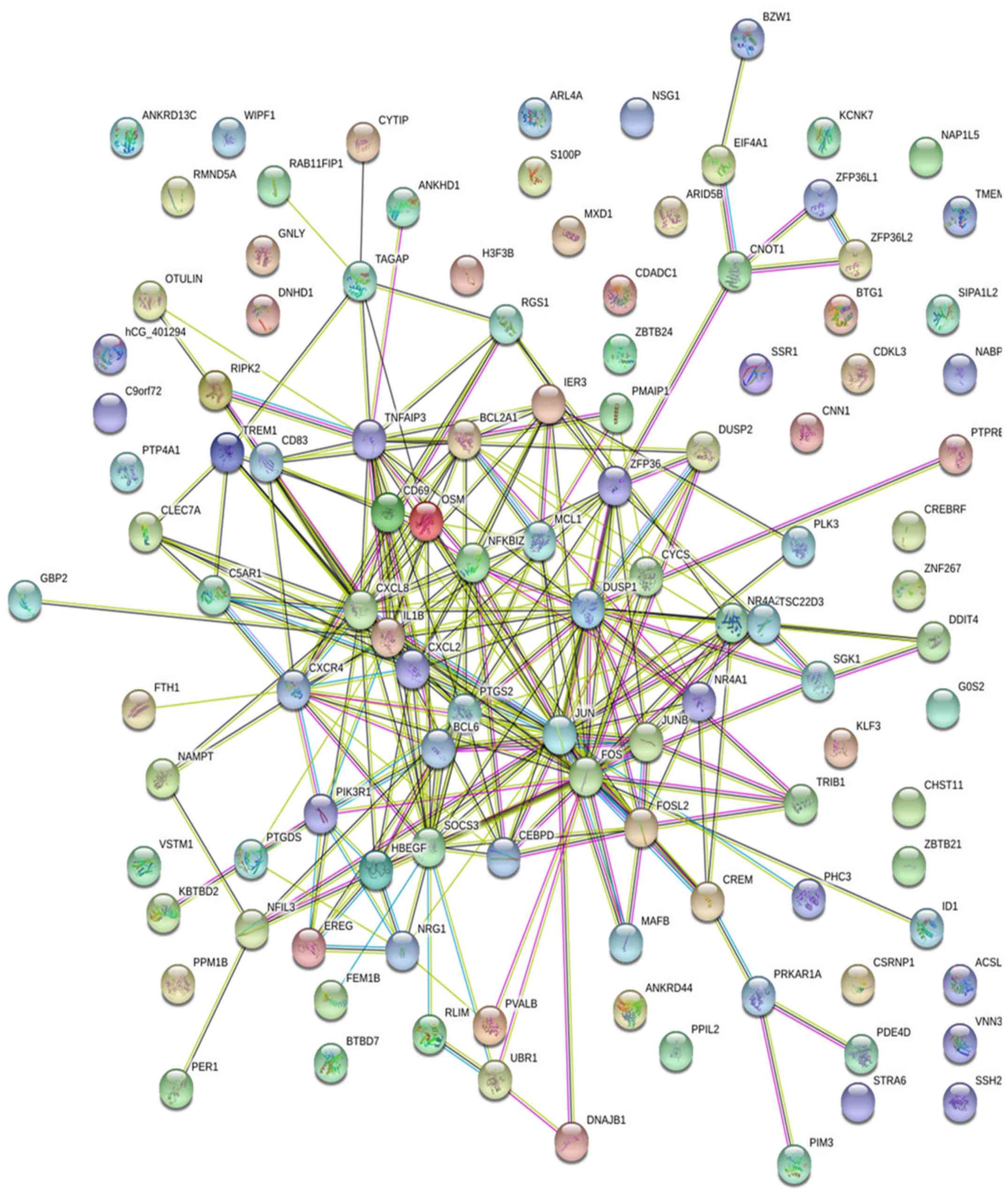
Open Access

In silico identification of common and specific signatures in coronary heart diseases

  • Authors:
    • Zhijia Yang
    • Haifang Ma
    • Wei Liu
  • View Affiliations

  • Published online on: August 13, 2020     https://doi.org/10.3892/etm.2020.9121
  • Pages: 3595-3614
  • Copyright: © Yang et al. This is an open access article distributed under the terms of Creative Commons Attribution License.

Metrics: Total Views: 0 (Spandidos Publications: | PMC Statistics: )
Total PDF Downloads: 0 (Spandidos Publications: | PMC Statistics: )


Abstract

Coronary heart disease (CHD) is on the increase in developing countries, where lifestyle choices such as smoking, bad diet, and no exercise contribute and increase the incidence of high blood pressure and high cholesterol levels to cause CHD. Through utilization of a biomarker-based approach for developing interventions, the aim of the study was to identify differentially expressed genes (DEGs) and their association and impact on various bio-targets. The microarray datasets of both healthy and CHD patients were analyzed to identify the DEGs and their interactions using Gene Ontology, PANTHER, Reactome, and STRING (for the possible associated genes with multiple targets). Our data mining approach suggests that the DEGs were associated with molecular functions, including protein binding (75%) and catalytic activity (56%); biological processes such as cellular process (83%), biological regulation (57%), and metabolic process (44%); and cellular components such as cell (65%) and organelle (58%); as well as other associations including apoptosis, inflammatory, cell development and metabolic pathways. The molecular functions were further analyzed, and protein binding in particular was analyzed using network analysis to determine whether there was a clear association with CHD and disease. The ingenuity pathway analysis revealed pathways related to cell cholesterol biosynthesis, the immune system including cytokinin signaling, in which, the understanding of DEGs is crucial to predict the advancement of preventive strategies. Results of the present study showed that, there is a need to validate the top DEGs to rule out their molecular mechanism in heart failure caused by CHD.

Introduction

Coronary heart disease (CHD), also known as coronary artery disease (CAD) is one of a group of diseases of the heart blood vessels affecting millions of individuals worldwide. According to the center for disease control (CDC) reports, each death out of four is related to heart diseases, leading to approximately 610,000 mortalities annually worldwide (1). Among the heart diseases, CAD is the most common, responsible for the death of 370,000 individuals annually worldwide (1). CAD occurs when the elasticity of arteries, as well as vein and vessel smoothing, become plaque in the inner wall, making them rigid and narrowed. This condition restricts the blood flow to the heart muscle, leading to oxygen starvation. The condition of plaque rupture leads to the heart failure or cardiac death (2).

Recently, there has been an increase in the incidence of CHD (also known as ischemic heart disease) in China (3). In addition, CHD has become the most common reason for death in middle and high-income countries (4). According to the data report by NHANES, CHD prevalence was higher in males than females across all ages (7.4 v/s 5.3%, respectively) (3). The American Heart Association explains ‘The important difference between sex and pathology’, clinical presentation and outcomes in CHD patients (5). Thus it is crucial to pay attention to sex disparities and subsequently to personalize treatment (6). Patients with CHD are also susceptible to more complicated clinical problems. Currently, the diagnosis and therapy of CHD is rare and costly as compared to coronary angiography, which is the most popular clinical management option (7). CHD is one of the leading causes of death, and markedly affects the immunity of the body, making it an economic burden worldwide (8,9). This is a complex disease involving multiple mechanisms and influenced by many risk factors, including physical activity, genetics, diet, and smoking (10,11). Recently, a genome-wide association study (GWAS) identified many candidate loci associated with CHD and myocardial infection (MI) (12-14). Although genetics play an important role, accounting for approximately 50% of CHD heritability, the exact mechanism and causative agent of CHD are not yet revealed clearly (15-17). In this regard, it is important to understand and address the candidate genome association in developing CHD.

MicroRNAs (miRNA) are small noncoding RNAs with a length of 22-25 nucleotide and which play a key role in the regulation of gene expression and have implications in many human disorders (18), including many biological processes such as cell differentiation, proliferation and apoptosis (19-21). To the best of the knowledge of the authors, the association pattern of miRNAs to CHD is lacking, leading to demand for specific CHD patients. Although relevant research has been undertaken to address DEGs associated with CHD, DEGs have only been used to check the expression pattern in case of CHD. In this study, we addressed the possible association of genes with CHD, which may be useful for the diagnosis and treatment of this disease in the near future. Additionally, analysis of gene expression data and network analysis were performed to gain a better understanding of CHD for the identification of differentially expressed genes (DEGs), biomarkers and therapeutic target options.

Materials and methods

Data availability

To identify key genes for the development of CHD biomarkers, we used gene expression datasets of 4 angiographically proven patients who were being treated for more than 3 months or from group-1 (n=100) compared to healthy control (n=50). This dataset was downloaded from the GEO module of National Center for Biotechnology Information (https://www.ncbi.nlm.nih.gov/geo/query/acc.cgi?acc=GSE56885). Microarray gene expression profiles were downloaded and further analyzed for the identification of DEGs. In this dataset, GSM1370681 and GSM1370682 represent the replicate samples of healthy individuals and GSM1370683 to GSM1370686 of four patients as baseline associated with CAD.

Differential Expression Analysis (DEG)

Using the default parameters, WEGO 2.0, and GEO2R (https://www.ncbi.nlm.nih.gov/geo/geo2r/) were used to analyze the GEO series (22). The Benjamini and Hochberg false discovery rate method was utilized to adjust the P-values. NCBI-generated annotations were employed to display the DEG list by comparing the overall common gene expression pattern as compared to the control. On the basis of this analysis, possible associations related to CHD were reported. Although inappropriate to consider the data for analysis on inter-datasets, the average value of LogFC for all four datasets was assessed to represent the expression level.

Gene ontology (GO) analysis

The major bioinformatics tool GO was used as an initiative to understand the function of genes and gene products of Homo sapiens. The PANTHER (Protein ANalysis THrough Evolutionary Relationships) classification database (23) was used to perform the GO analysis, and the pathway analysis was performed using Reactome (24).

Protein-Protein Interaction (PPI) network construction analysis

An online freely available software package, STRING, was utilized to establish the PPI network (25), and all the cut-off points were combined to analyze the topology property of networks. Gene edges of >15 degrees were defined as hub genes.

Results and Discussion

Screening of differentially expressed common genes from microarray data sets

Atherosclerosis is one of the leading causes of cardiovascular diseases such as CHD (26). Understanding of the key players in expression, regulation and function, of GWAS CHD genes will provide the options to treat this disease, leading to further developments of novel therapeutic interventions (27). In this study, the first compressive investigation was conducted to identify the expression profile of collected microarray data sets of CHD. The dataset of two controls in replicate and four baseline test samples were used. We report an overall expression and function of genes associated with different biological processes, which may lead to CHD during pathological conditions. The overall study design is shown in Fig. 1, which presents CHD data of Homo sapiens from the GEO database, with four series of test samples and two control study sets. First, we used WEGO to visualize the GO annotations and the percentage of genetic association of different functions in cells to address the possible association with CHD.

A total of 52,998 genes sharing different functions such as cellular (18,476), biological (17,307) and molecular (17,215) functions were identified

Out of those, the highest gene association to cell, cell part, organelle, organelle part, membrane, binding, cellular process, biological regulation, and metabolic were topmost in the metadata of the CHD-associated data set (Fig. 2). The different GO representing the 0-90% range of gene expression as compared to control data set is shown in Fig. 3. The principal findings of this study confirm the association of immune system, inflammation, and apoptosis as mediators in the development of CHD. The impact of the immune system plays a key role in the development of heart failure. A transcriptomic study reported the sustained activation of the adoptive immune system which may be a contributing factor in the progression of CHD (28). Another report suggests that the imbalance in inflammatory and anti-inflammatory cytokines may lead to the onset of extensive fibrosis (29).

From the GEO database, accession GSE56885 of CSD patients, who were being treated for more than 3 months was selected

From the included patients, two controls in replicate and four overall test samples were used to consolidate data refining. The GEO2R was used to analyze the control and test data series by normalizing the microarray data for high quality. DEGs with different fold change confirm their crucial role in CHD (Table I).

Table I

Up - and down regulated genes with associated function in CHD.

Table I

Up - and down regulated genes with associated function in CHD.

A, Upregulation
ID LogFCGene symbolGene ontology: Biological functionGene ontology: Cellular componentGene ontology: Molecular function
117256323.963NR4A2Negative regulation of transcription from RNA polymerase II promoterNucleusRna polymerase II regulatory region sequence-specific DNA binding
117167712.98LOC102724428Negative regulation of transcription from RNA polymerase II promoterIntracellularNucleotide binding
117198983.593HBEGFMAPK CasCHDe receptor bindingExtracellular regionEpidermal growth factor
117188416.035CXCL8AngiogenesisExtracellular regionCytokine activity
117612722.91BCL2A1Apoptotic processCytoplasmProtein binding
117439722.917DDIT4Response to hypoxiaIntracellular14-3-3 Protein binding
117327194.2EREGMAPK CasCHDe receptor bindingExtracellular regionEpidermal growth factor
117442196.925G0S2Apoptotic processMitochondrionProtein binding
117216953.385DUSP2Inactivation of MAPK activityNucleusPhosphoprotein phosphatase activity
117427654.1RGS1Immune responseCytoplasmGTPase activator activity
117240375.573PTGS2Prostaglandin biosynthetic processNucleusPeroxidase activity
117467212.647TREM1Positive regulation of defense response to virus by hostExtracellular regionReceptor activity
117597493.44KLF3Transcription, DNA-templatedNucleusNucleic acid binding
117448501.885SSH2Protein dephosphorylationExtracellular spaceDNA binding
117430002.982CD83Regulation of cytokine productionPlasma membraneProtein binding
117159313.478SGK1Regulation of cell growthNucleusNucleotide binding
117244472.157PDE4DRegulation of heart rateCytoplasmCyclic-nucleotide phosphodiesterase activity
117377503.272SGK1Regulation of cell growthNucleusNucleotide binding
117281902.058CXCR4Activation of MAPK activityCytoplasmVirus receptor activity
117431102.268NAMPTVitamin metabolic process (carboxylating) activityExtracellular region Nicotinate-nucleotide diphosphorylase
117631702.103FOSL2Keratinocyte development sequence-specific DNA bindingNucleusRNA polymerase II regulatory region
117336983.143SGK1Regulation of cell growthNucleusNucleotide binding
117449322.245CREBRFNegative regulation of transcription from RNA polymerase II promoterNucleusTranscription factor activity, Sequence-specific DNA binding
117577212.265CSRNP1Transcription, DNA-templated RNA polymerase II transcription regulatory region sequence-specific bindingNucleusTranscriptional activator activity,
117157662.552DUSP1Inactivation of MAPK activityNucleusPhosphoprotein phosphatase activity
117281912.465CXCR4Activation of MAPK activityCytoplasmVirus receptor activity
117192182.567SOCS3Response to hypoxiaIntracellularProtein kinase inhibitor activity
117395402.195PIK3R1Cellular glucose homeostasisNucleusTransmembrane receptor protein tyrosine kinase adaptor activity
117281892.54CXCR4Activation of MAPK activityCytoplasmVirus receptor activity
117435961.75PTPREProtein dephosphorylationNucleusPhosphoprotein phosphatase activity
117178301.69TSC22D3Negative regulation of transcription from RNA polymerase II promoterNucleusTranscription factor activity, sequence-specific DNA binding
117529933.005DUSP1Inactivation of MAPK activityNucleusPhosphoprotein phosphatase activity
117178971.79PTP4A1Protein dephosphorylationNucleusPhosphoprotein phosphatase activity
117520391.468PHC3Multicellular organismal developmentNucleusDNA Binding
117565872.1PTGDSProstaglandin biosynthetic processExtracellular regionProstaglandin-D synthase activity
117236792.607CD69Signal transductionIntegral component of plasma membraneTransmembrane signaling Receptor activity
117189392.723TNFAIP3B-1 B cell homeostasisNucleusProtease binding
117364672.2TAGAPSignal transductionCytosolGuanyl-nucleotide exchange factor activity
117390942.712CXCR4Activation of MAPK activityCytoplasmVirus receptor activity
117633671.535NABP1Double-strand break repair via homologous recombinationNucleusDNA binding
117431112.857NAMPTVitamin metabolic processExtracellular region Nicotinate-nucleotide diphosphorylase (Carboxylating) activity
117156732.002JUNBNegative regulation of transcription from RNA polymerase II pomoterChromatinRNA polymerase II regulatory region Sequence-specific DNA binding
117179942.812NR4A1Positive regulation of endothelial cell proliferationNucleusTranscriptional activator activity, RNA polymerase II core promoter proximal region sequence-specific binding
117156912.008ZFP36Negative regulation of transcription from RNA polymerase II promoterNucleusDNA binding
117330222.29BTG1Regulation of transcription, DNA-templatedNucleusTranscription cofactor activity
117244461.738PDE4DRegulation of heart rateCytoplasmCyclic-nucleotide phosphodiesterase activity
117371762.052C9ORF72EndocytosisExtracellular regionProtein binding
117154871.405MCL1Cell fate determinationIntracellularProtein binding
117226152.275HCAR2 /// HCAR3Neutrophil apoptotic process response to virus by hostPlasma membraneSignal transducer activity
117198622.093TREM1Positive regulation of defense response to virus by hostExtracellular regionReceptor activity
117347991.505RLIMNegative regulation of transcription from RNA polymerase II promoterNucleusTranscription corepressor activity
117631691.667FOSL2Keratinocyte developmentNucleusRNA polymerase II regulatory region sequence-specific DNA binding
117183943.535JUNAngiogenesisNuclear chromosomeRNA polymerase II core promoter proximal region sequence-specific DNA binding
117496521.69ZBTB21Transcription, DNA-templatedNucleusRNA polymerase II regulatory region sequence-specific DNA binding
117240384.715PTGS2Prostaglandin biosynthetic processNucleusPeroxidase activity
117538031.37CYCSResponse to reactive oxygen speciesProtein phosphatase type 2A complexProtein serine
117256313.285NR4A2Negative regulation of transcription from RNA polymerase II promoterNucleusRNA polymerase II regulatory region sequence-specific DNA binding
117512421.28FCGR2A /// FCGR2CImmune system processCytoplasmTransmembrane signaling receptor activity
117216292.755MAFBTranscription, DNA-templatedNucleusRNA polymerase II core promoter proximal region sequence-specific DNA binding
117333551.655C5AR1Activation of MAPK activityCytosolComplement component C5a binding
117596281.838WIPF1Actin cortical patch assemblyRuffleActin binding
117268891.505ZFP36L1Nuclear-transcribed mRNA catabolic process, deadenylation-dependent decayNucleusDNA binding
117507001.375ACSL1Long-chain fatty acid metabolic processMitochondrionNucleotide binding
117166021.895KBTBD2Protein ubiquitinationCul3-RING ubiquitin ligase complexUbiquitin-protein transferase activity
117514151.688TSC22D3Negative regulation of transcription from RNA polymerase II promoterNucleusTranscription factor activity, sequence-specific DNA binding
117447751.497BZW1Transcription, DNA-templatedCytoplasmBinding
117477361.2CNN1Regulation of smooth muscle contractionCytoskeletonActin binding
117277573.808OSMPositive regulation of acute inflammatory responseExtracellular regionCytokine activity
117587301.39DUSP1Inactivation of MAPK activityNucleusPhosphoprotein phosphatase activity
117448101.312ZBTB24Hematopoietic progenitor cell differentiationNucleusNucleic acid binding
117371471.357CLEC7AResponse to yeastNucleoplasmOpsonin Binding
117197131.662PPM1BProtein dephosphorylationCytoplasmMagnesium ion binding
117154451.248DNAJB1Protein foldingNucleusAtpase activator activity
117200622.98IER3Response to protozoanNucleusProtein binding
117575132.262NFKBIZTranscription, DNA-templatedNucleusTranscription cofactor activity
117585221.92CREMGlucose metabolic processNucleusCore promoter sequence-specific DNA binding
117248351.355HCAR2 /// HCAR3Neutrophil apoptotic processPlasma membraneSignal transducer activity
117624061.695GBP2Immune responseGolgi membraneNucleotide binding
117525771.438FTH1Iron ion transportCellFerroxidase activity
117441285.325CXCL2Response to molecule of bacterial originExtracellular regionCytokine activity
117606781.337PPIL2Protein polyubiquitinationNucleusPeptidyl-prolyl cis-trans isomerase activity
117275691.328OTULINAngiogenesisCytoplasmUbiquitin-specific protease activity
117199164.9IL1BNegative regulation of transcription from RNA polymerase II promoterExtracellular regionReceptor binding
117158171.18ZFP36L2Nuclear-transcribed mRNA catabolic process, deadenylation-dependent decayNucleusDNA binding
117434341.4CHST11Chondrocyte developmentGolgi membrane N-acetylgalactosamine 4-O-sulfotransferase activity
117242361.272RIPK2Activation of MAPK activityCytoplasmNucleotide binding
117207451.815BCL6Protein import into nucleus, translocationNucleusRNA polymerase II regulatory region sequence-specific DNA binding
117492911.75FOSConditioned taste aversionNucleusRNA polymerase II core promoter proximal region sequence-specific DNA binding
117640291.58CEBPDTranscription from RNA polymerase II promoterNucleusRNA polymerase II core promoter proximal region sequence-specific DNA binding
117183471.585S100PResponse to organic substanceNucleusMagnesium ion binding
117245092.365PMAIP1Release of cytochrome c from mitochondriaNucleusProtein binding
117346901.252CYTIPRegulation of cell adhesionNucleoplasmProtein binding
117367821.395RAB11FIP1TransportCytoplasmProtein binding
117640301.27CEBPDTranscription from RNA polymerase II promoterNucleusRNA polymerase II core promoter proximal region sequence-specific DNA binding
117433441.67RMND5A  Protein binding
117160482.357TRIB1Protein phosphorylationNucleusProtein kinase activity
117563871.558ARL4AIntracellular protein transportIntracellularNucleotide binding
117245102.298PMAIP1Release of cytochrome c from mitochondriaNucleusProtein binding
117326651.782VSTM1Immune system processExtracellular regionCytokine activity
117597801.348ANKRD13CProtein retention in ER lumenEndoplasmic reticulumReceptor binding
117371481.333CLEC7AResponse to yeastNucleoplasmOpsonin binding
117585931.325H3F3BChromatin silencing at rdnaNuclear chromosomeRNA polymerase II core promoter sequence-specific DNA binding
117639721.2SSR1TranslationEndoplasmic reticulumProtein binding
117275231.36ZNF267Transcription, DNA-templatedIntracellularNucleic acid binding
117183954.04JUNAngiogenesisNuclear chromosomeRNA polymerase II core promoter proximal region sequence-specific DNA binding
11727032-2.47NSG1Posittive regulation of transcription fromNucleusTranscription regulatory region RNA polymerase II promoter sequence-specific DNA binding
117180611.88PVALBCytosolic calcium ion homeostasisNucleusCalcium ion binding
117216302.138MAFBTranscription, DNA-templatedNucleusRNA polymerase II core promoter proximal region sequence-specific DNA binding
117637551.068GNLYCellular defense responseExtracellular region 
117579241.542SIPA1L2Positive regulation of gtpase activity Gtpase activator activity
117328591.03DNHD1Microtubule-based movementDynein complexMicrotubule motor activity
117500161.498MXD1Negative regulation of transcription from RNA polymerase II promoterNucleusRNA polymerase II core promoter proximal region sequence-specific DNA binding
117441274.107CXCL2Response to molecule of bacterial originExtracellular regionCytokine activity
117635561.777EIF4A1Nuclear-transcribed mrna catabolic process, deadenylation-dependent decayNucleusNucleotide binding
117454661.252CDADC1Metabolic process Catalytic activity
117563581.87PLK3G1ChromatinNucleotide binding
117183973.16JUNAngiogenesisNuclear chromosomeRNA polymerase II core promoter proximal region sequence-specific DNA binding
117639541.04SCARNA9L   
117306551.12CNOT1Negative regulation of transcription from RNA polymerase II promoterCytoplasmic mrna processing bodyPoly(A)-specific ribonuclease activity
117540744.195G0S2Apoptotic processMitochondrionProtein binding
117392301.132ARL4AIntracellular protein transportIntracellularNucleotide binding
117430102.372NFIL3Negative regulation of transcription from RNA polymerase II promoterNucleusRNA polymerase II regulatory region sequence-specific DNA binding
117240364.27PTGS2Prostaglandin biosynthetic processNucleusPeroxidase activity
117474741.64NR4A2Negative regulation of transcription from RNA polymerase II promoterNucleusRNA polymerase II regulatory region sequence-specific DNA binding
117408921KCNK7TransportPlasma membraneVoltage-gated ion channel activity
117331401.617ARL4AIntracellular protein transportIntracellularNucleotide binding
117247690.978FCGR2A /// FCGR2CImmune system processCytoplasmTransmembrane signaling receptor activity
117601920.987TMEM68Metabolic processMembraneTransferase activity, transferring acyl groups
117349881.285FEM1BEpithelial cell maturationNucleusUbiquitin-protein transferase activity
117577982.205MAFBTranscription, DNA-templatedNucleusRNA polymerase II core promoter proximal region sequence-specific DNA binding
117403471.2NRG1MAPK casCHDeExtracellular regionTranscription cofactor activity
117537912.535PRKAR1AMesoderm formationImmunological synapseNucleotide binding
117251141.45ANKHD1Regulation of translationNucleoplasmNucleic acid binding
117346591.54FOSConditioned taste aversionNucleusRNA polymerase II core promoter proximal region sequence-specific DNA binding
117160711.3PIM3Protein phosphorylationCytoplasmNucleotide binding
117206121.04NAP1L5Nucleosome assemblyNucleus 
117559870.98ANKRD44  Protein binding
117178951.285PTP4A1Protein dephosphorylationNucleusPhosphoprotein phosphatase activity
117251991.525BTBD7Multicellular organismal developmentNucleusProtein binding
117466811.88VNN3Nitrogen compound metabolic processExtracellular spaceHydrolase activity
117207261.413UBR1Ubiquitin-dependent protein catabolic processUbiquitin ligase complexUbiquitin-protein transferase activity
117608181.05CDKL3Cellular protein modification processCytoplasmNucleotide binding
117336861.052STRA6Retinoid metabolic processPlasma membraneReceptor activity
117247680.958FCGR2A /// FCGR2CImmune system processCytoplasmTransmembrane signaling receptor activity
117485160.932NAP1L5Nucleosome assemblyNucleus 
117189270.975ARID5BNegative regulation of transcription from RNA polymerase II promoterNucleusRNA polymerase II regulαtory region sequence-specific DNA binding
117161952.692ID1Negative regulation of transcription from RNA polymerase II promoterNucleusTranscription factor activity, sequence-specific DNA binding
B, Down regulation
11720657-3.328HLA-DRB5Immune system processGolgi membraneProtein binding
11724843-2.185CISHRegulation of cell growthCytoplasmprotein kinase inhibitor activity
11762593-2.075NUMA1G2 Structural molecule activity
11742832-1.783ASPMNeuron migrationSpindle poleBinding
11758261-2.223CEP55Mitotic cytokinesisCytoplasmProtein binding
11758089-1.885HMMRCarbohydrate metabolic processCytoplasmProtein binding
11721145-1.542MKI67DNA metabolic processChromosome, centromeric regionNucleotide binding
11743423-2.188NSG1Positive regulation of receptor recyclingGolgi membraneReceptor binding
11736674-1.588KLHL35  Protein binding
11759710-1.417TXNDC9Cell redox homeostasisCellProtein binding
11735385-1.752DACT1Negative regulation of transcription from RNA polymerase II promoterNucleusProtein kinase C binding
11748198-1.55NSG1Positive regulation of receptor recyclingGolgi membraneReceptor binding
11732363-1.775ZNF2Transcription, DNA-templatedIntracellularNucleic acid binding
11741074-1.407METTL18Methylation Methyltransferase activity
11722367-1.55DLGAP5Protein dephosphorylationNucleusPhosphoprotein phosphatase activity
11751805-1.83TYMSG1NucleusNucleotide binding
11764270-1.498PLGLB1 /// PLGLB2 Extracellular region 
11747230-1.518BUB1Mitotic cell cycleChromosome, centromeric regionNucleotide binding
11723209-1.732KBTBD6Protein ubiquitinationCul3-RING ubiquitin ligase complexUbiquitin-protein transferase activity
11732390-1.465CCR9ChemotaxisCytosolSignal transducer activity
11716666-1.623ID3Negative regulation of transcription from RNA polymerase II promoterNucleusTranscription factor activity, sequence-specific DNA binding
11763252-1.705PSPHProtein dephosphorylationCytoplasmMagnesium ion binding
11720240-1.307TMSB15AActin filament organizationCytoplasmActin binding
11716793-1.518CCNB2G2NucleusProtein binding
11717163-1.375CDC20Mitotic cell cycleSpindle poleProtein binding
11716427-1.71POMCGeneration of precursor metabolites and energyExtracellular regionG-protein coupled receptor binding
11755958-1.4ZNF691Regulation of transcription, DNA-templatedNucleusRNA polymerase II regulatory region sequence-specific DNA binding
11724022-2.403TRIM13Signal transductionIntracellularUbiquitin-protein transferase activity
11730821-1.295CDKN3Regulation of cyclin-dependent protein serineNucleusPhosphoprotein phosphatase activity
11726302-1.245DTLProtein polyubiquitinationNucleusUbiquitin-protein transferase activity
11744793-1.44DLGAP5Protein dephosphorylationNucleusPhosphoprotein phosphatase activity
11718599-1.502TM2D2 Membrane 
11760734-1.272GULP1TransportCytoplasmSignal transducer activity
11718058-1.782TYMSG1NucleusNucleotide binding
11734748-1.245LOC100507547 /// PRRT1Response to biotic stimulusPlasma membrane 
11730796-1.165PSPHProtein dephosphorylationCytoplasmMagnesium ion binding
11727968-1.148ESCO2Mitotic cell cycleChromatinLysine N-acetyltransferase activity, acting on acetyl phosphate as donor
11758219-1.48RRM2G1Nucleus Ribonucleoside-diphosphate reductase activity thioredoxin disulfide as acceptor
11755381-1.73PLGLA /// PLGLB1 /// PLGLB2 Extracellular region 
11762018-1.635DCLRE1CNucleotide-excision repair, telomeric regionNuclear chromosome,Single-stranded DNA
    DNA damage recognition endodeoxyribonuclease activity
11734263-1.97ZNF780ATranscription, DNA-templatedIntracellularNucleic acid binding
11733149-1.62DDX58Positive regulation of defense response to virus by hostCytoplasmNucleotide binding
11754360-1.762RRM2G1Nucleus Ribonucleoside-diphosphate reductase activity, thioredoxin disulfide as acceptor
11758872-2.118CDC37L1Protein foldingCytoplasmProtein binding
11728830-1.47RAB3IPProtein targeting to membraneNucleusGuanyl-nucleotide exchange factor activity
11759287-1.37DNAJB4Protein foldingNucleoplasmProtein binding
1172130009-1.157ZNF174Negative regulation of transcription from RNA polymerase II promoterNucleusRNA polymerase II transcription factor activity, sequence-specific DNA binding
11756778-1.195NEFLMAPK casCHDeCytoplasmStructural molecule activity
11740504-1.218ZNF680Transcription, DNA-templatedIntracellularRNA polymerase II core promoter proximal region sequence-specific DNA binding
11758615-1.177FRMD4BEstablishment of epithelial cell polarityRuffleProtein binding
11738883-1.055TNFSF14Apoptotic processExtracellular regionReceptor binding
11726443-1.183KCTD6Protein homooligomerization Protein binding
11727112-1.397SIT1Adaptive immune responsePlasma membraneProtein binding
11717301-1.218TACSTD2Cell cycleExtracellular spaceReceptor activity
11734218-1.38ZNF681Positive regulation of defense response to virus by hostIntracellularNucleic acid binding
11728339-1.667CENPBD1 NucleusDNA binding
11764234-1.865INTS7DNA damage checkpointNucleusBinding
11721932-1.09KIF23Mitotic spindle elongationNucleusNucleotide binding
11753763-1.18CDKN3Regulation of cyclin-dependent protein serineNucleusPhosphoprotein phosphatase activity
11721190-1.312TTC9CProtein peptidyl-prolyl isomerizationCytoplasmPeptidyl-prolyl cis-trans isomerase activity
11721418-1.2SH3RF3  Protein binding
11733069-1.825WDR5B  Protein binding
11730969-1.765THAP2 NucleolusNucleic acid binding
11719780-1.728TNFAIP8L2Immune system processCytoplasmProtein binding
11758125-1.32DEPDC1BSignal transductionIntracellularGtpase activator activity
11733695-1.055UBE2CMitotic cell cycleNucleusNucleotide binding
11756100-1.227TMEM60 Membrane 
11732295-1.455ZNF566Transcription, DNA-templatedIntracellularNucleic acid binding
11724984-1.255EXPH5Keratinocyte developmentIntracellularProtein binding
11726757-1.165CDC25ARegulation of cyclin-dependent protein serineIntracellularPhosphoprotein phosphatase activity
11727604-1.278EPB41L4A CytoplasmCytoskeletal protein binding
11716226-1.147LIMA1Negative regulation of actin filament depolymerizationStress fiberActin binding
11760155-1.105FUBP1Transcription, DNA-templatedNucleusTranscriptional activator activity, RNA polymerase II distal enhancer sequence-specific binding
11725833-1.087ALKBH1In utero embryonic developmentNucleusCatalytic activity
11723939-1.115CCNB1G1Spindle polePatched binding
11722160-1.218GRB10Signal transductionCytoplasmSh3
11759488-1.133EYA3DNA repairNucleusChromatin binding
11739828-1.188CYS1Kidney developmentCytoplasmProtein binding
11740637-1.258GPR19Signal transductionPlasma membraneSignal transducer activity
11717629-1.82KIF1BPMitochondrial transportCytoplasmProtein binding
11753965-1.15MSL3P1Transcription, DNA-templatedNucleus 
11758149-0.975RACGAP1Mitotic cytokinesisAcrosomal vesicleGtpase activator activity
11732175-1.44FANCFOvarian follicle developmentNucleusUbiquitin-protein transferase activity
11750856-1.062CCR2Blood vessel remodelingCytoplasmSignal transducer activity
11757036-1.278SAC3D1Cell cycleNucleusProtein binding
11758529-1.192CENPAEstablishment of mitotic spindle orientationChromosome, centromeric regionDNA binding
11732544-1.292GPR18Signal transductionPlasma membraneSignal transducer activity
11718213-1.118SLC27A2Very long-chain fatty acid metabolic processMitochondrionNucleotide binding
11725709-0.968WDHD1RNA processingChromosome, centromeric regionDNA binding
11739531-1.598PLGLB2 Extracellular region 
11745077-1.282CRNKL1Spliceosomal complex assemblyPrp19 complexRNA binding
11730590-1.198KCTD21Protein homooligomerization Protein binding
11727196-1.37ZNF202Negative regulation of transcription from RNA polymerase II promoterIntracellularRNA polymerase II transcription factor activity, sequence-specific DNA binding
11731676-1.567CCR2Blood vessel remodelingCytoplasmSignal transducer activity
11721473-1.388HCCSMetabolic processMitochondrionHolocytochrome-c synthase activity
11737395-1.292SOWAHD  Protein binding
11737108-1.05ACKR4Receptor-mediated endocytosisEndosomeSignal transducer activity
11728404-1.113SHCBP1Fibroblast growth factor receptor signaling pathway Protein binding
11760278-0.968 HCG8 /// ZNRD1-AS1  
11728360-1.655BCDIN3DRNA methylationNucleusProtein binding
11743686-1.005ZNF436Transcription, DNA-templatedIntracellularNucleic acid binding
11762571-1.425GNPDA2Carbohydrate metabolic processNucleus Glucosamine-6-phosphate deaminase activity
In our study, many immune response processes were significantly changed and DEGs are associated with the metabolic process, which is associated with CHD

The CHDs of the innate immune system were largely mediated through neutrophils and monocyte, and macrophages (30), to contribute to the process of the chronic inflammation process.

Functional enrichment and unified DEG analysis

To precisely understand the gene changes during CHD, the DEGs GO was performed using the online PANTHER database for high-throughput analysis to classify the proteins and their genes into family and subfamily, molecular function, biological process, and pathway (31). In the dataset analyzed, the two significant changes in molecular function were protein binding (75%) and catalytic activity (56%), followed by molecular regulator, molecular transducer activity, structural activity, transcription regulator activity, and transporter activity (Fig. 4A). In terms of the biological process, the three most significant classes of CHD were cellular process (83%), biological regulation (57%), and metabolic process (44%) (Fig. 4B). Additionally, in terms of cellular components, another two more significant components are cell (65%) and organelle (58%), which were found to be associated with CHD (Fig. 4C). Many other target-associated DEGs were involved in the biological process, molecular function and cellular components.

Analyzed potential DEGs of the CHD data set shows protein classes distributed among transcription factor (24%), enzyme modulator (20%), nucleic acid binding (18%), and signaling molecules (18%) (Fig. 5A)

The DEGs mainly associated with CHD key pathways showed the significance are inflammation mediated by chemokine and cytokine signaling pathway (11%), CCKR signaling map (11%), gonadotropin-releasing hormone receptor pathway (8%), apoptosis signaling pathway (6%), and p53 pathway (5%) (Fig. 5B). This result was consistent with GO analysis, confirming the classes of proteins associated with CHD. Many genes associated with inflammatory roles, and a previous study showed a conserved signature of dilated cardiomyopathy (DCM) plays an important role in cell survival promotion during end-stage of heart failure (32). In the present study, we also revealed the expression pattern of apoptotic or inflammatory genes (Fig. 4) (33,34).

Pathway analysis

To address the overview of data insight into the pathways, which are associated and connected for CHD development (35), we analyzed 164 DEGs involved in different functional pathways compared to reference and expected genes for those pathways. A total of 13 pathways were found to be associated with signaling-, immune-, and transcription-related pathways (36). Genes were confirmed in the uploaded list over the expected one (number in the list divided by the expected number). If >1, it indicated that the category is over-represented in the experiment. Conversely, the category is under-represented if <1. In the future, overexpressed genes are likely to serve as the marker selected in the development of CHD interventions. The P-value indicates the Fisher's exact test (37) or Binomial statistic in which the probability is the number of genes observed in this category occurred by chance (randomly), as determined by the reference list (Table II).

Table II

Pathway enrichment and reactome selected for CHD associated pathways.

Table II

Pathway enrichment and reactome selected for CHD associated pathways.

Reactome pathways Homo-sapiens REFLIST (20996)Client text box Input (212) Client text box input (expected) Client text box input (over/under)Client text box input (fold enrichment)Client text box input (raw P-value)Client text box input (FDR)
PI3K events in ERBB4 signaling (R-HSA-1250342)940.09+44.026.47E-063.54E-03
PI3K events in ERBB2 signaling (R-HSA-1963642)1340.13+30.472.09E-056.53E-03
ERBB2 activates PTK6 Signaling (R-HSA-8847993)1130.11+27.013.30E-044.53E-02
Chemokine receptors bind chemokines (R-HSA-380108)4860.48+12.381.61E-055.88E-03
Interleukin-10 signaling (R-HSA-6783783)4550.45+111.40E-042.79E-02
Interleukin-4 and Interleukin-13 signaling (R-HSA-6785807)111121.12+10.713.96E-098.68E-06
Peptide ligand-binding receptors (R-HSA-375276)18691.88+4.791.58E-042.66E-02
G alpha (i) signalling events (R-HSA-418594)392153.96+3.791.59E-056.98E-03
Signaling by Interleukins (R-HSA-449147)449174.53+3.754.90E-063.58E-03
Class A/1 (Rhodopsin-like receptors) (R-HSA-373076)321123.24+3.71.40E-042.56E-02
Cytokine signaling in Immune system (R-HSA-1280215)669236.76+3.44.96E-075.43E-04
Generic transcription Pathway (R-HSA-212436)1,0942611.05+2.356.61E-051.45E-02
RNA polymerase II transcription (R-HSA-73857)1,2162812.28+2.285.19E-051.26E-02
Gene expression (transcription) (R-HSA-74160)1,3512913.64+2.131.95E-043.05E-02
Immune system (R-HSA-168256)2,0354120.55+22.09E-055.73E-03
Signal transduction (R-HSA-162582)2,6674626.93+1.712.66E-043.89E-02
PPI analysis

To address the PPI of the CHD dataset in this study, STRING online suits was used to address the possible interaction of protein of CHD associated DEGs. A total of 112 nodes, 257 edges, 4.59 average node edge, 0.387 average clustering coefficient, 77 expected edge number, and <1.0e-16 PPI enrichment value were observed, and shown the network was significantly interacted than expected. Previous studies investigated the rare variants through targeted expression profiling across CHD relevant tissues from appropriate cases and controls (38,39). The PPI indicates the interaction of genes associated with multiple genes for outcome. In the present study, we identified 422 GO for biological process, 31 GO for molecular function, 12 GO for cellular component, 33 pathways, 30 reactome pathways, 13 UniProt keywords, 11 PFAM protein domains, 29 INTERO protein domains, and 3 SMART protein domains in the analysis of CHD microarray data set. In those findings, associated edges shows physically binding protein and some of them were associated with but did not have physical binding. Of these, only the top-ranking ones have been presented (Fig. 6 and Table III).

Table III

Protein-protein interaction network of CHD associated genes.

Table III

Protein-protein interaction network of CHD associated genes.

A, Biological process (GO).
Sl. NoGO-termDescriptionCount in gene setFalse discovery rate
1GO:0050789Regulation of biological process100 of 11,1162.85e-09
2GO:0065007Biological regulation101 of 11,7402.13e-08
3GO:0050794Regulation of cellular process95 of 10,4842.13e-08
4GO:0048523Negative regulation of cellular process59 of 4,4542.13e-08
5GO:0048519Negative regulation of biological process62 of 4,9532.13e-08
B, Molecular function (GO).
Sl. NoGO-termDescriptionCount in gene setFalse discovery rate
1GO:0000977RNA polymerase II regulatory region sequence-specific DNA binding16 of 6470.00061
2GO:0005515Protein binding62 of 6,6050.00065
3GO:0043565Sequence-specific DNA binding19 of 1,0470.00083
4GO:0140110Transcription regulator activity28 of 2,0690.0011
5GO:0005488binding89 of 11,8780.0026
C, Cellular components (GO).
Sl. NoGO-termDescriptionCount in gene setFalse discovery rate
1GO:0005634Nucleus67 of 6,8928.05e-05
2GO:0035976Transcription factor AP-1 complex3 of 50.0015
3GO:0005622Intracellular102 of 14,2860.0015
4GO:0044424Intracellular part99 of 13,9960.0064
5GO:0043227Membrane-bounded organelle85 of 11,2440.0067
D, KEGG pathways.
Sl. NoGO-termDescriptionCount in gene setFalse discovery rate
1hsa04668TNF signaling pathway9 of 1084.47e-06
2hsa04380Osteoclast differentiation8 of 1248.58e-05
3hsa04657IL-17 signaling pathway7 of 929.94e-05
4hsa04621NOD-like receptor signaling pathway8 of 1660.00034
5hsa05210Colorectal cancer6 of 850.00051
E, Reactome pathways.
Sl. NoGO-termDescriptionCount in gene setFalse discovery rate
1HSA-6785807Interleukin-4 and Interleukin-13 signaling11 of 1063.07e-08
2HSA-449147Signaling by Interleukins14 of 4396.23e-05
3HSA-1280215Cytokine Signaling in Immune system17 of 6546.23e-05
4HSA-1250342PI3K events in ERBB4 signaling4 of 97.21e-05
5HSA-1963642PI3K events in ERBB2 signaling4 of 130.00019
F, UniPort PFAM Protein domains
Sl. NoDomainDescriptionCount in gene setFalse discovery rate
1PF07716Basic region leucine zipper7 of 442.23e-06
2PF03131bZIP Maf transcription factor5 of 330.00017
3PF00170bZIP transcription factor5 of 360.00017
4PF04553Tis11B like protein, N terminus2 of 20.0061
5PF00782Dual specificity phosphatase, catalytic domain4 of 450.0061
G, INTERPRO Protein Domains and Features
Sl. NoDomainDescriptionCount in gene setFalse discovery rate
1IPR004827Basic-leucine zipper domain8 of 547.06e-07
2IPR029021Protein-tyrosine phosphatase-like5 of 1010.0181
3IPR008917Transcription factor, Skn-1-like, DNA-binding domain superfamily3 of 160.0181
4IPR007635Tis11B-like protein, N-terminal2 of 20.0181
5IPR005643Jun-like transcription factor2 of 30.0181
SMART Protein Domains
Sl. NoDomainDescriptionCount in gene setFalse discovery rate
1SM00338Basic region leucin zipper8 of 531.47e-07
2SM00195Dual specificity phosphatase, catalytic domain3 of 280.0246
3SM00356Zinc finger3 of 420.0488
Understanding and ruling the mechanism

There are several challenges to identifying the genetic basis of CHD that are also the determinants of this complex disease, including phenotypic and genetic heterogeneity, gene-environment, and etiological spectrum range and their effect. Considering research efforts involved in determining the genetic basis of this CHD, there is a need to understand the fine complexity of genetic association leading to mortality in developing countries. There is a need to focus on clinical manifestation rather than factors which influence or are heritable by genetic factors. There are many challenges in determining the genetic association of CHDs, such as phenotypic heterogeneity, genetic heterogeneity, small gene effects, gene-gene and gene-environment interactions and rare variants causing complex diseases. Some of the key points to be undertaken such as mortality, challenge in identifying the genetic determinants, studying linkage mapping through conventional approaches, and cataloguing of human diseases variation at single-nucleotide polymorphism (SNP), as well as genotyping will increase the likelihood of success.

In conclusion, we studied a comprehensive gene expression profile of microarray data of CHD. During the progression of CHD, there was a significant change in the expression of genes involved in the immune system, inflammation, and cell signaling through protein binding. This analysis provides valuable information for future research and in understanding the mechanism of CHD as well as identification of novel interventions for therapeutic application.

Acknowledgements

Not applicable

Funding

This research received no specific grants from any funding agencies.

Availability of data and materials

The datasets used and/or analyzed during the current study are available from the corresponding author on reasonable request.

Authors' contributions

ZY conceived and designed the study. WL provided study materials. ZY, HM and WL were responsible for the collection and assembly of data, data analysis and interpretation. ZY was involved in writing the manuscript. All authors read and approved the final manuscript.

Ethics approval and consent to participate

Not applicable

Patient consent for publication

Not applicable

Competing interests

The authors declare that they have no competing interests.

References

1 

Underlying Cause of Death 1999-2017. Centers for Disease Control and Prevention 24/7.

2 

Haukilahti MAE, Holmström L, Vähätalo J, Kenttä T, Tikkanen J, Pakanen L, Kortelainen ML, Perkiömäki J, Huikuri H, Myerburg RJ, et al: Sudden cardiac death in women. Circulation. 139:1012–1021. 2019.PubMed/NCBI View Article : Google Scholar

3 

Ding D, Wang M, Su D, Hong C, Li X, Yang Y, Zhang Y, Hu G and Ling W: Body mass index, high-sensitivity C-reactive protein and mortality in Chinese with coronary artery disease. PLoS One. 10(e0135713)2015.PubMed/NCBI View Article : Google Scholar

4 

Yusuf S, Rangarajan S, Teo K, Islam S, Li W, Liu L, Bo J, Lou Q, Lu F, Liu T, et al: Cardiovascular risk and events in 17 low-, middle-, and high-income countries. N Engl J Med. 371:818–827. 2014.PubMed/NCBI View Article : Google Scholar

5 

Benjamin EJ, Blaha MJ, Chiuve SE, Cushman M, Das SR, Deo R, de Ferranti SD, Floyd J, Fornage M, Gillespie C, et al: American Heart Association Statistics Committee and Stroke Statistics Subcommittee: Heart disease and stroke statistics-2017 Update: A report from the American Heart Association. Circulation. 135:e146–e603. 2017.PubMed/NCBI View Article : Google Scholar

6 

Mehta LS, Beckie TM, DeVon HA, Grines CL, Krumholz HM, Johnson MN, Lindley KJ, Vaccarino V, Wang TY, Watson KE, et al: American Heart Association Cardiovascular Disease in Women and Special Populations Committee of the Council on Clinical Cardiology, Council on Epidemiology and Prevention, Council on Cardiovascular and Stroke Nursing, and Council on Quality of Care and Outcomes Research: Acute myocardial infarction in women: A scientific statement from the American Heart Association. Circulation. 133:916–947. 2016.PubMed/NCBI View Article : Google Scholar

7 

Fyyaz S, Rauf A, Hudson J and Olabintan O: 107 Nice 2016 stable chest pain guidelines: Improved yield of severe coronary artery disease at invasive coronary angiography. Heart. 105(88)2019.

8 

Timmis A, Townsend N, Gale C, Grobbee R, Maniadakis N, Flather M, Wilkins E, Wright L, Vos R, Bax J, et al: ESC Scientific Document Group: European Society of Cardiology: Cardiovascular Disease Statistics 2017. Eur Heart J. 39:508–579. 2018.PubMed/NCBI View Article : Google Scholar

9 

Erbel R, Aboyans V, Boileau C, Bossone E, Bartolomeo RD, Eggebrecht H, Evangelista A, Falk V, Frank H, Gaemperli O, et al: ESC Committee for Practice Guidelines: 2014 ESC Guidelines on the diagnosis and treatment of aortic diseases: Document covering acute and chronic aortic diseases of the thoracic and abdominal aorta of the adult. Eur Heart J. 35:2873–2926. 2014.PubMed/NCBI View Article : Google Scholar

10 

Piepoli MF, Hoes AW, Agewall S, Albus C, Brotons C, Catapano AL, Cooney MT, Corrà U, Cosyns B, Deaton C, et al: ESC Scientific Document Group: 2016 European guidelines on cardiovascular disease prevention in clinical practice: The Sixth Joint Task Force of the European Society of Cardiology and Other Societies on Cardiovascular Disease Prevention in Clinical Practice (constituted by representatives of 10 societies and by invited experts) Developed with the special contribution of the European Association for Cardiovascular Prevention & Rehabilitation (EACPR). Eur Heart J. 37:2315–2381. 2016.PubMed/NCBI View Article : Google Scholar

11 

Kwak BR, Bäck M, Bochaton-Piallat ML, Caligiuri G, Daemen MJAP, Davies PF, Hoefer IE, Holvoet P, Jo H and Krams R: Biomechanical factors in atherosclerosis: Mechanisms and clinical implications. Eur Heart J. 35:3013–3020. 2014.PubMed/NCBI View Article : Google Scholar

12 

CARDIoGRAMplusC4D Consortium. Deloukas P, Kanoni S, Willenborg C, Farrall M, Assimes TL, Thompson JR, Ingelsson E, Saleheen D and Erdmann J: Large-scale association analysis identifies new risk loci for coronary artery disease. Nat Genet. 45:25–33. 2013.PubMed/NCBI View Article : Google Scholar

13 

Nikpay M, Goel A, Won H-H, Hall LM, Willenborg C, Kanoni S, Saleheen D, Kyriakou T, Nelson CP, Hopewell JC, et al: A comprehensive 1,000 genomes-based genome-wide association meta-analysis of coronary artery disease. Nat Genet. 47:1121–1130. 2015.PubMed/NCBI View Article : Google Scholar

14 

Kessler T, Vilne B and Schunkert H: The impact of genome-wide association studies on the pathophysiology and therapy of cardiovascular disease. EMBO Mol Med. 8:688–701. 2016.PubMed/NCBI View Article : Google Scholar

15 

Roberts R: Genetics of coronary artery disease. Circ Res. 114:1890–1903. 2014.PubMed/NCBI View Article : Google Scholar

16 

Zhao Y, Chen J, Freudenberg JM, Meng Q, Rajpal DK and Yang X: Network-based identification and prioritization of key regulators of coronary artery disease loci. Arterioscler Thromb Vasc Biol. 36:928–941. 2016.PubMed/NCBI View Article : Google Scholar

17 

Miller CL, Pjanic M and Quertermous T: From locus association to mechanism of gene causality: The devil is in the details. Arterioscler Thromb Vasc Biol. 35:2079–2080. 2015.PubMed/NCBI View Article : Google Scholar

18 

Cheng Y and Zhang C: MicroRNA-21 in cardiovascular disease. J Cardiovasc Transl Res. 3:251–255. 2010.PubMed/NCBI View Article : Google Scholar

19 

Chen CZ, Li L, Lodish HF and Bartel DP: MicroRNAs modulate hematopoietic lineage differentiation. Science. 303:83–86. 2003.PubMed/NCBI View Article : Google Scholar

20 

Johnson CD, Esquela-Kerscher A, Stefani G, Byrom M, Kelnar K, Ovcharenko D, Wilson M, Wang X, Shelton J, Shingara J, et al: The let-7 microRNA represses cell proliferation pathways in human cells. Cancer Res. 67:7713–7722. 2007.PubMed/NCBI View Article : Google Scholar

21 

Cheng AM, Byrom MW, Shelton J and Ford LP: Antisense inhibition of human miRNAs and indications for an involvement of miRNA in cell growth and apoptosis. Nucleic Acids Res. 33:1290–1297. 2005.PubMed/NCBI View Article : Google Scholar

22 

Smyth GK, Michaud J and Scott HS: Use of within-array replicate spots for assessing differential expression in microarray experiments. Bioinformatics. 21:2067–2075. 2005.PubMed/NCBI View Article : Google Scholar

23 

Mi H, Muruganujan A, Casagrande JT and Thomas PD: Large-scale gene function analysis with the PANTHER classification system. Nat Protoc. 8:1551–1566. 2013.PubMed/NCBI View Article : Google Scholar

24 

Jupe S, Fabregat A and Hermjakob H: Expression data analysis with reactome. Curr Protoc Bioinformatics 49: 8.20.1-8.20.9, 2015. https://doi.org/10.1002/0471250953.bi0820s49.

25 

Szklarczyk D, Morris JH, Cook H, Kuhn M, Wyder S, Simonovic M, Santos A, Doncheva NT, Roth A, Bork P, et al: The STRING database in 2017: Quality-controlled protein-protein association networks, made broadly accessible. Nucleic Acids Res. 45:D362–D368. 2017.PubMed/NCBI View Article : Google Scholar

26 

Rana JS, Nieuwdorp M, Jukema JW and Kastelein JJ: Cardiovascular metabolic syndrome - an interplay of, obesity, inflammation, diabetes and coronary heart disease. Diabetes Obes Metab. 9:218–232. 2007.PubMed/NCBI View Article : Google Scholar

27 

Kessler T, Vilne B and Schunkert H: The impact of genome-wide association studies on the pathophysiology and therapy of cardiovascular disease. EMBO Mol Med. 8:688–701. 2016.PubMed/NCBI View Article : Google Scholar

28 

Torre-Amione G: Immune activation in chronic heart failure. Am J Cardiol. 95 (11A):3C–8C; discussion 38C-40C. 2005.PubMed/NCBI View Article : Google Scholar

29 

Brenes-Castro D, Castillo EC, Vázquez-Garza E, Torre-Amione G and García-Rivas G: Temporal frame of immune cell infiltration during heart failure establishment: Lessons from animal models. Int J Mol Sci. 19(19)2018.PubMed/NCBI View Article : Google Scholar

30 

Flores-Arredondo JH, García-Rivas G and Torre-Amione G: Immune modulation in heart failure: Past challenges and future hopes. Curr Heart Fail Rep. 8:28–37. 2011.PubMed/NCBI View Article : Google Scholar

31 

Gu X, Li B, Jiang M, Fang M, Ji J, Wang A, Wang M, Jiang X and Gao C: RNA sequencing reveals differentially expressed genes as potential diagnostic and prognostic indicators of gallbladder carcinoma. Oncotarget. 6:20661–20671. 2015.PubMed/NCBI View Article : Google Scholar

32 

Barth AS, Kuner R, Buness A, Ruschhaupt M, Merk S, Zwermann L, Kääb S, Kreuzer E, Steinbeck G, Mansmann U, et al: Identification of a common gene expression signature in dilated cardiomyopathy across independent microarray studies. J Am Coll Cardiol. 48:1610–1617. 2006.PubMed/NCBI View Article : Google Scholar

33 

Satterthwaite G, Francis SE, Suvarna K, Blakemore S, Ward C, Wallace D, Braddock M and Crossman D: Differential gene expression in coronary arteries from patients presenting with ischemic heart disease: Further evidence for the inflammatory basis of atherosclerosis. Am Heart J. 150:488–499. 2005.PubMed/NCBI View Article : Google Scholar

34 

Sinnaeve PR, Donahue MP, Grass P, Seo D, Vonderscher J, Chibout SD, Kraus WE, Sketch M Jr, Nelson C, Ginsburg GS, et al: Gene expression patterns in peripheral blood correlate with the extent of coronary artery disease. PLoS One. 4(e7037)2009.PubMed/NCBI View Article : Google Scholar

35 

Gotoh T, Endo M and Oike Y: Endoplasmic reticulum stress-related inflammation and cardiovascular diseases. Int J Inflam. 2011:1–9. 2011.PubMed/NCBI View Article : Google Scholar

36 

Haas MJ and Mooradian AD: Regulation of high-density lipoprotein by inflammatory cytokines: Establishing links between immune dysfunction and cardiovascular disease. Diabetes Metab Res Rev. 26:90–99. 2010.PubMed/NCBI View Article : Google Scholar

37 

Costa THR, de Figueiredo Neto JA, de Oliveira AEF, Lopes e Maia MF and de Almeida AL: Association between chronic apical periodontitis and coronary artery disease. J Endod. 40:164–167. 2014.PubMed/NCBI View Article : Google Scholar

38 

Jensen MK, Pers TH, Dworzynski P, Girman CJ, Brunak S and Rimm EB: Protein interaction-based genome-wide analysis of incident coronary heart disease. Circ Cardiovasc Genet. 4:549–556. 2011.PubMed/NCBI View Article : Google Scholar

39 

Duan S, Luo X and Dong C: Identification of susceptibility modules for coronary artery disease using a genome wide integraded network analysis. Gene:. 531:347–357. 2013.PubMed/NCBI View Article : Google Scholar

Related Articles

Journal Cover

October-2020
Volume 20 Issue 4

Print ISSN: 1792-0981
Online ISSN:1792-1015

Sign up for eToc alerts

Recommend to Library

Copy and paste a formatted citation
x
Spandidos Publications style
Yang Z, Ma H and Liu W: In silico identification of common and specific signatures in coronary heart diseases. Exp Ther Med 20: 3595-3614, 2020
APA
Yang, Z., Ma, H., & Liu, W. (2020). In silico identification of common and specific signatures in coronary heart diseases. Experimental and Therapeutic Medicine, 20, 3595-3614. https://doi.org/10.3892/etm.2020.9121
MLA
Yang, Z., Ma, H., Liu, W."In silico identification of common and specific signatures in coronary heart diseases". Experimental and Therapeutic Medicine 20.4 (2020): 3595-3614.
Chicago
Yang, Z., Ma, H., Liu, W."In silico identification of common and specific signatures in coronary heart diseases". Experimental and Therapeutic Medicine 20, no. 4 (2020): 3595-3614. https://doi.org/10.3892/etm.2020.9121