Introduction
The outcome of in vitro fertilization (IVF)
is influenced by a number of factors. One of the most important
factors is oocyte quality. The microenvironment of the follicular
fluid is important for oocyte development (1). The analysis of oocyte-derived
products, including follicular fluid and cumulus cells, has
provided novel candidates that can be used to assess oocyte
competence and embryo implantation ability (2-6).
In the endometrium, prokineticin (PROK) 1 protein expression is
highest at the midluteal phase of the menstrual cycle and this
corresponds to the window of embryo implantation and has been
reported to be a biomarker for human endometrial receptivity
(7-9).
PROKs promote the contractile function of ileal longitudinal
muscles (10). PROK1 and PROK2
share 44% homology but exhibit different effects (10). PROK2 is mainly expressed in the
central nervous system and in primary spermatocytes. PROK2 (-/-)
mice display hypoplasia of olfactory bulb as well as an
insufficient number and a reduced function of gonadotropin (Gn)
releasing hormone (GnRH) neurons, which reduces GnRH secretion and
affects the initiation of reproductive axis, which is related to
the occurrence of Kallmann Syndrome (11). Additionally, PROK2 plays an
important role in the final stage of spermatogenesis (12). The selection of dominant follicles
often requires more complex capillary networks and angiogenesis is
closely associated with follicular development (13). PROK1 has been revealed to promote
angiogenesis and is also known as endocrine gland (EG)-derived
vascular endothelial growth factor (VEGF) (10). The PROK1 precursor protein contains
19 signal peptides that are formed by amino acids (AA). Mature
PROK1 is composed of 86 AA but does not belong to the VEGF family.
However, mature PROK1 shares homology with mamba intestinal toxin
and bombina variegate 8(14). Human
PROK1 is expressed mainly in the glands that produce natural
steroids (10,14-19).
Kisliouk et al (18)
demonstrated that the PROK mRNA was expressed in luteinizing
granulosa cells. Treatment with denylate cyclase activator
forskolin was revealed to upregulate PROK1 mRNA expression in SVOG
cells in a dose-dependent manner (18). By contrast, classical angiogenic
factors, including hypoxia and thrombin, inhibit PROK1 expression
in SVOG cells (18). Studies have
indicated that PROK1 expression in human, chimpanzee and bovine
ovaries exhibited clear spatial and temporal specificity (8,14,18).
It has also been revealed that PROK1 is dynamically distributed
during folliculogenesis, meaning that the expression of PROK1 in
primordial and primary follicles is significantly higher compared
with expression in the antral follicles (19). This suggests that the differential
regulation of PROK1 expression by hormones exists during follicular
maturation (19). It has been
revealed that human chorionic Gn (HCG) can increase PROK1
expression in luteinizing granulosa cells and the placenta by
binding with luteinizing hormone/choriogonadotropin receptor
(20). This indicates that the
increase in PROK1 secretion and expression is a response to peaks
in physiological luteinizing hormone (LH) and that exogenous HCG
and is an important factor in the final maturation of oocytes
(20). A previous study has
demonstrated that the level of PROK1 in the follicular fluid of the
implantation group was significantly higher compared with the
non-implantation group. Implantation potential could also be
predicted if PROK1 >5.7 ng/follicle (19). Furthermore, it has also been
reported that no significant difference in PROK1 level in the
follicular fluid was observed between the pregnant group with a
normal ovarian response and the non-pregnant group with a normal
ovarian response (21).
Anti-Mullerian hormone (AMH) is a glycoprotein in the transforming
growth factor-β superfamily and is secreted by granular cells in
the sinusoidal and preantral follicles (22). After HCG injection, the granular
cells are highly luteinized and the secretion of AMH may therefore
be reduced (23). It remains
undetermined whether changes in follicular fluid or peripheral
blood PROK1 during an ovulation induction cycle are closely
associated with vascularization and follicular development and if
this can represent the degree of follicle maturation or predict
pregnancy outcome. In the present study, PROK1, AMH and estradiol
(E2) was assessed in follicular fluid and the present study aimed
to assess if these can predict the outcome of clinical pregnancy.
Assessments were performed using follicular fluid secreted by
highly luteinizing granulosa cells during the induction of
ovulation. The current study also aimed to assess whether there was
a correlation among E2, AMH and PROK1 levels in in follicular fluid
and whether E2, AMH and PROK1 levels were associated with
fertilization rate, good embryo rate and the number of usable
blastocysts, which indirectly reflected the potential success rate
of embryo implantation.
Materials and methods
Patients
A total of 81 infertile female patients (53 with
primary infertility and 28 with secondary infertility) who received
routine IVF-ET or intracytoplasmic sperm injection (ICSI) between
May and July 2016 at the Reproductive Medical Center of the
Yuhuangding Hospital (Yantai, China) were included in the present
study. The mean age of the patients was 32.30±3.67 years, mean body
mass index (BMI) was 24.05±3.98 kg/m2, infertile period
was 4.57±2.8 years, basal follicle stimulating hormone (FSH) level
was 6.56±2.16 mIU/ml and basal AMH level prior to downregulation
was 4.12±2.98 ng/ml. Among all patients included in the current
study, 63 underwent routine IVF (including 1 patient who received a
sperm donation), 15 underwent ICSI and 3 underwent IVF + rescue
ICSI. Patients aged >45 years, patients who exhibited karyotype
abnormalities, had a history of ovarian hyperstimulation syndrome
(OHSS), endometriosis, repeated embryo implantation failures,
ectopic pregnancy, ovarian surgery, ovarian benign or malignant
tumors or smoking were excluded from the present study. No eligible
follicular fluid was obtained from 4 of the patients and the
oocytes were not mature after ovum retrieval in 2 patients. A total
of 75 cases underwent IVF/ICSI and embryo culture. Among these 75
cases, 22 cases cancelled the embryo transfer. As a result, a total
of 53 cases underwent embryo transfer and their pregnancy outcomes
were traced. All procedures performed in the current study were
approved by the Ethics Committee of Yuhuangding Hospital. Written
informed consent was obtained from all patients or their
families.
Downregulation, ovulation induction
and follicular monitoring
Among the 81 patients, 62 received downregulation
using a long regimen with luteal phase Gn releasing hormone (GnRH)
agonist (24). A total of 7 days
after ovulation, 0.05 mg triptorelin (Ipsen Pharma Biotech S.A.S.)
was given to patients for a period of 16 days of downregulation.
The starting dose and timing of Gn were subsequently determined
according to the number of antral follicles, body mass index and
size of follicles in patients (25,26).
Transvaginal ultrasound scans were performed on day 6 of ovarian
stimulation. Follicle diameters <10 mm and E2 levels <200
pg/ml on day 6 were defined as a slow response. If the slow
response occurred, the dose of Gn was increased. Gn included
recombinant human follicle stimulating hormone
(Gonal-F®; Merck KGaA) and human menopausal Gn (Lizhu
Pharmaceutical Trading Co., Ltd.). Transvaginal ultrasound scan was
performed on day 6 of ovarian stimulation. If the low response
occurred, the dose of Gn needed to be increased. The remaining 19
patients underwent GnRH-antagonist regimen (27). During the menstrual period, a
transvaginal ultrasound was used to confirm that no follicle
exceeded 10 mm in diameter. According to the ovarian reserve, body
mass index and follicle size, the starting dose of Gn was
determined (26). Patients with
follicle diameters >14 mm or luteinizing hormone (LH) expression
levels >10 mIU/ml, received injections of 0.25 mg ganirelix
acetate (Orgalutran; Merck Sharp & Dohme-Hoddesdon) prior to
the injection of human chorionic Gn (HCG). The time of HCG
injection (8000 IU; Lizhu Pharmaceutical Trading Co., Ltd.) was
determined by follicular size (the diameter of at least 2 dominance
follicles ≥18 mm) and levels of E2 (at least 300 pg/ml per dominant
follicle) (28).
Oocyte retrieval and
fertilization
At 34-36 h after HCG injection, oocytes were
retrieved under the guidance of a vaginal ultrasound. Oocytes were
hatched 4 h prior to in vitro fertilization. Embryos were
transferred on day 3 (cleavage stage) or day 5/day 6 (blastocyst
stage). Only fertilized oocytes (2 pronuclei; 2PN) were further
cultured to the blastocyst stage for 5 or 6 days, and unfertilized
oocytes were discarded. Luteal support was provided after
transplantation. Progesterone capsules (200 mg; UTROGESTAN; Besins
Healthcare) were given via vaginal administration, q8 h, until week
10 of gestation. Transplantation was cancelled if (OHSS) occurred,
the thickness of endometrium was <7 mm or no transplantable
embryo was available. The total fertilization rate of IVF was
calculated using the following formula: N=number of 2PN oocytes +
number of 1PN oocytes + number of multi-PN oocytes + number of 0PN
cleavage oocytes)/total number of sperm oocytes x100. Total
fertilization rate of ICSI was calculated using the following
formula: N=(number of 2PN oocytes + number of 1PN oocytes + number
of multi-PN oocytes)/total number of oocytes injected with
MetaphaseII x100. These results were combined together to get the
overall fertilization rate, The formula for calculating the good
embryos rate is as follows: Good embryo rate=the number of good
embryos/the number of cleavage stage embryos. Cleavage embryos with
seven or eight cells on day 3 after oocyte retrieval that contained
<20% anucleate fragments and no apparent morphological
abnormalities were classified as good embryo (29).
Sample collection
On the day of egg retrieval, follicular puncture and
follicular fluid extraction were performed on patients using double
lumen needles under the guidance of a vaginal ultrasound.
Follicular fluid was collected from the follicle with the largest
diameter (single follicular fluid) prior to centrifugation at 3,000
x g for 15 min at 4˚C to separate cells. Fasting elbow vein blood
(2 ml) was collected in anticoagulant blood tubes on the morning of
egg retrieval. The blood was centrifuged at 450 x g for 30 min in
25˚C prior to serum collection.
Grouping standard
A total of 2 weeks after transplantation, patients
with blood β-HCG <10 mIU/ml were included in the absolute
non-pregnancy group (n=27). A total of 34 days after
transplantation, patients with blood β-HCG >10 mIU/ml received
ultrasonography and those with a gestational sac were included in
the absolute pregnancy group (n=20). If blood β-HCG was >10
mIU/ml but no gestational sac was observed, the patients were
included in the possible pregnancy group (n=6), which was not
included in the analysis of the current study.
Electrochemiluminescence
A Cobas e Immunoassay Analyzer (Roche Diagnostics)
was used to determine E2 and β-HCG levels in the follicular fluid
according to the manufacturer's protocol. The dilution ratio of
follicular fluid ranged from 1:100 to 1:400. The lower and upper
limits of E2 detection were 18.4 pmol/l (5 pg/ml) and 11,010 pmol/l
(3,000 pg/ml), respectively.
ELISA
The levels of PROK1 in serum and follicular fluid
were determined using a PROK1 ELISA kit (cat. no. 900-k244;
PeproTech Inc.), and the detection concentration range was 6-2000
pg/ml. The sample was diluted if the concentration was out of the
aforementioned range. AMH level in both serum and follicular fluid.
was determined using an AMH ELISA kit (cat. no. KR-001; Guangzhou
Kangrun Biotech Co., Ltd.) according to manufacturer's protocol.
The detection range was 0.06-18 ng/ml.
Statistical analysis
All results were analyzed using SPSS software
(version 21.0; IBM Corp.). Each measurement was repeated three
times. The means of two groups of data with a normal distribution
were compared using a Student's t-test. A Mann-Whitney U rank sum
test was used to compare the difference between two groups of
non-normal distribution. When the two variables were normally
distributed, a Pearson correlation analysis was used. When the
variables did not conform to the assumption of a normal
distribution of bivariate variables, a Spearman's rank correlation
analysis was performed. Linear regression was used to analyze the
association between the dose of Gn and the levels of E2 and PROK1.
A χ2 test was used for the comparison of frequencies.
The area under the receiver operating characteristic (ROC) curve
(AUC) was used to evaluate the accuracy of follicular fluid E2, AMH
and PROK1 levels in predicting pregnancy rate. P<0.05 was
considered to indicate a statistically significant difference.
Results
General characteristics of
participants
A total of 81 patients met the inclusion criteria
and 77 follicular fluid specimens that met the requirements were
obtained (Table I).
 | Table IBaseline characteristics of enrolled
patients. |
Table I
Baseline characteristics of enrolled
patients.
| Characteristic | Statistics, median
(interquartile range)/mean ± SD/% rate |
|---|
| Gn dosage (IU) | 1,575
(350-2,025) |
| E2 on HCG day
(pg/ml) | 2,425
(1,373.75-3,767.75) |
| Endometrium
thickness on HCG day (mm) | 11 (6-15) |
| E2 concentration in
peripheral blood on day of OR (pg/ml) | 1,415
(942.55-1,992) |
| PROK1 concentration
in peripheral blood on day of OR (pg/ml) | 39.55
(28-63.34) |
| AMH concentration
in peripheral blood on day of OR (ng/ml) | 0.866
(0.42-1.36) |
| Follicular fluid
specimen volume (ml) | 3.4 (1.2-5.7) |
| E2 concentration in
follicular fluid (pg/ml) |
64,0349±291,957 |
| AMH concentration
in follicular fluid (ng/ml) | 1.52
(0.96-2.65) |
| PROK1 concentration
in follicular fluid (pg/ml) | 833
(462-1,376) |
| No. of retrieved
oocytes | 8.40±4.41 |
| No. of mature
oocytes | 6.44±3.67 |
| No. of fertilized
oocytes | 6 (3-9) |
| Good embryo
rate | 57.03% |
Comparison between the absolute
pregnancy group and absolute non-pregnancy group
Among 53 patients with embryo transfer, 20 cases
exhibited an absolute pregnancy, 27 cases exhibited an absolute
non-pregnancy, and 6 cases exhibited a possible pregnancy. Among
the 20 cases with absolute pregnancy, 13 had a single fetus and 7
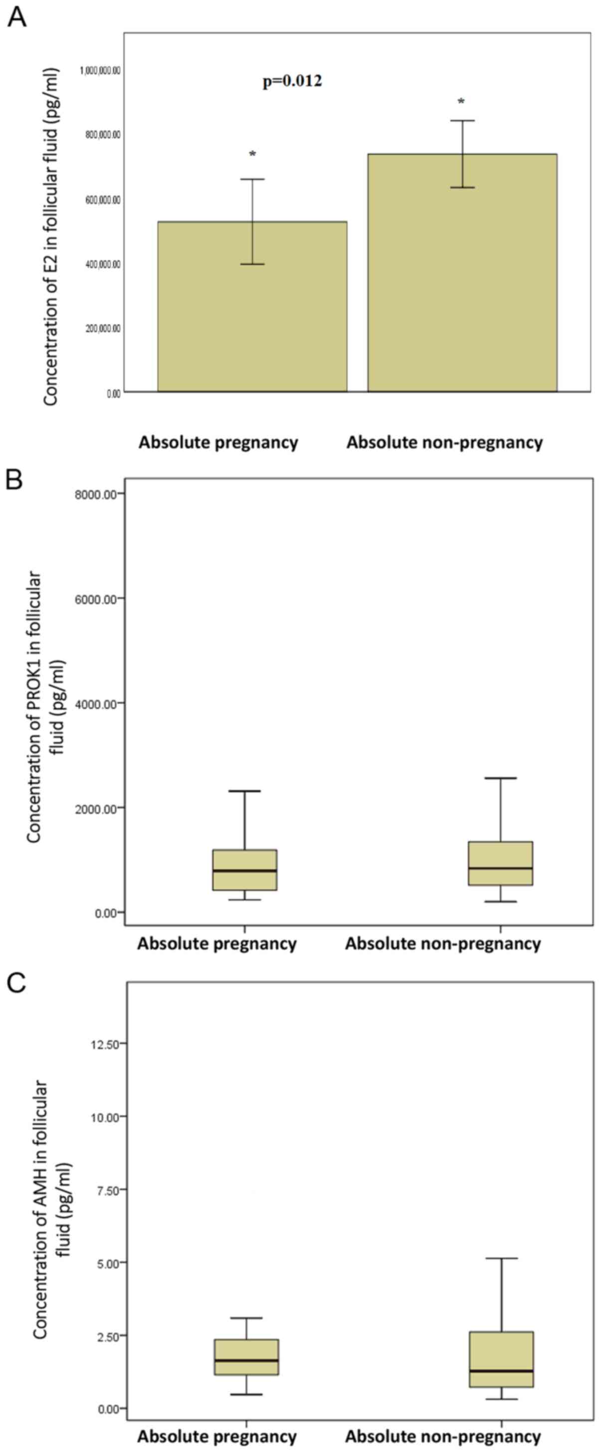
had a double fetus. The concentration of E2 in the follicular fluid
from the largest follicles in the absolute pregnancy group
(526419±289944 pg/ml) was significantly lower compared with the
absolute non-pregnancy group (736085±261885 pg/ml; t=-2.742;
P=0.012; Fig. 1A; Table II). The concentrations of PROK1 and
AMH in the follicular fluid from the largest follicles in absolute
pregnancy group were not significantly different from those in the
absolute non-pregnancy group (Fig.
1B and C; Table II). In addition, the concentrations
of peripheral blood PROK1 and AMH in the absolute pregnancy group
were not significantly different from those in the absolute
non-pregnancy group on the day of egg retrieval (Table II). The fertilization rate of
absolute pregnancy group was significantly higher than that of
absolute non-pregnancy group (χ2=5.861; P=0.015;
Table II).
| Table IIComparison of variables between
absolute pregnancy group and non-pregnancy group. |
Table II
Comparison of variables between
absolute pregnancy group and non-pregnancy group.
| Variable | Absolute pregnancy
group group (n=21) | Absolute
non-pregnancy (n=27) | Statistical
value | P-value |
|---|
| Age (years) | 32.29±2.59 | 32.26±4.27 | 0.027a | 0.979 |
| Years of
infertility | 4.95±3.09 | 4.87±2.98 | 0.093a | 0.926 |
| Constituent ratio
of primary infertility | 47.62% (10/21) | 70.37% (19/27) | 2.557b | 0.110 |
| Body mass index
(kg/m2) | 24.84±3.94 | 23.87±3.41 | 0.909a | 0.368 |
| Basal FSH
(mIU/ml) | 6.54
(2.95-10.04) | 6.56
(3.25-12.45) | -0.753c | 0.451 |
| Basal AMH
(ng/ml) | 4.17±2.69 | 3.67±2.25 | 0.707a | 0.483 |
| Gn dosage (IU) | 1,855±421 | 1,628±448 | 1.797a | 0.079 |
| E2 on HCG day
(pg/ml) | 2,232±1,317 | 2,640±1,974 | 0.108a | 0.421 |
| Endometrium
thickness on HCG day (mm) | 11 (9-12) | 7 (4.25-10.75) | -0.274c | 0.784 |
| No. of retrieved
oocytes | 9.00±3.45 | 7.28±3.56 | 1.789a | 0.079 |
| E2 concentration on
day of OR (pg/ml) | 1,415
(985-1,673) | 1,199
(718-2336) | 0.322c | 0.747 |
| PROK1 concentration
on day of OR (pg/ml) | 44.33 (16-285) | 41.03 (13-314) | -1.091c | 0.275 |
| AMH concentration
on day of OR (ng/ml) | 0.798
(0.15-4.14) | 0.835
(0.66-4.8) | -0.200c | 0.841 |
| PROK1 concentration
in follicular fluid (pg/ml) | 797.91
(235.99-2,653.33) | 836.78
(201.61-7,468.00) | -0.785c | 0.433 |
| AMH concentration
in follicular fluid (ng/ml) | 1.63
(0.47-6.96) | 1.27
(0.31-13.69) | -1.232c | 0.218 |
| E2 concentration in
follicular fluid (pg/ml) |
526,419±289,944 |
736,085±261,885 | -2.626a | 0.012 |
| Fertilization
rate | 88.4%
(137/155) | 78.5%
(146/186) | 5.861b | 0.015 |
| No. of usable
blastocysts | 2.62±2.31 | 1.58±2.04 | 1.596a | 0.118 |
Concentration of E2 in follicular
fluid is associated with the Gn dose
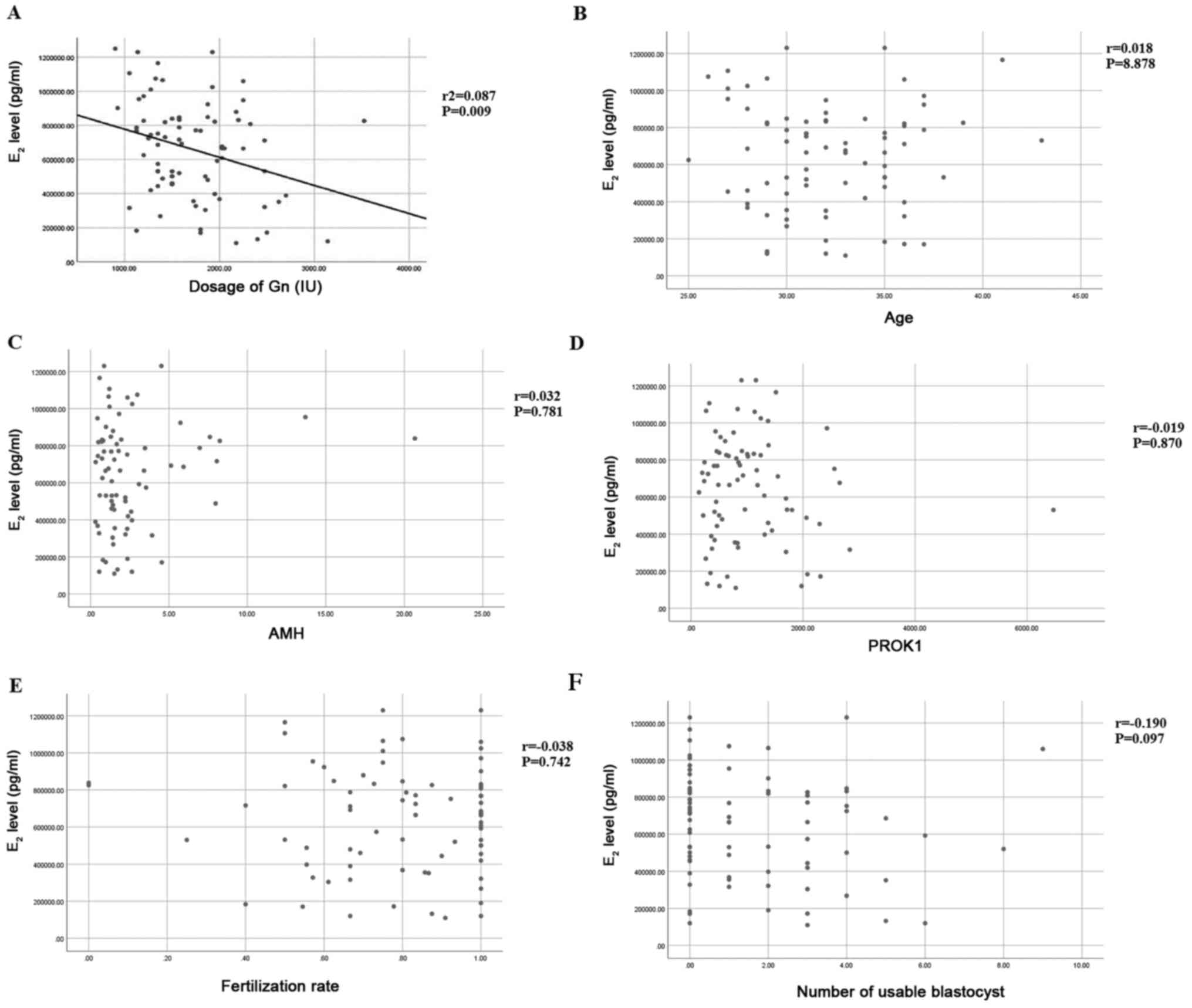
The relationship between Gn dose and E2
concentration in follicular fluid was analyzed by linear regression
analysis. The regression coefficient is -164.865, constant
942432.470. T statistic value of regression coefficient t-test is
-2.670, r2=0.087, P=0.009 (Fig. 2A). Therefore, regression coefficient
can be considered to be significant, from which the linear
regression equation can be obtained: Y=942432.470-164.865X. To
examine if E2 was correlated with other variables, a correlation
analysis was performed. The concentration of E2 was not associated
with age (r=0.018, P=0.878), AMH level in follicular fluid
(r=0.032, P=0.781), PROK1 level in follicular fluid (r=-0.019,
P=0.870), fertilization rate (r=-0.038, P=0.742) or number of
usable blastocysts (r=-0.109, P=0.097) (Fig. 2B-F).
Follicular fluid PROK1 level is not
correlated with Gn dosage but age
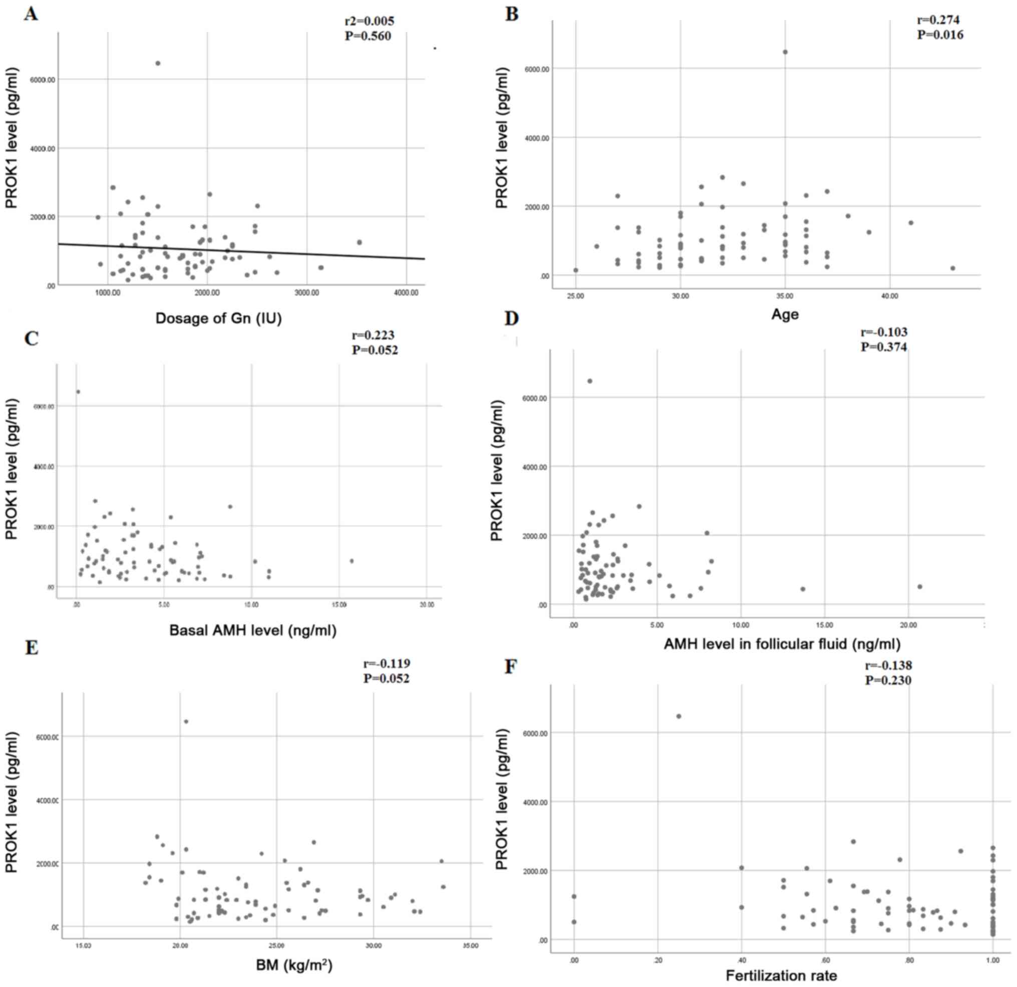
The relationship between the dose of Gn and the
concentration of PROK1 in follicular fluid was analyzed using a
linear regression analysis. The dose of Gn is the independent
variable on the x-axis, the concentration of PROK1 in follicular
fluid is the dependent variable on the y-axis, the determination
coefficient, r2=0.005, the regression coefficient is
-0.119, the constant is 1263, the observation value of T statistics
of regression coefficient T test is -0.585, and the P value of
t-test is 0.560. Therefore, the regression coefficient displayed no
significance (Fig. 3A). To examine
if PROK1 was correlated with other variables, a correlation
analysis was performed. The levels of PROK1 in follicular fluid was
correlated with age (r=0.274, P=0.016) (Fig. 3B). There was no significant
correlation between PROK1 in follicular fluid and basal AMH
(r=0.223, P=0.052), AMH in follicular fluid (r=-0.103, P=0.374),
body mass index (BMI) (r=-0.119, P=0.301) and fertilization rate
(r=-0.138, P=0.230) (Fig. 3C-F)
E2 level does not affect the number of
retrieved oocytes, fertilization rate and good embryo rate
Using the median of E2 levels in final fertilized
follicular fluid (676610 pg/ml) from the 75 cases as the cut-off
point, these cases were divided into the low E2 group (≤676610
pg/ml; 37 cases) and high E2 group (>676610 pg/ml; 38 cases).
The number of retrieved oocytes, fertilization rate and good embryo
rate were not significantly different between the two groups
(Table III). The result suggests
that E2 level did not affect the number of retrieved oocytes,
fertilization rate and good t embryo rate.
| Table IIINumber of retrieved eggs,
fertilization rate and good embryo rate in the low and high E2
groups. |
Table III
Number of retrieved eggs,
fertilization rate and good embryo rate in the low and high E2
groups.
| Characteristic | Low E2 group (mean
± SD) | High E2 group (mean
± SD) |
χ2/t-value | P-value |
|---|
| No. of retrieved
eggs | 9.03±4.23 | 8.37±4.59 | 0.565a | 0.520 |
| Fertilization
rate | 81.99%
(264/322) | 80.74%
(239/296) | 0.158b | 0.691 |
| Good embryo
rate | 70.89%
(168/237) | 65.78%
(148/225) | 1.393b | 0.238 |
E2 level in follicular fluid can
predict the outcome of pregnancy
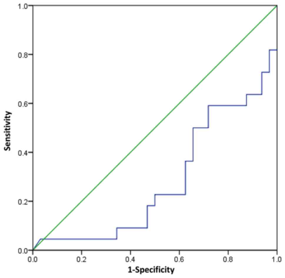
To evaluate the predictive value of E2 level in
follicular fluid, a ROC curve was plotted and AUC was calculated.
The AUC was 0.283 (95% CI, 0.144-0.423; P=0.007; Fig. 4). The AUC was significant which
indicates that E2 levels can discriminate between pregnancy
outcomes. The optimum cut off value of E2 level in follicular fluid
is 689345 pg/ml, which was shown to predict pregnancy outcome with
a sensitivity of 27.3%, and specificity of 37.5%.
Discussion
The concentration of E2 in the largest single
follicular fluid (657425±275979 pg/ml) of the present study was
10-50 times greater than has been reported previously (30,31).
This may be due to the high number of granulosa cell layers in the
largest follicle, which increases E2 secretion (32). Additionally, E2 concentration in
peripheral blood on the day of egg retrieval was 1565±1001 pg/ml,
400 times lower than that in the follicular fluid. The median level
of PROK1 in the follicular fluid on the day of egg retrieval was
only 21 times greater than that in the peripheral blood,
demonstrating that vascular endothelial cells underwent rapid
proliferation from luteinized granular cells to well-functioning
granular corpus luteum and membranous corpus luteum. PROK1
vasculogenic factor is associated with this aforementioned process,
in which PROK1 aggregates in a short amount of time to exhibit an
increased PROK1 level compared with the peripheral blood (29,33).
By contrast, the median level of AMH in the follicular fluid on the
day of egg retrieval was only slightly higher than levels in the
peripheral blood. This result may be due to AMH secreted by small
antral follicles being the main source of AMH in the peripheral
blood, instead of follicular fluid AMH (34).
E2 level in the follicular fluid of the absolute
pregnancy group was demonstrated to be significantly lower than
that in the absolute non-pregnancy group, suggesting that high
levels of estrogen had a negative effect on the pregnancy outcome.
It has been previously reported that high estrogen levels in the
peripheral blood can lead to poor pregnancy outcomes by affecting
endometrial receptivity (35,36).
In the present study, high E2 levels in the follicular fluid
exhibited an adverse effect on pregnancy outcome, suggesting that
abnormally high estrogen levels might affect the development
potential of adjacent oocytes via a variety of autocrine paracrine
signaling pathways. One previous study has revealed that E2
concentration in single follicular fluid was negatively correlated
with good embryo rate (7). However,
the current study failed to demonstrate this correlation (data not
shown). The results indicated that a high E2 level in the
follicular fluid was negatively associated with Gn dose. The ROC
curve analyzed the diagnostic accuracy of E2 levels to discriminate
between absolute pregnancy per transfer and absolute non pregnancy
per transfer. In the ROC model, E2 levels were predictive of
pregnancy outcome. However, the AUC was <0.5 and the sensitivity
and specificity of the cut off value was low.
The selection of dominant follicles often requires
complex capillary networks, and angiogenesis is closely associated
with follicular development (9). In
the ovaries, PROK1 not only promotes angiogenesis, but also affects
ovarian function (19,29). However, its specific roles have not
yet been clearly determined. A previous study has demonstrated that
PROK1 secretion in the follicular fluid is significantly different
between pregnant and non-pregnant patient groups (19); however, contradictory results have
also been reported (21). In the
present study, no significant differences were observed between
PROK1 expression in the follicular fluid of the pregnant and
non-pregnant group. The promoter of the PROK1 gene has a cAMP
response element (CRE) binding site (20). It has been previously reported that
AMH inhibited the binding of follicle-stimulating hormone to
follicle-stimulating hormone receptor through autocrine paracrine
and affects the transcription of estrogen synthetase (37). The results in the present study
revealed that PROK1 expression and AMH in the follicular fluid
exhibited no correlation. The level of PROK1 in follicular fluid
was correlated with age but not correlated with basal AMH. The
level of PROK1 in follicular fluid of older people showed an
increasing trend However, this possible correlation between the two
requires further study.
In the present study, no significant differences
were exhibited in the baseline characteristics between the absolute
pregnancy group and the absolute non-pregnancy group, suggesting
that the two groups were comparable. However, the limitation of the
present study is that a small number of cases was used for
analysis, which may affect the results. Additionally, the upper
limit of the electrochemiluminescence reagent for the determination
of E2 concentration was 3,000 pg, and some samples required
dilution 400 times, which may have also influenced the results.
In conclusion, the present study demonstrated that
E2 level in follicular fluid is a better predictor of the outcome
of IVF-ET compared with AMH or PROK1 level determination.
Acknowledgements
The authors wish to thank Dr Jiangfeng Lv from
Clinical Laboratory of Jinan People's Hospital (Jinan, China) and
Dr Xiumei Yu from Affiliated Yuhuangding Hospital of Qingdao
University (Yantai, China) for their help in the hormone tests.
Funding
The current study was supported by the National
Natural Science Foundation of China (grant. no. 81671416).
Availability of data and materials
The datasets used and/or analyzed during the current
study are available from the corresponding author on reasonable
request.
Authors' contributions
Each author believes that the manuscript represents
honest work. YL and CH collaborated to design the study. YL and XH
were responsible for performing experiments. YL and SD analyzed the
data. All authors collaborated to interpret results and develop the
manuscript. All authors read and approved the final manuscript.
Ethics approval and consent to
participate
All procedures performed in the current study were
approved by the Ethics Committee of Affiliated Yuhuangding Hospital
of Qingdao University. Written informed consent was obtained from
all patients or their families.
Patient consent for publication
Not applicable.
Competing interests
The authors declare that they have no competing
interests.
References
|
1
|
Durán Reyes G, Rosales AM and Hicks Gómez
JJ: Participation of the follicular fluid in follicular
development, oocyte maturation and spermatic function. Ginecol
Obstet Mex. 65:349–356. 1997.(In Spanish). PubMed/NCBI
|
|
2
|
Hammadeh ME, Ertan AK, Zeppezauer M,
Baltes S, Georg T, Rosenbaum P and Schmidt W: Immunoglobulins and
cytokines level in follicular fluid in relation to etiology of
infertility and their relevance to IVF outcome. Am J Reprod
Immunol. 47:82–90. 2002.PubMed/NCBI View Article : Google Scholar
|
|
3
|
Lédée N, Petitbarat M, Rahmati M,
Dubanchet S, Chaouat G, Sandra O, Perrier-d'Hauterive S, Munaut C
and Foidart JM: New pre-conception immune biomarkers for clinical
practice: Interleukin-18, interleukin-15 and TWEAK on the
endometrial side, G-CSF on the follicular side. J Reprod Immunol.
88:118–123. 2011.PubMed/NCBI View Article : Google Scholar
|
|
4
|
Vercammen M, Verloes A, Haentjens P and
Van de Velde H: Can soluble human leucocyte antigen-G predict
successful pregnancy in assisted reproductive technology? Curr Opin
Obstet Gynecol. 21:285–290. 2009.PubMed/NCBI View Article : Google Scholar
|
|
5
|
Hammadeh ME, Fischer-Hammadeh C, Amer AS,
Rosenbaum P and Schmidt W: Relationship between cytokine
concentration in serum and preovulatory follicular fluid and in
vitro fertilization/intracytoplasmic sperm injection outcome. Chem
Immunol Allergy. 88:80–97. 2005.PubMed/NCBI View Article : Google Scholar
|
|
6
|
Ovayolu A, Özdamar Ö, Gün İ, Arslanbuğa
CY, Kutlu T, Tunalı G and Uluhan R: The assesment of follicular
fluid presepsin levels in poor ovarian responder womenandits
relationship with the reproductive outcomes. Int J Clin Exp Med.
8:9961–9966. 2015.PubMed/NCBI
|
|
7
|
Mehta BN, Chimote MN, Chimote NN, Nath NM
and Chimote NM: Follicular-fluid anti-Mullerian hormone (FF AMH) is
a plausible biochemical indicator of functional viability of oocyte
in conventional in vitro fertilization (IVF) cycles. J Hum Reprod
Sci. 6:99–105. 2013.PubMed/NCBI View Article : Google Scholar
|
|
8
|
Sallam HN, Sallam NH and Sallam SH:
Non-invasive methods for embryo selection. Facts Views Vis Obgyn.
8:87–100. 2016.PubMed/NCBI
|
|
9
|
Koo YA, Lee B, Park HJ, Choi J, Lee E and
Choi D: Altered vascular endothelial growth factor expression
during GnRH antagonist protocol in women of reproductive age with
normal baseline hormone profiles. Fertil Steril. 91:744–748.
2009.PubMed/NCBI View Article : Google Scholar
|
|
10
|
Li M, Bullock CM, Knauer DJ, Ehlert FJ and
Zhou QY: Identification of two prokineticin cDNAs: Recombinant
proteins potently contract gastrointestinal smooth muscle. Mol
Pharmacol. 59:692–698. 2001.PubMed/NCBI View Article : Google Scholar
|
|
11
|
Matsumoto S, Yamazaki C, Masumoto KH,
Nagano M, Naito M, Soga T, Hiyama H, Matsumoto M, Takasaki J,
Kamohara M, et al: Abnormal development of the olfactory bulb and
reproductive system in mice lacking prokineticin receptor PKR2.
Proc Natl Acad Sci USA. 103:4140–4145. 2006.PubMed/NCBI View Article : Google Scholar
|
|
12
|
Tu LH, Yu LL, Xiong CL and Zhang HP:
Potential role of prokineticin 2 in experimental varicoceleinduced
rat testes. Urology. 80:952.e15–19. 2012.PubMed/NCBI View Article : Google Scholar
|
|
13
|
Kim KH, Oh DS, Jeong JH, Shin BS, Joo BS
and Lee KS: Follicular blood flow is a better predictor of the
outcome of in vitro fertilization-embryo transfer than follicular
fluid vascular endothelial growth factor and nitric oxide
concentrations. Fertil Steril. 82:586–592. 2004.PubMed/NCBI View Article : Google Scholar
|
|
14
|
LeCouter J, Kowalski J, Foster J, Hass P,
Zhang Z, Dillard-Telm L, Frantz G, Rangell L, DeGuzman L, Keller
GA, et al: Identification of an angiogenic mitogen selective for
endocrine gland endothelium. Nature. 412:877–884. 2001.PubMed/NCBI View
Article : Google Scholar
|
|
15
|
Lin R, LeCouter J, Kowalski J and Ferrara
N: Characterization of endocrine gland-derived vascular endothelial
growth factor signaling in adrenal cortex capillary endothelial
cells. J Biol Chem. 277:8724–8729. 2002.PubMed/NCBI View Article : Google Scholar
|
|
16
|
Kisliouk T, Friedman A, Klipper E, Zhou
QY, Schams D, Alfaidy N and Meidan R: Expression pattern of
prokineticin 1 and its receptors in bovine ovaries during the
estrous cycle: Involvement in corpus luteum regression and
follicular atresia. Biol Reprod. 76:749–758. 2007.PubMed/NCBI View Article : Google Scholar
|
|
17
|
Lee KF, Lee YL, Chan RW, Cheong AW, Ng EH,
Ho PC and Yeung WS: Up-regulation of endocrine gland-derived
vascular endothelial growth factor but not vascular endothelial
growth factor in human ectopic endometriotic tissue. Fertil Steril.
93:1052–1060. 2010.PubMed/NCBI View Article : Google Scholar
|
|
18
|
Kisliouk T, Levy N, Hurwitz A and Meidan
R: Presence and regulation of endocrine gland vascular endothelial
growth factor/prokineticin-1 and its receptors in ovarian cells. J
Clin Endocrinol Metab. 88:3700–3707. 2003.PubMed/NCBI View Article : Google Scholar
|
|
19
|
Alfaidy N, Hoffmann P, Gillois P,
Gueniffey A, Lebayle C, Garçin H, Thomas-Cadi C, Bessonnat J,
Coutton C, Villaret L, et al: PROK1 level in the follicular
microenvironment: A new noninvasive predictive biomarker of embryo
implantation. J Clin Endocrinol Metab. 101:435–444. 2016.PubMed/NCBI View Article : Google Scholar
|
|
20
|
Brouillet S, Hoffmann P, Chauvet S,
Salomon A, Chamboredon S, Sergent F, Benharouga M, Feige JJ and
Alfaidy N: Revisiting the role of hCG: New regulation of the
angiogenic factor EG-VEGF and its receptors. Cell Mol Life Sci.
69:1537–1550. 2012.PubMed/NCBI View Article : Google Scholar
|
|
21
|
Gao MZ, Zhao XM, Lin Y, Sun ZG and Zhang
HQ: Effects of EG-VEGF, VEGF and TGF-β1 on pregnancy outcome in
patients undergoing IVF-ET treatment. J Assist Reprod Genet.
29:1091–1096. 2012.PubMed/NCBI View Article : Google Scholar
|
|
22
|
Visser JA and Themmen AP: Anti-Müllerian
hormone and folliculogenesis. Mol Cell Endocrinol. 234:81–86.
2005.PubMed/NCBI View Article : Google Scholar
|
|
23
|
Fanchin R, Louafi N, Méndez Lozano DH,
Frydman N, Frydman R and Taieb J: Per-follicle measurements
indicate that anti-müllerian hormone secretion is modulated by the
extent of follicular development and luteinization and may reflect
qualitatively the ovarian follicular status. Fertil Steril.
84:167–173. 2005.PubMed/NCBI View Article : Google Scholar
|
|
24
|
Qin Y, Zhao Z, Sun M, Geng L, Che L and
Chen ZJ: Association of basal serum testosterone levels with
ovarian response and in vitro fertilization outcome. Reprod Biol
Endocrinol. 9(9)2011.PubMed/NCBI View Article : Google Scholar
|
|
25
|
Hassan AMA, Kotb MMM, AwadAllah AMA,
Shehata NAA and Wahba A: Follicular sensitivity index (FSI): A
novel tool to predict clinical pregnancy rate in IVF/ICSI cycles. J
Assist Reprod Genet. 34:1317–1324. 2017.PubMed/NCBI View Article : Google Scholar
|
|
26
|
La Marca A, Grisendi V, Giulini S, Argento
C, Tirelli A, Dondi G, Papaleo E and Volpe A: Individualization of
the FSH starting dose in IVF/ICSI cycles using the antral follicle
count. J Ovarian Res. 6(11)2013.PubMed/NCBI View Article : Google Scholar
|
|
27
|
Kolibianakis EM, Venetis CA,
Kalogeropoulou L, Papanikolaou E and Tarlatzis BC: Fixed versus
flexible gonadotropin-releasing hormone antagonist administration
in in vitro fertilization: A randomized controlled trial. Fertil
Steril. 95:558–562. 2011.PubMed/NCBI View Article : Google Scholar
|
|
28
|
Huang X, Hao C, Shen X, Liu X, Shan Y,
Zhang Y and Chen L: Differences in the transcriptional profiles of
human cumulus cells isolated from MI and MII oocytes of patients
with polycystic ovary syndrome. Reproduction. 145:597–608.
2013.PubMed/NCBI View Article : Google Scholar
|
|
29
|
Vural F, Vural B, Doğer E, Çakıroğlu Y and
Çekmen M: Perifollicular blood flow and its relationship with
endometrial vascularity, follicular fluid EG-VEGF, IGF-1, and
inhibin-a levels and IVF outcomes. J Assist Reprod Genet.
33:1355–1362. 2016.PubMed/NCBI View Article : Google Scholar
|
|
30
|
Ruiz de Assín R, Clavero A, Gonzalvo MC,
Ramírez JP, Zamora S, Fernández A, Martínez L and Castilla JA:
Comparison of methods to determine the assigned value in an
external quality control programme for embryo evaluation. Reprod
Biomed Online. 19:824–829. 2009.PubMed/NCBI View Article : Google Scholar
|
|
31
|
Nardo LG, Gelbaya TA, Wilkinson H, Roberts
SA, Yates A, Pemberton P and Laing I: Circulating basal
anti-Mullerian hormone levels as predictor of ovarian response in
women undergoing ovarian stimulation for in vitro fertilization.
Fertil Steril. 92:1586–1593. 2009.PubMed/NCBI View Article : Google Scholar
|
|
32
|
Rodgers RJ and Irving-Rodgers HF:
Formation of the ovarian follicular antrum and follicular fluid.
Biol Reprod. 82:1021–1029. 2010.PubMed/NCBI View Article : Google Scholar
|
|
33
|
Fraser HM, Bell J, Wilson H, Taylor PD,
Morgan K, Anderson RA and Duncan WC: Localization and
quantification of cyclic changes in the expression of endocrine
gland vascular endothelial growth factor in the human corpus
luteum. J Clin Endocrinol Metab. 90:427–434. 2005.PubMed/NCBI View Article : Google Scholar
|
|
34
|
Durlinger AL, Visser JA and Themmen AP:
Regulation of ovarian function: The role of anti-Mullerian hormone.
Reproduction. 124:601–609. 2002.PubMed/NCBI View Article : Google Scholar
|
|
35
|
Kosmas IP, Kolibianakis EM and Devroey P:
Association of estradiol levels on the day of hCG administration
and pregnancy achievement in IVF: A systematic review. Hum Reprod.
19:2446–2453. 2004.PubMed/NCBI View Article : Google Scholar
|
|
36
|
Yu Ng EH, Yeung WS, Yee Lan Lau E, So WW
and Ho PC: High serum oestradiol concentrations in fresh IVF cycles
do not impair implantation and pregnancy rates in subsequent
frozen-thawed embryo transfer cycles. Hum Reprod. 15:250–255.
2000.PubMed/NCBI View Article : Google Scholar
|
|
37
|
Kanakkaparambil R, Singh R, Li D, Webb R
and Sinclair KD: B-vitamin and homocysteine status determines
ovarian response to gonadotropin treatment in sheep. Biol Reprod.
80:743–752. 2009.PubMed/NCBI View Article : Google Scholar
|