Introduction
Obesity, which is associated with hormonal and
metabolic imbalances, is a common problem among pregnant women. It
frequently leads to adverse pregnancy outcomes, such as premature
delivery, dystocia, fetal macrosomia (1). Based on accumulating evidence by both
clinical and animal studies, an intrauterine high-fat environment
plays a crucial role in the development of diseases in offspring,
such as hypertension, diabetes and nervous system diseases such as
schizophrenia (1,2). Generally, the response of embryos to
an adverse intrauterine environment consists of adaptive changes.
These adjustments to stress or stimulation continuously alter the
physiological and metabolic functions of the body. Even after the
stimulus has been removed, the effect remains and becomes a key
factor contributing to chronic diseases in adults (2,3).
Animal experiments have shown that obesity leads to lower fertility
through alterations in various metabolic pathways, including
insulin metabolism, sex hormones signaling and follicular
development (4). After performing
hematoxylin-eosin (H&E) staining of ovarian tissue from
pregnant mice fed a high-fat diet (HFD), researchers observed
significantly decreased numbers of follicles at all levels in the
offspring of mothers fed a HFD compared with the offspring of
mothers fed a control diet (5).
However, the mechanisms by which a maternal HFD leads to
reproductive abnormalities in offspring remain unclear.
Reactive oxygen species (ROS) play important roles
in mediating hormonal signaling, oocyte formation, steroidogenesis,
follicle formation, ovulation, corpus luteum formation and germ
cell function, thus regulating the female reproductive system.
However, when ROS are overproduced, oxidative stress (OS) is
initiated, causing cytotoxic effects and irreversible damage to the
organism (6). Superoxide dismutase
(SOD) is the primary antioxidant enzyme in the body, and
malondialdehyde (MDA) is produced by lipid peroxidation. Both of
these molecules serve as indicators of OS in vivo (7,8). The
present study established an HFD model in female mice by
establishing four groups fed various HFDs during the preconception
period (4 weeks) and the gestation-lactation period, and OS levels
(SOD and MDA) and insulin resistance in the offspring were analyzed
to investigate the effects and mechanisms of maternal HFD on the
reproductive function of offspring. Additionally, the developmental
status of follicles was observed using H&E staining. Reverse
transcription-quantitative (RT-q)PCR and western blot analyses were
performed to analyze the expression of DNA damage markers, such as
phosphorylated form of H2AX histone variant (γH2AX), forkhead box
protein O3a (FOXO3a), Bcl-2-like protein 11 (Bim) and insulin-like
growth factor (IGF)-1, in ovarian tissue and to further explore the
pathogenic mechanism. In addition, a dietary intervention was added
to experimental subgroups. The present study aimed to determine the
effect of an HFD on otherwise healthy mice during pregnancy on the
reproductive function of offspring and to determine whether
reproductive function was improved by adjusting the diet program in
the HFD group. These results may provide guidance for pregnancy
preparation and for clinical interventions during pregnancy.
Materials and methods
Animal treatment
All animals were provided by the Laboratory Animal
Center of Capital Medical University, and the study protocol was
approved by the Local Ethics Review Committee of Capital Medical
University Beijing Obstetrics and Gynecology Hospital (approval no.
AEEI-2019-111). Female C57BL/6J mice (n=24; 8 weeks old; weight,
18-20 g) were randomly divided into four groups, housed under a
12-h light/dark cycle (lights on at 07:00 h) at a constant
temperature of 22±2˚C, with food and water available ad
libitum. A total of 12 male mice were mated with female mice in
estrus state at a ratio of 1:2. The HF/HF group was fed an HFD (60%
kcal fat; Research Diets, Inc.) during the preconception period (4
weeks) and the gestation-lactation period. The HF/C group was fed a
HFD at preconception (4 weeks) and a normal diet during the
gestation-lactation period. The C/HF group was fed a normal diet in
the preconception period (4 weeks) and a HFD in the
gestation-lactation period. Finally, the C/C group was fed a
regular diet in both the preconception and gestation-lactation
periods. A total of 90 female offspring were group housed after
weaning at 28 days of age. We collected 16 of the offspring from
each group that were fed a normal diet until 10 weeks of age. The
offspring were sacrificed at diestrus, as identified by a vaginal
smear. Mice were fasted for 12 h before been sacrificed. For
ethical reasons, mice were exposed to 3% isoflurane for 15 min
before sampling. Skin pinch reaction and stimulate toe response
were used to confirm the depth of anesthesia. The mice were
euthanized by decapitation. Fresh blood samples (1 ml) were
obtained from the mother and offspring, and the serum was collected
and stored at -80˚C. Sixty-four pairs of ovaries were collected.
The left ovary was fixed with 10% neutral buffered formaldehyde for
24 h at 25˚C and processed for histological analysis,
while the right ovary was immediately frozen in liquid nitrogen and
stored at -80˚C until the gene expression analysis.
Analysis of the OS levels
The SOD and MDA levels in serum samples from the
mother and offspring were analyzed to evaluate OS. All steps were
performed strictly in accordance with the test kit instructions
[Malondialdehyde (MDA) assay kit (cat. no. A003-1-2) and Superoxide
Dismutase (SOD) assay kit (cat. no. A001-3-2)] provided by the
Nanjing Jiangcheng Bioengineering Institute Co., Ltd. The blood
glucose and total cholesterol levels were also measured using an
automatic biochemical analyzer. Serum insulin, testosterone and
follicle-stimulating hormone (FSH) levels were measured using an
ELISA kit (Nanjing Jiangcheng Bioengineering Institute Co., Ltd.)
according to the manufacturer's instructions (follice stimulating
hormone assay kit, cat. no. cH101; insulin assay kit, cat. no.
H203; testosterone assay kit, cat. no. H090). Furthermore, the
insulin resistance index was calculated based on fasting glucose
and insulin levels [plasma glucose levels (GLU, mmol/l) x serum
insulin levels (mIU/l)/22.5].
Histological analysis of the
ovaries
One ovary from each offspring was fixed with 10%
neutral buffered formaldehyde (24 h, 25˚C), embedded in
paraffin, and serially sectioned to a thickness of 4-µm at 50-mm
intervals at fixed points in all ovaries. Sections were stained
with H&E at 25˚C for the morphometric analysis.
Additionally, some sections were stained with an anti-Mullerian
hormone antibody (Abcam) overnight at 4˚C to assist with
the identification of granulosa tissue in the follicles. Only
follicles with a visible oocyte nuclei were included, and follicles
showing evidence of atresia were excluded. All morphometric
analyses were performed by the same observer, and an independent
researcher was blinded to the experimental group from which the
sections were obtained during the analysis. The total area of each
section was calculated (area x thickness of section), and the
number of follicles for each animal were counted and the total area
of the ovarian tissue was calculated. The number of primordial
follicles was reported as the number of follicles per
mm2, while the numbers of antral and graafian follicles
are reported on a per ovary basis.
Gene expression analysis
The expression of IGF-1, FOXO3a,
γH2AX and Bim mRNA was measured using RT-qPCR. Total
RNA was extracted from the ovary tissues using TRIzol®
reagent (Sigma-Aldrich; Merck KGaA). Reverse transcription was
performed using a SuperScript First-Strand Synthesis system
(Invitrogen; Thermo Fisher Scientific; 42˚C for 50 min,
95˚C for 5 min). qPCR was carried out on a 7900HT
real-time PCR system with SYBR Premix Ex Taq II (Takara Bio, Inc.).
the thermocycling conditions were as follows: 50˚C for 2
min, 95˚C for 10 min, (95˚C for 15 sec,
60˚C for 1 min) x40 cycles (generate amplification
curve), 95˚C for 15 sec, 60˚C for 15 sec,
95˚C for 15 sec (generate dissolution curve). The
2-ΔΔCq method was used for expression analysis (9). Gene-specific primers and probes were
designed using Prime Express software (version 3.0.1, Thermo Fisher
Scientific, Inc; cat. no. 4363991) and purchased from Shanghai
GenePharma Co., Ltd. The sequences of the primers used in the
present study are as shown in Table
SI. All reactions were performed at least three times, and the
products were analyzed using a Roche Light Cycler 480. β-actin was
included in all reactions as an internal housekeeping control.
Western blot analysis
Total cellular protein was extracted using lysis
buffer (50 mmol HEPES, 1 mmol MgCl2, 10 mmol
ethylenediaminetetraacetic acid, 1% Triton X-100, pH 6.4). The
protein levels were quantified using a BCA Protein Assay kit
(Beyotime Institute of Biotechnology). The protein expression
levels of IGF-1, FOXO3a, γH2AX, Bim and β-actin in ovaries were
analyzed using western blotting. In total, 30 µg protein was loaded
per lane onto a 12% gel and resolved using SDS-PAGE. After
electrophoresis, proteins were transferred to a nitrocellulose
membrane. After being blocked with 5% lipid-free milk solution for
2 h at room temperature (RT). The membrane was probed with primary
antibodies against IGF-1, FOXO3a, γH2AX, Bim and β-actin [1:1,000;
Abcam, Anti-Bim antibody, cat. no. ab32158; Anti-IGF1 antibody,
cat. no. ab9572; Anti-gamma H2A.X (phospho S139) antibody (9F3),
cat. no. ab26350; Anti-FOXO3a antibody, cat. no. ab23683] at RT for
2 h. The secondary horseradish peroxidase-conjugated antibody was
anti-rabbit (1:10,000, Santa Cruz Biotechnology, Inc.) at RT for 2
h. Protein bands were visualized using the diaminobenzidine
detection kit (cat. no. SW2010; Beijing Solarbio Science &
Technology Co., Ltd.). The densities of sample bands were analyzed
with Quantity One analysis software 1709600 (Bio-Rad Laboratories,
Inc.).
Statistical analysis
The experimental data were analyzed using SPSS
version 21 (IBM Corp) for MacBook. All data are presented as mean ±
standard deviation from at least three independent repetitions.
One-way ANOVA was performed for between-group comparisons. If data
showed homogeneity of variance, Tukey's post hoc test was used,
otherwise Dunnett's T3 test was used. P<0.05 was considered to
indicate a statistically significant difference.
Results
Serum glucose, total cholesterol,
insulin, testosterone and FSH levels in female mouse offspring
Higher glucose and total cholesterol levels were
observed in the HF/HF and C/HF groups compared with those in the
C/C group (P<0.05), and the levels measured in the HF/HF group
were higher compared with those in the C/HF group. Significantly
higher insulin levels were measured in the HF/HF group compared
with those in the HF/C, C/HF and C/C groups (all P<0.05), which
showed a decreasing trend (Table I,
HOMA-IR). Furthermore, the insulin resistance index was calculated
based on fasting glucose and insulin levels [plasma glucose levels
(GLU, mmol/l) x serum insulin levels (mIU/l)/22.5]. The index value
also showed a decreasing trend: HF/HF>HF/C>C/HF >C/C. It
demonstrated that a high-fat diet during pregnancy increased
insulin levels and the tendency towards insulin resistance in
offspring mice. Reducing the intake of fat during the pregnancy and
lactation periods effectively improved the level of insulin and
insulin resistance. The FSH levels showed the following increasing
trend: HF/HF < HF/C < C/HF < C/C. The testosterone levels
in these groups also showed a decreasing trend: Showed a decreasing
trend: HF/HF>HF/C>C/HF>C/C (all, Table I).
 | Table ISerum Glu, TC, insulin, T and FSH
levels in female offspring mice (mean ± SD, n=16). |
Table I
Serum Glu, TC, insulin, T and FSH
levels in female offspring mice (mean ± SD, n=16).
| | Diet group |
|---|
| Marker | HF/HF | HF/C | C/HF | C/C |
|---|
| Glu, mmol/l |
11.72±1.44a-c |
8.22±1.01a |
7.45±0.95a | 5.82±0.88 |
| TC, mmol/l |
4.94±0.83a-c |
3.63±0.66a |
3.24±0.20a | 2.70±0.33 |
| Insulin, mU/l |
29.39±2.79a-c |
25.97±2.05a |
20.11±3.36a,b | 11.33±1.72 |
| HOMA-IR |
15.48±3.28a-c |
9.57±1.86a |
6.80±2.08a,b | 2.99±0.89 |
| T, nmol/l |
21.54±0.65a-c |
16.92±0.66a |
7.48±0.54a,b | 5.48±0.41 |
| FSH, mIU/ml |
5.53±0.37a-c |
7.02±0.45a |
10.47±0.47a,b | 12.53±0.47 |
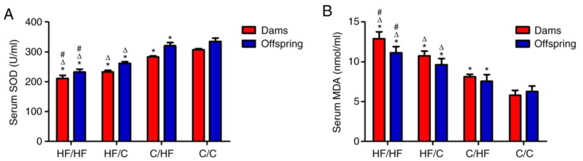
Serum levels of SOD and MDA
Among the groups, significantly lower SOD levels
were measured in both the mothers and offspring from the HF/HF
group compared with those in the HF/C, C/HF and C/C groups (vs.
HF/HF group, all P<0.05), which indicated an increasing trend.
The highest MDA levels were detected in both the mothers and
offspring from the HF/HF group, and the following decreasing trend
was observed: HF/C>C/HF>C/C (vs. HF/HF group, all P<0.05).
This effect persisted even when the offspring were fed a normal
diet. OS was significantly improved by reducing fat intake, as
shown in SOD levels and in MDA levels compared with the HF/HF group
(Fig. 1A and B, respectively).
Ovarian histology and analysis
The morphometric analyses of ovarian tissue are
shown in Fig. 2. The number of
primordial follicles, follicular development and number of
follicles were increased in the ovaries of offspring from the C/C
group compared with offspring from the HF/HF group. Significantly
fewer primordial follicles and preantral follicles were observed in
the HF/HF group compared with the C/C group (P<0.05). The number
of primordial follicles and preantral follicles in the HF/C group
was lower compared with the C/HF group (P<0.05). The HF/HF and
HF/C groups showed no significant difference in primordial
follicles and preantral follicles number (P>0.05). Moreover,
significant differences in the number of antral and graafian
follicles were not observed among the groups (P>0.05). The
result showed that HFD reduced the number of primordial and
preantral follicles but had little effect on antral and graafian
follicles. In addition, exposure to the HFD in the preconception
period exerted a greater effect on the number of primordial and
preantral follicles in offspring compared with a HFD in the
pregnancy and lactation periods, as presented in HF/C and C/HF.
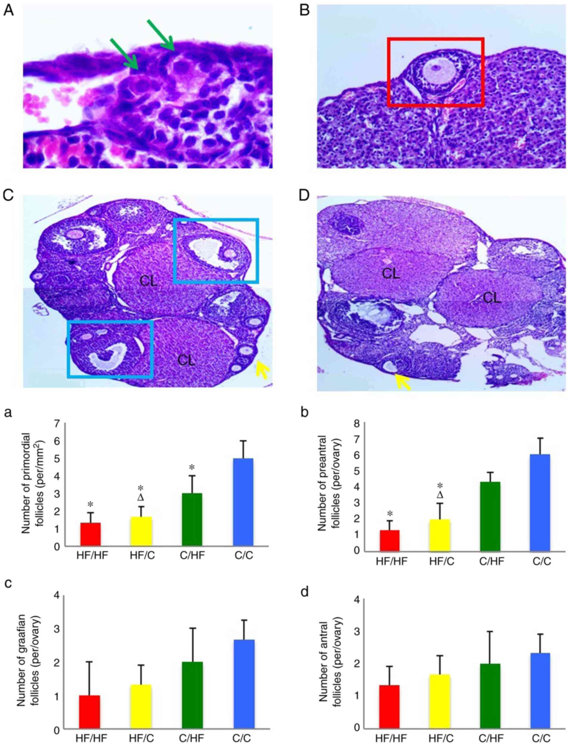 | Figure 2The random photomicrographs of
cross-sections of ovaries from (A-C) C/C and (D) HF/HF) female mice
stained with hematoxylin and eosin. The green arrows indicate the
primordial follicles (scale bar, 50 µm). The red square indicates
the preantral follicles (scale bar, 100 µm). The blue squares
indicate the graafian follicles (scale bar, 200 µm). The yellow
arrows indicate the antral follicles (scale bar, 200 µm). The
numbers of (a) primordial follicles, (b) preantral follicles, (c)
graafian follicles and (d) antral follicles in 10-week old
offspring. The offspring were from the HF/HF, HF/C, C/HF and C/C
groups (n=16). All values are reported as means ± SEM.
*P<0.05 vs. the C/C group; ΔP<0.05 vs.
the C/HF group (one-way ANOVA). CL, corpus lutem; HF, high fat; C,
control. |
Gene and protein expression
studies
The expression of γH2AX mRNA was increased in
the HF/HF group compared with the C/C group and HF/C group (both
P<0.05; Fig. 3A). The expression
of FOXO3a mRNA in the ovaries of offspring in the HF/HF,
HF/C, C/HF and C/C groups showed a decreasing trend (vs. C/C group,
all P<0.05) (Fig. 3B). Higher
expression of IGF-1 and Bim mRNAs was detected in the
HF/HF group compared with the HF/C group, and higher expression was
observed in the HF/C compared with the C/HF group (both P<0.05;
Fig. 3C-D). In addition, the
expression of each protein was analyzed using western blot
analysis. The results of the western blot analyses were consistent
with those of the RTq-PCR analysis (Fig. 4). Significantly higher BIM and FOXO
levels were measured in the HF/HF group compared with those in the
HF/C, C/HF and C/C groups (all P<0.05).
 | Figure 3The expression of the (A)
γH2AX, (B) FOXO3a, (C) IGF-1 and (D)
Bim mRNAs in ovaries from 10-week old offspring. The
offspring were from the HF/HF, HF/C, C/HF and C/C groups (n=16).
The values are presented as the relative fold changes normalized to
the C/C group, with 1 representing the value obtained for the C/C
group. All values are reported as means ± SEM.
*P<0.05 vs. the C/C group; #P<0.05 vs.
the HF/C group; ΔP<0.05 vs. the C/HF group (one-way
ANOVA). FOXO3a, forkhead box protein O3a; IGF-1, insulin-like
growth factor 1; γH2AX, phosphorylated form of H2AX histone
variant; Bim, Bcl-2-like protein 11; HF, high fat; C, control. |
Discussion
The Developmental Origins of Health and Disease
concept has emerged over the past 50 years, describing how maternal
and environmental factors potentially affect the child's growth and
development (10). The environment
of the maternal uterus not only affects the growth of embryos but
also exerts a long-term effect on the health of the offspring, a
phenomenon known as fetal programming (11). An mice model study revealed that
maternal HFD decreases the number of follicles in the ovaries of
offspring (12,13). In the present study, the offspring
mice of a mother fed an HFD had fewer follicles compared with the
mice born to a mother who was fed a regular diet. Moreover,
exposure to an HFD during pregnancy affected the reproductive
function of offspring, based on results obtained from the C/HF and
C/C groups. A reduction in fat intake significantly reduced the
associated adverse outcomes, as demonstrated by the difference in
results obtained from the HF/HF and HF/C groups. Exposure to an HFD
during the preconception period exerted a greater effect on the
offspring compared with exposure to an HFD in the gestational and
lactation periods, as shown in the HF/C and C/HF groups. Thus, an
HFD during the preconception period resulted in much greater impact
on follicular growth and development in the ovaries of offspring.
It is speculated that these changes might be caused by several
factors, such as changes in oocyte quality, maternal obesity and
metabolic changes, which require further study. Since obesity may
lead to lower fertility in women through alterations in various
metabolic pathways, including insulin metabolism, sex hormones and
follicular development (13), the
current study detected serum levels of insulin, testosterone and
FSH in mothers and their female mice offspring. Both the dams and
offspring in the HF/HF group presented an increased levels of
insulin and a tendency towards insulin resistance. A reduction in
fat intake during the pregnancy and lactation periods effectively
improved the insulin levels and insulin resistance, as the insulin
level showed a decreasing trend: HF/HF>HF/C >C/HF>C/C.
Insulin resistance has been shown to stimulate the ovaries to
secrete excessive androgens, causing FSH-grain cell axis
dysfunction (14). Consistent with
the present study, the analysis of mouse serum samples showed that
HFD-exposed offspring had increased levels of testosterone and
decreased levels of FSH.
The current study detected OS markers (SOD and MDA)
and analyzed the expression of OS-related genes in the ovaries of
adult offspring mice to further explore the underlying pathogenic
mechanisms of an HFD. OS was substantially higher in the HFD group
HF/HF, HF/C, C/HF) compared with in the control group, as
demonstrated by lower SOD and higher MDA levels. SOD levels showed
an increasing trend among the HF/C, C/HF and C/C groups
(P<0.05), while MDA levels showed the opposite trend. A higher
level of OS was observed in the offspring of the HFD groups
compared with the offspring of the control group, suggesting that
even after removing pathogenic factors, the negative effect on the
offspring persisted. Fortunately, OS was significantly improved by
reducing fat intake, as shown by the increase in SOD levels
(HF/HF<HF/C<C/HF<C/C) and the decrease in MDA levels
(HF/HF>HF/C>C/HF>C/C) among groups. Usually, OS occurs
when the body produces excess ROS, causing imbalances with
antioxidants. The consumption of an HFD usually results in a large
number of fatty acids. The consumption of SOD during fatty acid
oxidation causes the accumulation of the peroxide free radical MDA
(6,15). With the increased production of free
radicals, an OS response is more likely to occur (7). One of the limitations of the present
study was that there was no analysis the levels of superoxide
anions and hydrogen peroxide. The increased OS level may be caused
by DNA damage, while OS further increased DNA damage, causing a
vicious cycle (16).
Regarding the gene expression analysis, H2AX,
a member of the histone H2A family, is necessary for OS-induced DNA
damage. When damage occurs, H2AX is quickly phosphorylated at
Ser139 to produce γH2AX, creating a marker of DNA damage (17). In the present study, γH2AX
expression increased in offspring of the HFD groups. Based on these
findings, cellular DNA in the offspring of mothers fed an HFD is
more vulnerable to damage (16,18).
FOXO3a has critical functions in the repair of damaged cellular DNA
and participates in the regulation of primordial follicle
formation, follicle development and oocyte apoptosis (19). The present study revealed increased
expression of FOXO3a in the ovaries of offspring from the HFD
groups. FOXO expression is modulated by the insulin/PI3K/Akt
pathway, the main pathway involved in insulin signal transduction.
In the absence of insulin, FOXO is located in the nucleus and
functions as a transcription factor by binding to the target gene
promoter. When insulin activates the PI3K/Akt pathway, FOXO is
phosphorylated. Phosphorylated FOXO does not bind the target gene
promoter and is transferred from the nucleus to the cytoplasm,
losing its transcriptional function. When insulin resistance is
present, the sensitivity of insulin is reduced (19,20).
As the signal transmission of the PI3K/Akt pathway is weakened,
FOXO transcription increases, thereby affecting glucose metabolism,
resulting insulin resistance (19-21).
Consistent with these findings, both the HFD groups and
corresponding offspring exhibited increased levels of insulin and a
tendency towards insulin resistance in the current study. In
addition, the DNA damage and subsequent OS also induced the
expression of FOXO3a to repair the damaged cells. Thus, it was
speculated that the increase in FOXO3a expression in offspring was
modulated by the insulin/PI3K/Akt pathway. This change may be
associated with insulin resistance and increased levels of OS
caused by the HFD. Further studies are needed to validate this
conclusion.
In summary, an HFD during the preconception period
resulted in long-term consequences on follicular growth and
development in the ovaries of adult offspring mice. HFD exposure
during the preconception period exerted a greater effect on
offspring compared with HFD exposure during the gestational and
lactation periods. A reduction in HF intake during the
preconception, gestation and lactation periods improved the
reproductive ability of female offspring. The present study
revealed the adverse effects of the HFD on the reproductive
function of offspring. It was speculated that DNA was more
vulnerable to damage in mice fed the HFD, causing OS and insulin
resistance. OS, DNA damage and insulin resistance may have
increased the expression of FOXO3a, which protects cells from
OS-induced damage (22); however,
this adaptation to an adverse intrauterine environment affected the
number of the ovaries. The long-term effects on follicular growth
and development might be involved in increasing OS and the
activation of the insulin/PI3K/Akt pathway. Further studies are
needed to confirm these conclusion.
Supplementary Material
The sequences of the PCR primers used
in the present study.
Acknowledgements
Not applicable.
Funding
This study was funded by the National Natural
Science Foundation of China (grant no. 81471521).
Availability of data and materials
The datasets used and/or analyzed in the current
study are available from the corresponding author on reasonable
request.
Authors' contributions
All authors contributed to the study conception and
design. QS was responsible for the integrity of the work as a whole
from inception to the published article, such as performing animal
experiments and writing the manuscript. SY and FW collected the
data and performed the statistical analysis. All authors read and
approved the final manuscript.
Ethics approval and consent to
participate
Ethics committee approval was obtained from the
Institutional Ethics Committee of Capital Medical University
(Beijing, China). All animals were provided by the Laboratory
Animal Center of Capital Medical University, and the study protocol
was approved by the Capital Medical University Ethics Review
Committee (Capital Medical University Beijing Obstetrics and
Gynecology Hospital; approval no. AEEI-2019-111).
Patient consent for publication
Not applicable.
Competing interests
The authors declare that they have no competing
interests.
References
|
1
|
McIntyre HD, Catalano P, Zhang C, Desoye
G, Mathiesen ER and Damm P: Gestational diabetes mellitus. Nat Rev
Dis Primers. 5(47)2019.PubMed/NCBI View Article : Google Scholar
|
|
2
|
Poston L, Caleyachetty R, Cnattingius S,
Corvalán C, Uauy R, Herring S and Gillman MW: Preconceptional and
maternal obesity: Epidemiology and health consequences. Lancet
Diabetes Endocrinol. 4:1025–1036. 2016.PubMed/NCBI View Article : Google Scholar
|
|
3
|
Stephenson J, Heslehurst N, Hall J,
Schoenaker DA, Hutchinson J, Cade JE, Poston L, Barrett G, Crozier
SR, Barker M, et al: Before the beginning: Nutrition and lifestyle
in the preconception period and its importance for future health.
Lancet. 391:1830–1841. 2018.PubMed/NCBI View Article : Google Scholar
|
|
4
|
Hohos NM and Skaznik-Wikiel ME: High-fat
diet and female fertility. Endocrinology. 158:2407–2419.
2017.PubMed/NCBI View Article : Google Scholar
|
|
5
|
Cheong Y, Sadek KH, Bruce KD, Macklon N
and Cagampang FR: Diet-induced maternal obesity alters ovarian
morphology and gene expression in the adult mouse offspring. Fertil
Steril. 102:899–907. 2014.PubMed/NCBI View Article : Google Scholar
|
|
6
|
Lu J, Wang Z, Cao J, Chen Y and Dong Y: A
novel and compact review on the role of oxidative stress in female
reproduction. Reprod Biol Endocrinol. 16(80)2018.PubMed/NCBI View Article : Google Scholar
|
|
7
|
Di Segni C, Silvestrini A, Fato R,
Bergamini C, Guidi F, Raimondo S, Meucci E, Romualdi D, Apa R,
Lanzone A and Mancini A: Plasmatic and intracellular markers of
oxidative stress in normal weight and obese patients with
polycystic ovary syndrome. Exp Clin Endocrinol Diabetes.
125:506–513. 2017.PubMed/NCBI View Article : Google Scholar
|
|
8
|
Catalano PM and Shankar K: Obesity and
pregnancy: Mechanisms of short term and long term adverse
consequences for mother and child. BMJ. 356(j1)2017.PubMed/NCBI View
Article : Google Scholar
|
|
9
|
Livak KJ and Schmittgen TD: Analysis of
relative gene expression data using real-time quantitative PCR and
the 2(-Delta Delta C(T)) method. Methods. 25:402–408.
2001.PubMed/NCBI View Article : Google Scholar
|
|
10
|
Mochizuki K, Hariya N, Honma K and Goda T:
Relationship between epigenetic regulation, dietary habits, and the
developmental origins of health and disease theory. Congenit Anom
(Kyoto). 57:184–190. 2017.PubMed/NCBI View Article : Google Scholar
|
|
11
|
Hanson M, Barker M, Dodd JM, Kumanyika S,
Norris S, Steegers E, Stephenson J, Thangaratinam S and Yang H:
Interventions to prevent maternal obesity before conception, during
pregnancy, and post partum. Lancet Diabetes Endocrinol. 5:65–76.
2017.PubMed/NCBI View Article : Google Scholar
|
|
12
|
Da Broi MG, Giorgi VSI, Wang F, Keefe DL,
Albertini D and Navarro PA: Influence of follicular fluid and
cumulus cells on oocyte quality: Clinical implications. J Assist
Reprod Genet. 35:735–751. 2018.PubMed/NCBI View Article : Google Scholar
|
|
13
|
Hohos NM, Cho KJ, Swindle DC and
Skaznik-Wikiel ME: High-fat diet exposure, regardless of induction
of obesity, is associated with altered expression of genes critical
to normal ovulatory function. Mol Cell Endocrinol. 470:199–207.
2018.PubMed/NCBI View Article : Google Scholar
|
|
14
|
Petrakis D, Vassilopoulou L, Mamoulakis C,
Psycharakis C, Anifantaki A, Sifakis S, Docea AO, Tsiaoussis J,
Makrigiannakis A and Tsatsakis AM: Endocrine disruptors leading to
obesity and related diseases. Int J Environ Res Public Health.
14(1282)2017.PubMed/NCBI View Article : Google Scholar
|
|
15
|
Radin L, Šimpraga M, Vince S, Kostelić A
and Milinković-Tur S: Metabolic and oxidative status of Saanen
goats of different parity during the peripartum period. J Dairy
Res. 82:426–433. 2015.PubMed/NCBI View Article : Google Scholar
|
|
16
|
Bisht S, Faiq M, Tolahunase M and Dada R:
Oxidative stress and male infertility. Nat Rev Urol. 14:470–485.
2017.PubMed/NCBI View Article : Google Scholar
|
|
17
|
Turinetto V and Giachino C: Multiple
facets of histone variant H2AX: A DNA double-strand-break marker
with several biological functions. Nucleic Acids Res. 43:2489–2498.
2015.PubMed/NCBI View Article : Google Scholar
|
|
18
|
Gao T, Diaz-Hirashi Z and Verdeguer F:
Metabolic signaling into chromatin modifications in the regulation
of gene expression. Int J Mol Sci. 19(4108)2018.PubMed/NCBI View Article : Google Scholar
|
|
19
|
Lee S and Dong HH: FoxO integration of
insulin signaling with glucose and lipid metabolism. J Endocrinol.
233:R67–R79. 2017.PubMed/NCBI View Article : Google Scholar
|
|
20
|
Ma J, Matkar S, He X and Hua X: FOXO
family in regulating cancer and metabolism. Semin Cancer Biol.
50:32–41. 2018.PubMed/NCBI View Article : Google Scholar
|
|
21
|
Manning BD and Toker A: AKT/PKB signaling:
Navigating the network. Cell. 169:381–405. 2017.PubMed/NCBI View Article : Google Scholar
|
|
22
|
Fasano C, Disciglio V, Bertora S, Lepore
Signorile M and Simone C: FOXO3a from the nucleus to the
mitochondria: A round trip in cellular stress response.
Cells-Basel. 8(1110)2019.PubMed/NCBI View Article : Google Scholar
|