Introduction
IBS-D is a common chronic functional
gastrointestinal disorder characterized by chronically recurring
abdominal pain, diarrhea, discomfort that is relieved by
defecation, or altered bowel habits, which correlate with impaired
intestinal mucosal barrier function (1). Previous studies have demonstrated that
the prevalence of IBS-D is 5-10% in the general population, and
that it may result from immune activation, intestinal barrier
dysregulation and low-grade inflammation (2-4).
IBS-D severely affects quality of life; however, the
pathophysiology of IBS-D is poorly understood.
Accumulating evidence indicates that intestinal
barrier function is impaired in IBS-D (5). Tight junctions (TJs) regulate the
paracellular permeability of the intestinal barrier. The cytosolic
protein tight junction protein ZO-1 (ZO-1) is a vital protein in
TJs; the transmembrane proteins, including claudin-1 (CLDN1), and
the cytosolic protein bind to the actin cytoskeleton to control
paracellular permeability (6).
Recent evidence suggests that the impaired function of the
intestinal mucosal barrier may be implicated in the pathological
and pathogenic processes of IBS-D, indicated by an increase in
intestinal permeability (7).
MicroRNAs (miRNAs) are small non-coding
single-stranded RNA molecules, which serve a vital role in cell
proliferation, apoptosis and differentiation (8). miRNA binding sites are usually located
in the 3' untranslated region (UTR) of their target mRNAs and
regulate translation (9). Numerous
studies have demonstrated that miRNAs have been extensively
involved in abundant physiological and pathological processes,
which serve important roles in inflammation and the immune response
(10,11). Recently, the correlation between
miRNAs and IBS-D have showed that the change of miRNAs have
affected IBS-D, including the function of intestinal mucosal
barrier (12). miRNA-29a is an
important member of the miRNA-29 family. A recent study showed that
miRNA-29a regulates intestinal membrane permeability via the
glutamine synthetase gene (GLUL) in IBS-D (13). Proteins regulated by miRNA-29a
remain to be fully elucidated. Decreased ZO-1 and CLDN1 expression
are observed in the colonic tract of patients with IBS-D, which
contributes to weakening of the intestinal barrier (14). However, whether miRNA-29a is
involved in the intestinal barrier pathophysiology of IBS-D by
regulating ZO-1 and CLDN1 expression remains unclear.
In the present study, to evaluate the role of
miRNA-29a in intestinal mucosal barrier function, specimens were
collected from patients with IBS-D and IBS-D mouse models were
established, to compare the changes in miRNA-29a, ZO-1 and CLDN1 in
IBS-D.
Materials and methods
Patients
The study included 21 patients with IBS-D admitted
to the Department of Gastroenterology at the First Affiliated
Hospital of Guangzhou University of Chinese Medicine (Guangzhou,
China) between April 2017 and January 2018. There were 13 males and
8 females aged 21-56 years old (mean age, 35 years old; median age,
29 years old). All patients met the Rome IV criteria for IBS-D
(15). There were 7 male and 9
female healthy volunteers (mean age, 30 years old; median age 28,
years old) recruited as a control group. The human specimens
collected were sigmoid colon mucosa. All patients underwent
screening colonoscopies and provided written informed consent
before the study. The experiment was approved by the Medical
Research Ethics committee of The First Affiliated Hospital of
Guangzhou University of Chinese Medicine (approval no.
AF/JD-02/02), and was conducted in accordance with the principles
expressed in the Declaration of Helsinki. The specimens collected
from humans were stored at -80˚C.
Ultrastructural observation by
TEM
The colonoscopic biopsies from three patients with
IBS-D and three healthy volunteers were cut into 1 mm3
strips, and immediately fixed in 2.5% glutaraldehyde for 4 h at
4˚C. The strips were post-fixed in 2% osmium tetroxide in 0.1 mol/l
PBS (pH 7.4) for 2 h. Sample strips were dehydrated in graded
ethanol and subsequently embedded in Epon 812. Samples were cut
into ultrathin sections (75 nm) and observed in a JEM-1400 electron
microscope (Hitachi).
Animals
This study was approved by The Animal Experimental
Ethics Committee of the First Affiliated Hospital of Guangzhou
University of Chinese Medicine (approval no. TCMF1-2017009). The
experimental procedure was performed according to the Guide for the
Use and Care of Laboratory Animals. A total of 40 specific
pathogen-free male C57BL/6J mice (weighing 20-25 g) were purchased
from the Guangzhou Experimental Animals Center (Guangzhou, China;
Certificate no. SCXK [Yue] 2013-0092). The mice were housed at a
constant temperature of 20-22˚C in sawdust-lined plastic cages and
maintained on a 12:12-h light-dark cycle. The standard chow diet
and water were provided ad libitum.
Immunohistochemistry for ZO-1 and
CLDN1 expression
The immunohistochemistry study was performed as
described previously (16). The
mouse tissue sections (4 µm thick) were immersed in PBS and placed
in polycarbonate staining jars (Kartell) filled with 10 mM sodium
citrate buffer solution (pH 6.0). Following exposure to microwaves
for 15 min for antigen retreival, the tissues were incubated in a
solution of 10% bovine serum albumin at room temperature for 10
min. Sections were incubated with rabbit polyclonal ZO-1 antibody
(cat. no. 21773-1-AP; 187 µg/150 µl; 1:300; Proteintech Group,
Inc.) and rabbit polyclonal antibody CLDN1 (cat. no. 4933, 1:200,
Cell Signaling Technology, Inc.), respectively, incubated at 4˚C
overnight. Following washing in PBS, polyperoxidase rabbit IgG
secondary antibody (cat. no. SP-9001; 1:1,000; OriGene
Technologies, Inc.) was incubated for 2 h at room temperature, and
the sections were rinsed and cover-slipped. Subsequently, the
sections were examined with a microscope (Nikon 80). All areas were
analyzed under the same sensitivity captured with x200
magnification using Image-Pro Plus 6.0 software (Media Cybernetics,
Inc.). The positive staining area was selected in the colon mucosa
and the optical density was automatically calculated. The average
optical density values (IOD/area) in the colonic mucosa were
calculated from 8 random fields per section. In total, 10 samples
in each group were analyzed.
Establishment of the IBS-D mouse
model
An experimental mouse model of IBS-D was established
as per previous study (17,18). Then, 40 mice were randomly assigned
into four groups: Control, model, miRNA-29a negative control and
miRNA-29a inhibitor groups. A total of 10 mice were used in each
group. Following a period of fasting, not including water, for 24
h, the mice were anesthetized with 5% diethyl ether (19,20).
The use of ether was approved by the ethics committee. Due to the
animal individual differences, the tolerance of anesthesia varies.
We observed body reaction of mice during the process, including
breath, heart rate, muscular tension and corneal reflex. These
parameters were monitored to ensure the animals were fully
anesthetized following the administration of ether (21). Under anesthesia, the mouse model of
IBS-D was established by administration of TNBS into the proximal
colon. Following treatment with TNBS (1 mg/mouse in 50% ethanol),
the mice were subsequently maintained in a headstand for 2 min. The
mice in the control group were administrated an equal volume of 50%
ethanol instead of TNBS. Before the collection of tissue, the mice
were euthanized by cervical dislocation under anesthesia, induced
by the inhalation of 5% isoflurane. Cardio-respiratory arrest,
absence of reflex was monitored to ensure the euthanasia.
ELISA of D-LA and DAO
Mouse blood was extracted from the abdominal aorta,
and separated at 1,000 x g for 20 min at 4˚C. The serum D-LA and
DAO were determined according to the protocols of the D-LA (cat.
no. AE91431mo; AMEKO) and DAO ELISA kits (cat. no. AE90824mo;
AMEKO). Samples were analyzed in duplicate in accordance with the
product specification. A total of 10 samples in each group were
analyzed. The experiment was repeated three times.
Intraperitoneal injection with
miRNA-29a inhibitor
The experimental protocol follows previous study and
some improvements were made (22,
23), 10 μg of miRNA-29a inhibitor
(cat. no: miR200029-1-5, RiboBio, Guangzhou, China) or negative
control (cat. no: miR2N00003-1-5, RiboBio, Guangzhou, China) in 100
μl sterile saline was administered by intraperitoneal injection of
10 mice in each group for one time. All mice were sacrificed 24 h
after the administration of miRNA-29a inhibitor. The aseptic
operation was needed. The sequence of miRNA-29a inhibitor:
3'-AUCGUGGUAGACUUUAGCCAAU-5'.
Reverse transcription-quantitative PCR
(RT-qPCR)
The total miRNA from the tissues was extracted using
a miRcute miRNA isolation kit (cat. no. DP501; Tiangen Biotech Co.,
Ltd.) and used as a template for reverse transcription with a cDNA
miRcute Plus miRNA First-strand cDNA Synthesis kit (cat. no. KR211;
Tiangen Biotech Co., Ltd.). Subsequently, PCR amplification was
conducted with the miRcute miRNA RT-qPCR Detection kit (cat. no.
FP411-01; Tiangen Biotech Co., Ltd.), with U6 as an internal
reference.
The total mRNA from the tissues was extracted with
TRIzol® (cat. no. 15596026; Invitrogen; Thermo Fisher
Scientific, Inc.) and used as a template for reverse transcription
into cDNA (cat. no. RR820A; Takara Biotechnology Co., Ltd.).
Subsequently, PCR amplification was conducted in accordance with
the protocol of the RT-qPCR kit, with β-actin as an internal
reference. RT-PCR and quantitative analysis was conducted using the
Bio-Rad real-time PCR detection system (Bio-Rad Laboratories,
Inc.). The reaction conditions for miRNA-29a were as follows: 94˚C
for 2 min (one cycle), then 94˚C for 20 sec, and 60˚C for 34 sec
(35 cycles). The reaction conditions for ZO-1 and CLDN1 were as
follows: 95˚C for 30 sec (one cycle), 95˚C for 5 sec and then 60˚C
for 30 sec (40 cycles). The relative expression levels of miRNA-29a
and ZO-1 and CLDN1 mRNA were calculated using the
2-ΔΔCq method (24). All the primer sequences for
miRNA-29a, ZO-1, CLDN1, U6 and β-actin were synthesized by Sangon
Biotech Co., Ltd. The primer sequences were 5'-TAGCACCATCTG
AAATCGGTTA-3' for miRNA-29a; 5'-AGAGTGAACCAC GAGACGCTG-3' and
5'-TCTACTGTCCGTGCTATACATT GA-3' for ZO-1; 5'-CTGCCCCAGTGGAGGATT-3'
and 5'-CA GCCCAGCCAGTGAAGA-3' for CLDN1. The mean miRNA and mRNA
expression levels of the three RT-qPCR experiments were calculated
for each case.
Western blotting
Protein levels of ZO-1 and CLDN1 were quantified by
western blotting. Intestinal mucosa were lysed with
radioimmunoprecipitation assay buffer (Beyotime Institute of
Biotechnology, Beijing China) at the ratio of 80 mg tissue/ml. The
total protein concentration was measured using a bicinchoninic acid
concentration determination reagent kit (Beyotime Institute of
Biotechnology), according to the protocol. Equal amounts of protein
(50 µg/well) were separated by SDS-PAGE and transferred to PVDF
membranes (EMD Millipore). The membranes were blocked at room
temperature and incubated overnight at 4˚C using the following
primary antibodies: Rabbit polyclonal ZO-1 antibody (0.028 µg/150
µl; 1:2,000; cat. no. 21773-1-AP; Proteintech Group, Inc.) and
rabbit polyclonal CLDN1 antibody (1:1,000; cat. no. 4933; Cell
Signaling Technology, Inc.) and rabbit polyclonal β-actin antibody
(0.013 µg/150 µl; 1:5,000; cat. no. 20536-1-AP; Proteintech Group,
Inc.). All the antibodies were diluted with 5% bovine serum albumin
in 1X TBST. The membranes were subsequently washed, followed by
incubation with horseradish peroxidase-conjugated (HRP) goat
anti-rabbit secondary antibodies (1:2,000; cat. no. SA00001-2;
Proteintech Group, Inc.) for 1 h at room temperature. The bands
were observed using a chemiluminescent HRP kit (cat. no. WBKLS0500;
EMD Millipore) on a ChemiDoc™ imaging system (Bio-Rad Laboratories,
Inc.) in accordance with the protocol.
Statistical analysis
Experimental data were analyzed with SPSS 17.0
software (SPSS, Inc.). The results are stated as the mean ±
standard deviation. The differences between two groups were
analyzed by Student's t-test. Multiple groups were analyzed using
ANOVA (parametric) followed by Tukey's multiple comparison post hoc
test. P<0.05 was considered to indicate a statistically
significant difference.
Results
Comparisons of miRNA-29a and ZO-1 and
CLDN1 mRNA in the colon mucosa of patients with IBS-D
To investigate whether the colon mucosa of patients
with IBS-D have different molecular expression patterns than
healthy tissues, the present study analyzed the expression of
miRNA-29a, ZO-1 and CLDN1 in the colon mucosa from patients with
IBS-D by RT-qPCR. It was observed that the relative content of
miRNA-29a in the IBS-D group was significantly upregulated compared
with the control group (P<0.01; Fig.
1A). In addition, the ZO-1 and CLDN1 mRNA expression levels
were significantly downregulated compared with the IBS-D group
(P<0.01; Fig. 1B). These results
suggested that miRNA-29a, ZO-1 and CLDN1 may serve important roles
in the pathogenesis of IBS-D.
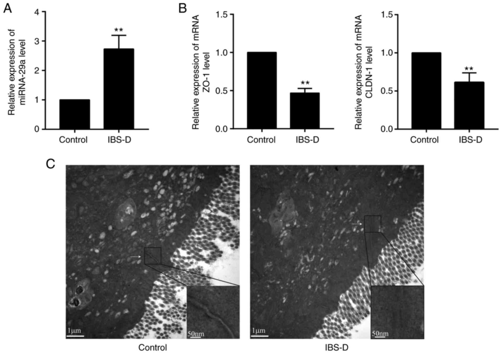 | Figure 1Alterations in miRNA-29a, ZO-1, CLDN1
and the JC in the colon mucosa of patients with IBS-D. Relative
expression of (A) miRNA-29a, (B) ZO-1 and CLDN1 was detected by
reverse transcription-quantitative PCR. **P<0.01 vs.
control. (C) Staining of the JC between colonic epithelium observed
by transmission electron microscopy. n=3. Magnification, x20,000.
IBS-D, diarrhea-predominant irritable bowel syndrome; miRNA,
microRNA; ZO-1, tight junction protein ZO-1; CLDN1, claudin-1; JC,
junctional complex. |
Alterations in the JC in the colon
mucosa of patients with IBS-D
To further assess the morphological alterations in
the colon mucosa of patients with IBS-D, TEM was used to visualize
the staining of the JC among colonic enterocytes. It was identified
that the staining was strong and continuous in the control group,
while the signals were faint and discontinuous in the IBS-D group
(Fig. 1C). These results suggested
that alterations in the JC may be implicated in the pathogenesis of
IBS-D.
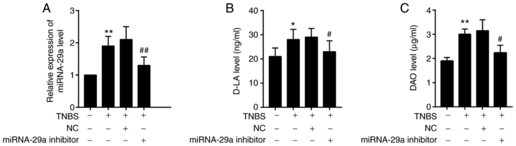
Comparisons of miRNA-29a, D-LA and DAO
in IBS-D mice
To analyze the content of miRNA-29a, RT-qPCR was
performed (Fig. 2A). To further
analyze the content of D-LA and DAO, ELISA was used (Fig. 2B and C). It was observed that miRNA-29a, D-LA
and DAO were significantly upregulated in the IBS-D mouse model
group (with TNBS treatment) compared with the control group
(P<0.01, P<0.05 and P<0.01, respectively). The miRNA-29a,
D-LA and DAO levels in IBS-D mice were significantly decreased by
the miRNA-29a inhibitor, but remained higher compared with the
control group (P<0.01, P<0.05 and P<0.05,
respectively).
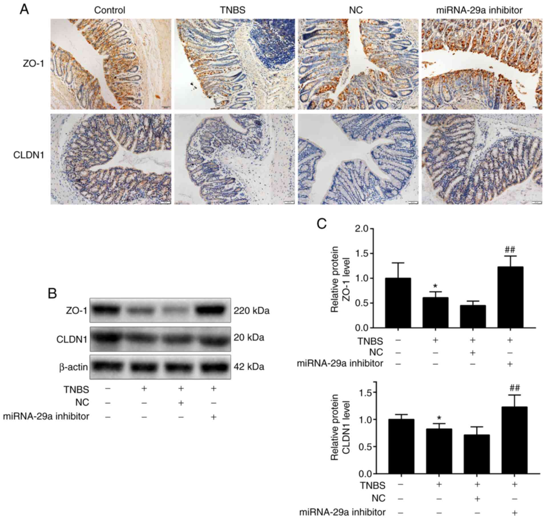
miRNA-29a downregulation alleviates
the impairment of the intestinal mucosal barrier and upregulates
the presence of ZO-1 and CLDN1 in IBS-D mice
To further investigate the presence of miRNA-29a,
ZO-1 and CLDN1 following administration of miRNA-29a inhibitor,
immunohistochemistry, RT-qPCR and western blot analysis were used.
The immunohistochemistry results (Fig.
3A) demonstrated that the presence of ZO-1 staining was
distributed on the edges of intestinal epithelial cells in brown,
while CLDN1 staining was distributed widely within the intestinal
epithelial cells in brown. In the IBS-D mouse group, ZO-1 and CLDN1
staining was unevenly distributed or faded, and the presence of
ZO-1 and CLDN1 was markedly reduced. However, following
administration of miRNA-29a inhibitor, the downregulation of ZO-1
and CLDN1 presence heightened significantly. As presented in
Table I, the average
immunohistochemistry optical density values of ZO-1 and CLDN1
decreased significantly (P<0.01 and P<0.05, respectively) in
the IBS-D group, compared with the control group. Nevertheless, the
values of ZO-1 and CLDN1 in the miRNA-29a inhibitor group were
significantly increased compared with the IBS-D group (P<0.05
and P<0.01, respectively). ZO-1 and CLDN1 were further evaluated
by western blotting. As presented in Fig. 3B and C, compared with the control group, ZO-1
and CLDN1 protein levels were downregulated significantly
(P<0.01 and P<0.05, respectively) in the IBS-D model group.
Furthermore, the effect of miRNA-29a on the expression of ZO-1 and
CLDN1 in IBS-D mice was evaluated by the administration of
miRNA-29a inhibitor. Compared with the IBS-D model group, ZO-1 and
CLDN1 protein levels were upregulated significantly (P<0.01) in
the miRNA-29a inhibitor group. These results suggested that
miRNA-29a reversed the TNBS-induced decrease in ZO-1 and CLDN1
protein levels.
 | Table IComparison of ZO-1 and CLDN1
immunohistochemistry average optical density values. |
Table I
Comparison of ZO-1 and CLDN1
immunohistochemistry average optical density values.
| Group | ZO-1 | CLDN1 |
|---|
| Control |
183,692±66,541.2 |
138,695.2±36,541.6 |
| TNBS |
95,120.5±26,458.3b |
68,745.9±12,548.3a |
| NC |
105,750.9±43,257.6 |
59,457.2±11,563.1 |
| miRNA-29a
inhibitor |
144,815.4±25,493.8c |
104,560.2±26,981.9d |
Discussion
The present study demonstrated that miRNA-29a
expression was upregulated in intestinal mucosal of IBS-D patients
and mice. Following administration of miRNA-29a inhibitor, the
impairment of the intestinal mucosal barrier was alleviated.
miRNA-29a inhibitor also decreased the expression of ZO-1 and
CLDN1. This may be implicated in the role of the intestinal mucosal
barrier in IBS-D.
miRNAs have been widely reported for their role in
various human disorders (8,25,26).
Recently, increasing evidence has demonstrated that miRNAs serve an
important role in the pathogenesis of IBS-D (27-29).
However, knowledge of the function of miRNAs in the progression of
IBS-D remains limited. miRNA-29 is highly expressed in glioma
tumorigenesis (30), pancreatic
ductal adenocarcinoma (31) and
chronic liver damage (32). It is
noteworthy that miRNA-29 has been reported to be involved in the
regulation of intestinal permeability and epithelial barrier
function (33,34). Consistent with early research
(35), the present results
demonstrated that miRNA-29a was significantly increased in patients
with IBS-D compared with the control, indicating that miRNA-29a
serves a role in IBS-D.
CLDN1 is a key TJ protein that maintains and
regulates intestinal permeability (36). ZO-1 has a vital role in maintaining
the appropriate structure and function of TJs. ZO-1 and CLDN1 are
the key proteins that form TJs, and their role in barrier
permeability has been thoroughly examined and demonstrated to be
crucial for the regulation of TJs (37,38).
Studies have indicated that the administration of ZO-1 siRNA may
affect and promote intestinal mucosal permeability (39). Moreover, although evident
histomorphological changes in the colon mucosa were not observed in
patients with IBS-D (40), the
change in ultrastructure was observed by TEM in the present study.
The present results demonstrated that the JC was discontinuous in
IBS-D, and that ZO-1 and CLDN1 expression was significantly
downregulated. Therefore, it may be speculated that the
ultrastructural change is associated with the degradation of ZO-1
and CLDN1.
D-LA is a metabolite of bacterial fermentation, the
level of which reflects the function of the intestinal mucosal
epithelial barrier. The level of DAO in serum can be used as a
marker to evaluate the integrity of the intestinal mucosa. To
further determine the role of miRNA-29a, intraperitoneal injection
with miRNA-29a inhibitor was performed, indicating that serum D-LA
and DAO were decreased (P<0.05) in the model group and
suggesting that intestinal mucosal barrier function may be improved
by reducing the expression of miRNA-29a. Furthermore, increased
ZO-1 and CLDN1 expression was also detected in IBS-D mice following
administration of miRNA-29a inhibitor, suggesting that miRNA-29a
serves an important role in the pathogenesis of IBS-D through
regulation of ZO-1 and CLDN1 expression; this indicated that the
regulation of TJs by ZO-1 and CLDN1 is important in maintaining the
integrity of the epithelial barrier in response to miRNA-29a
inhibitor.
There are several limitations to this study. First,
colonoscopic biopsies from IBS-D patients were sent for RNA
extraction and TEM. However, only the discontinuous distribution of
JC was observed by TEM, and the alterations of various junctional
proteins in IBS-D require further study and confirmation. Secondly,
since at least two investigators evaluated and agreed to the
clinical symptoms, the study was single blinded. Thirdly, a
previous study showed that CLDN-1 is the direct target of miRNA-29a
(34). We were unable to
conclusively determine whether ZO-1 is a direct target of
miRNA-29a. Nevertheless, we found that ZO-1 was significantly
upregulated after administering the intraperitoneal injection of
miRNA-29a inhibitor. Whether miRNA-29a regulates intestinal barrier
function in IBS-D by upregulating an important pathway including
ZO-1 requires verification in the future study.
In conclusion, the results of the present study
further suggested that miRNA-29a may be involved in the
pathogenesis of IBS-D by regulating intestinal mucosal barrier
function. Thus, the results provide evidence that miRNA-29a is a
possible molecular target for the treatment of IBS-D. Furthermore,
the present results indicated that the expression of ZO-1 and CLDN1
in the colonic mucosa of IBS-D mice was significantly increased
following intraperitoneal injection with miRNA-29a inhibitor.
Acknowledgements
The authors would like to thank Dr Xiaofei Li of the
Karolinska Institute for manuscript correction.
Funding
The present study was supported by The General
Program of National Nature Science Foundation of China (grant nos.
81673842 and 81703955) and The Special Clinical Program of the
First Affiliated Hospital of Guangzhou University of Chinese
Medicine (grant no. 2019ⅡT08).
Availability of data and materials
The datasets used and/or analyzed during the current
study are available from the corresponding author on reasonable
request.
Authors' contributions
HT and YC designed the study and approved the final
version of the manuscript. HZ and XX conducted the experiments. HZ
wrote the manuscript. HZ, YS and YW contributed to the acquisition,
analysis and interpretation of the case information, and obtained
intestinal patient samples. HZ and XX established the IBS-D mouse
model. DL and FX analyzed the patient data. YH and GH contributed
to the study conception and drafting the manuscript. All authors
read and approved the final manuscript.
Ethics approval and consent to
participate
The patient study was approved by The Medical
Research Ethics Committee of the First Affiliated Hospital of
Guangzhou University of Chinese Medicine (approval no.
AF/JD-02/02). All patients signed the informed consent. The animal
study was approved by The Animal Experimental Ethics Committee of
the First Affiliated Hospital of Guangzhou University of Chinese
Medicine (approval no. TCMF1-2017009)
Patient consent for publication
Not applicable.
Competing interests
The authors declare that they have no competing
interests.
References
|
1
|
Piche T, Barbara G, Aubert P, Bruley des
Varannes S, Dainese R, Nano JL, Cremon C, Stanghellini V, De
Giorgio R, Galmiche JP, et al: Impaired intestinal barrier
integrity in the colon of patients with irritable bowel syndrome:
Involvement of soluble mediators. Gut. 58:196–201. 2009.PubMed/NCBI View Article : Google Scholar
|
|
2
|
Martínez C, Lobo B, Pigrau M, Ramos L,
González-Castro AM, Alonso C, Guilarte M, Guilá M, de Torres I,
Azpiroz F, et al: Diarrhoea-predominant irritable bowel syndrome:
An organic disorder with structural abnormalities in the jejunal
epithelial barrier. Gut. 62:1160–1168. 2013.PubMed/NCBI View Article : Google Scholar
|
|
3
|
Matricon J, Meleine M, Gelot A, Piche T,
Dapoigny M, Muller E and Ardid D: Review article: Associations
between immune activation, intestinal permeability and the
irritable bowel syndrome. Aliment Pharmacol Ther. 36:1009–1031.
2012.PubMed/NCBI View Article : Google Scholar
|
|
4
|
Martínez C, González-Castro A, Vicario M
and Santos J: Cellular and molecular basis of intestinal barrier
dysfunction in the irritable bowel syndrome. Gut Liver. 6:305–315.
2012.PubMed/NCBI View Article : Google Scholar
|
|
5
|
Camilleri M, Lasch K and Zhou W: Irritable
bowel syndrome: Methods, mechanisms, and pathophysiology. The
confluence of increased permeability, inflammation, and pain in
irritable bowel syndrome. Am J Physiol Gastrointest Liver Physiol.
303:G775–G785. 2012.PubMed/NCBI View Article : Google Scholar
|
|
6
|
Bertiaux-Vandaële N, Youmba SB, Belmonte
L, Lecleire S, Antonietti M, Gourcerol G, Leroi AM, Déchelotte P,
Ménard JF, Ducrotté P, et al: The expression and the cellular
distribution of the tight junction proteins are altered in
irritable bowel syndrome patients with differences according to the
disease subtype. Am J Gastroenterol. 106:2165–2173. 2011.PubMed/NCBI View Article : Google Scholar
|
|
7
|
Bjorkman DJ and Popp JW: Mucosal Barrier
Defects in Irritable Bowel Syndrome. Who Left the Door Open? Am J
Gastroenterol. 101:864–865. 2006.PubMed/NCBI View Article : Google Scholar
|
|
8
|
Esteller M: Non-coding RNAs in human
disease. Nat Rev Genet. 12:861–874. 2011.PubMed/NCBI View
Article : Google Scholar
|
|
9
|
Ha M and Kim VN: Regulation of microRNA
biogenesis. Nat Rev Mol Cell Biol. 15:509–524. 2014.PubMed/NCBI View
Article : Google Scholar
|
|
10
|
He L and Hannon GJ: MicroRNAs: Small RNAs
with a big role in gene regulation. Nat Rev Genet. 5:522–531.
2004.PubMed/NCBI View
Article : Google Scholar
|
|
11
|
Xu XJ, Zhang YL, Liu L, Pan L and Yao SK:
Increased expression of nerve growth factor correlates with
visceral hypersensitivity and impaired gut barrier function in
diarrhoea-predominant irritable bowel syndrome: A preliminary
explorative study. Aliment Pharmacol Ther. 45:100–114.
2017.PubMed/NCBI View Article : Google Scholar
|
|
12
|
Martínez C, Rodiño-Janeiro BK, Lobo B,
Stanifer ML, Klaus B, Granzow M, González-Castro AM, Salvo-Romero
E, Alonso-Cotoner C, Pigrau M, et al: miR-16 and miR-125b are
involved in barrier function dysregulation through the modulation
of claudin-2 and cingulin expression in the jejunum in IBS with
diarrhoea. Gut. 66:1537–1538. 2017.PubMed/NCBI View Article : Google Scholar
|
|
13
|
Zhou Q, Souba WW, Croce CM and Verne GN:
MicroRNA-29a regulates intestinal membrane permeability in patients
with irritable bowel syndrome. Gut. 59:775–784. 2010.PubMed/NCBI View Article : Google Scholar
|
|
14
|
Wilcz-Villega E, McClean S and O'Sullivan
M: Reduced E-cadherin expression is associated with abdominal pain
and symptom duration in a study of alternating and diarrhea
predominant IBS. Neurogastroenterol Motil. 26:316–325.
2014.PubMed/NCBI View Article : Google Scholar
|
|
15
|
Drossman DA: Functional Gastrointestinal
Disorders: History, Pathophysiology, Clinical Features and Rome IV.
Gastroenterology. 150:1262–1279. 2016.PubMed/NCBI View Article : Google Scholar
|
|
16
|
Liu Y, Liu W, Peng QX, Peng JL, Yu LZ and
Hu JL: Protective effect of huoxiang zhengqi oral liquid on
intestinal mucosal mechanical barrier of rats with postinfectious
irritable bowel syndrome induced by acetic acid. Evid Based
Complement Alternat Med. 2014(218383)2014.PubMed/NCBI View Article : Google Scholar
|
|
17
|
Du C, Peng L, Kou G, Wang P, Lu L and Li
Y: Assessment of Serum sTREM-1 as a Marker of Subclinical
Inflammation in Diarrhea-Predominant Patients with Irritable Bowel
Syndrome. Dig Dis Sci. 63:1182–1191. 2018.PubMed/NCBI View Article : Google Scholar
|
|
18
|
Chao G, Wang Y, Ye F and Zhang S:
Regulation of Colonic Mucosal MicroRNA Expression via Multiple
Targets in Visceral Hypersensitivity Rats by Tongxieyaofang. Yonsei
Med J. 59:945–950. 2018.PubMed/NCBI View Article : Google Scholar
|
|
19
|
Raju D, Ilango K, Chitra V and Ashish K:
Evaluation of Anti-ulcer activity of methanolic extract of
Terminalia chebula fruits in experimental rats. J Pharm Sci Res.
1:101–107. 2009.
|
|
20
|
van Herck H, Baumans V, Brandt CJWM, Hesp
APM, Sturkenboom JH, van Lith HA, van Tintelen G and Beynen AC:
Orbital sinus blood sampling in rats as performed by different
animal technicians: The influence of technique and expertise. Lab
Anim. 32:377–386. 1998.PubMed/NCBI View Article : Google Scholar
|
|
21
|
van Herck H, Baumans V, de Boer SF, van
der Gugten J, van Woerkom AB and Beynen AC: Endocrine stress
response in rats subjected to singular orbital puncture while under
diethyl-ether anaesthesia. Lab Anim. 25:325–329. 1991.PubMed/NCBI View Article : Google Scholar
|
|
22
|
Liao XJ, Mao WM, Wang Q, Yang GG, Wu WJ
and Shao SX: MicroRNA-24 inhibits serotonin reuptake transporter
expression and aggravates irritable bowel syndrome. Biochem Biophys
Res Commun. 469:288–293. 2016.PubMed/NCBI View Article : Google Scholar
|
|
23
|
Deng Y, Zhou X, Xiang X, Ou Y and He J:
Effect of miRNA-19a on gastrointestinal motility in rats with
functional dyspepsia. Exp Ther Med. 15:4875–4879. 2018.PubMed/NCBI View Article : Google Scholar
|
|
24
|
Livak KJ and Schmittgen TD: Analysis of
relative gene expression data using real-time quantitative PCR and
the 2(-Delta Delta C(T)) method. Methods. 25:402–408.
2001.PubMed/NCBI View Article : Google Scholar
|
|
25
|
Viswambharan V, Thanseem I, Vasu MM,
Poovathinal SA and Anitha A: miRNAs as biomarkers of
neurodegenerative disorders. Biomarkers Med. 11:151–167.
2017.PubMed/NCBI View Article : Google Scholar
|
|
26
|
Issler O and Chen A: Determining the role
of microRNAs in psychiatric disorders. Nat Rev Neurosci.
16:201–212. 2015.PubMed/NCBI View
Article : Google Scholar
|
|
27
|
Hou Q, Huang Y, Zhu S, Li P, Chen X, Hou Z
and Liu F: miR-144 Increases Intestinal Permeability in IBS-D Rats
by Targeting OCLN and ZO1. Cell Physiol Biochem. 44:2256–2268.
2017.PubMed/NCBI View Article : Google Scholar
|
|
28
|
Wohlfarth C, Schmitteckert S, Härtle JD,
Houghton LA, Dweep H, Fortea M, Assadi G, Braun A, Mederer T, et
al: miR-16 and miR-103 impact 5-HT4 receptor signalling
and correlate with symptom profile in irritable bowel syndrome. Sci
Rep. 7(14680)2017.PubMed/NCBI View Article : Google Scholar
|
|
29
|
Kapeller J, Houghton LA, Mönnikes H,
Walstab J, Möller D, Bönisch H, Burwinkel B, Autschbach F, Funke B,
Lasitschka F, et al: First evidence for an association of a
functional variant in the microRNA-510 target site of the serotonin
receptor-type 3E gene with diarrhea predominant irritable bowel
syndrome. Hum Mol Genet. 17:2967–2977. 2008.PubMed/NCBI View Article : Google Scholar
|
|
30
|
Liu Y, Duan N and Duan S: MiR-29a Inhibits
Glioma Tumorigenesis through a Negative Feedback Loop of TRAF4/Akt
Signaling. BioMed Res Int. 2018(2461363)2018.PubMed/NCBI View Article : Google Scholar
|
|
31
|
Liang C, Shi S, Meng Q, Liang D, Hua J,
Qin Y, Zhang B, Xu J, Ni Q and Yu X: MiR-29a, targeting caveolin 2
expression, is responsible for limitation of pancreatic cancer
metastasis in patients with normal level of serum CA125. Int J
Cancer. 143:2919–2931. 2018.PubMed/NCBI View Article : Google Scholar
|
|
32
|
Huang YH, Yang YL and Wang FS: The Role of
miR-29a in the Regulation, Function, and Signaling of Liver
Fibrosis. Int J Mol Sci. 19(1889)2018.PubMed/NCBI View Article : Google Scholar
|
|
33
|
Zhou Q, Costinean S, Croce CM, Brasier AR,
Merwat S, Larson SA, Basra S and Verne GN: MicroRNA 29 targets
nuclear factor-κB-repressing factor and Claudin 1 to increase
intestinal permeability. Gastroenterology. 148:158–169.e8.
2015.PubMed/NCBI View Article : Google Scholar
|
|
34
|
Xue H, Zhang G, Geurts AM, Usa K, Jensen
DM, Liu Y, Widlansky ME and Liang M: Tissue-specific effects of
targeted mutation of Mir29b1 in rats. EBioMedicine. 35:260–269.
2018.PubMed/NCBI View Article : Google Scholar
|
|
35
|
Lin X, Zhou X, Liu D, Yun L, Zhang L, Chen
X, Chai Q and Li L: MicroRNA-29 regulates high-glucose-induced
apoptosis in human retinal pigment epithelial cells through PTEN.
In Vitro Cell Dev Biol Anim. 52:419–426. 2016.PubMed/NCBI View Article : Google Scholar
|
|
36
|
Pope JL, Bhat AA, Sharma A, Ahmad R,
Krishnan M, Washington MK, Beauchamp RD, Singh AB and Dhawan P:
Claudin-1 regulates intestinal epithelial homeostasis through the
modulation of Notch-signalling. Gut. 63:622–634. 2014.PubMed/NCBI View Article : Google Scholar
|
|
37
|
Hadj-Rabia S, Baala L, Vabres P,
Hamel-Teillac D, Jacquemin E, Fabre M, Lyonnet S, De Prost Y,
Munnich A, Hadchouel M, et al: Claudin-1 gene mutations in neonatal
sclerosing cholangitis associated with ichthyosis: A tight junction
disease. Gastroenterology. 127:1386–1390. 2004.PubMed/NCBI View Article : Google Scholar
|
|
38
|
Masahiko I, Mikio F, Kazumasa M and Koji
K: Mitinori Saitou and Tsukita AS: Direct Binding of Three Tight
Junction-associated MAGUKs, ZO-1, ZO-2, and ZO-3, with the COOH
Termini of Claudins. J Cell Biol. 147:1351–1363. 2000.PubMed/NCBI View Article : Google Scholar
|
|
39
|
Wang N, Han Q, Wang G, Ma WP, Wang J, Wu
WX, Guo Y, Liu L, Jiang XY, Xie XL, et al: Resveratrol Protects
Oxidative Stress-Induced Intestinal Epithelial Barrier Dysfunction
by Upregulating Heme Oxygenase-1 Expression. Dig Dis Sci.
61:2522–2534. 2016.PubMed/NCBI View Article : Google Scholar
|
|
40
|
Liu DR, Xu XJ and Yao SK: Increased
intestinal mucosal leptin levels in patients with
diarrhea-predominant irritable bowel syndrome. World J
Gastroenterol. 24:46–57. 2018.PubMed/NCBI View Article : Google Scholar
|