Open Access

Performance evaluation of two fundus oculi angiographic imaging system: Optos 200Tx and Heidelberg Spectralis

  • Authors:
    • Shuang Li
    • Jing-Jing Wang
    • Hong-Yang Li
    • Wei Wang
    • Meng Tian
    • Xu-Qiang Lang
    • Kang Wang
  • View Affiliations

  • Published online on: November 5, 2020     https://doi.org/10.3892/etm.2020.9451
  • Article Number: 19
  • Copyright: © Li et al. This is an open access article distributed under the terms of Creative Commons Attribution License.

Metrics: Total Views: 0 (Spandidos Publications: | PMC Statistics: )
Total PDF Downloads: 0 (Spandidos Publications: | PMC Statistics: )


Abstract

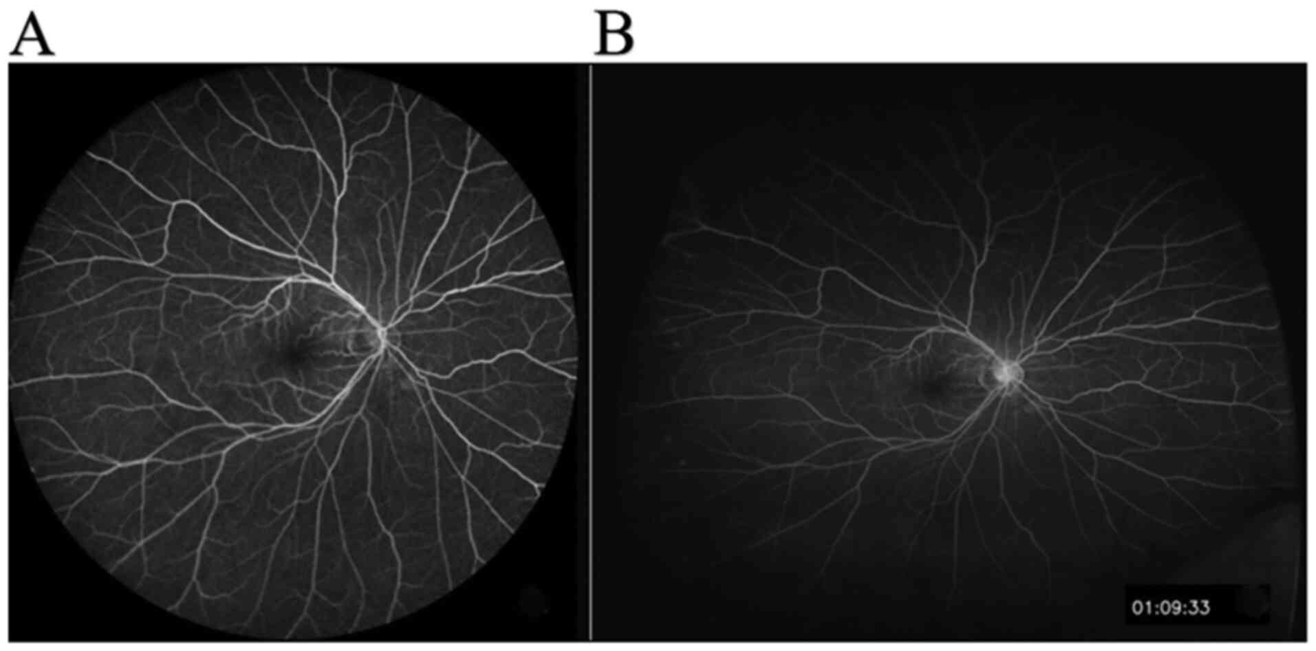
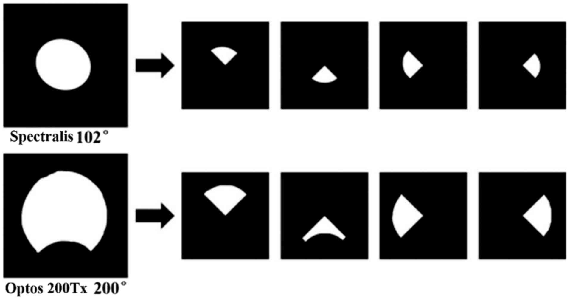
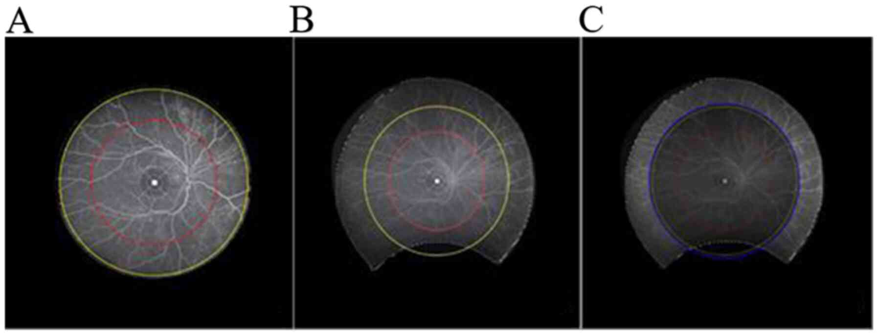
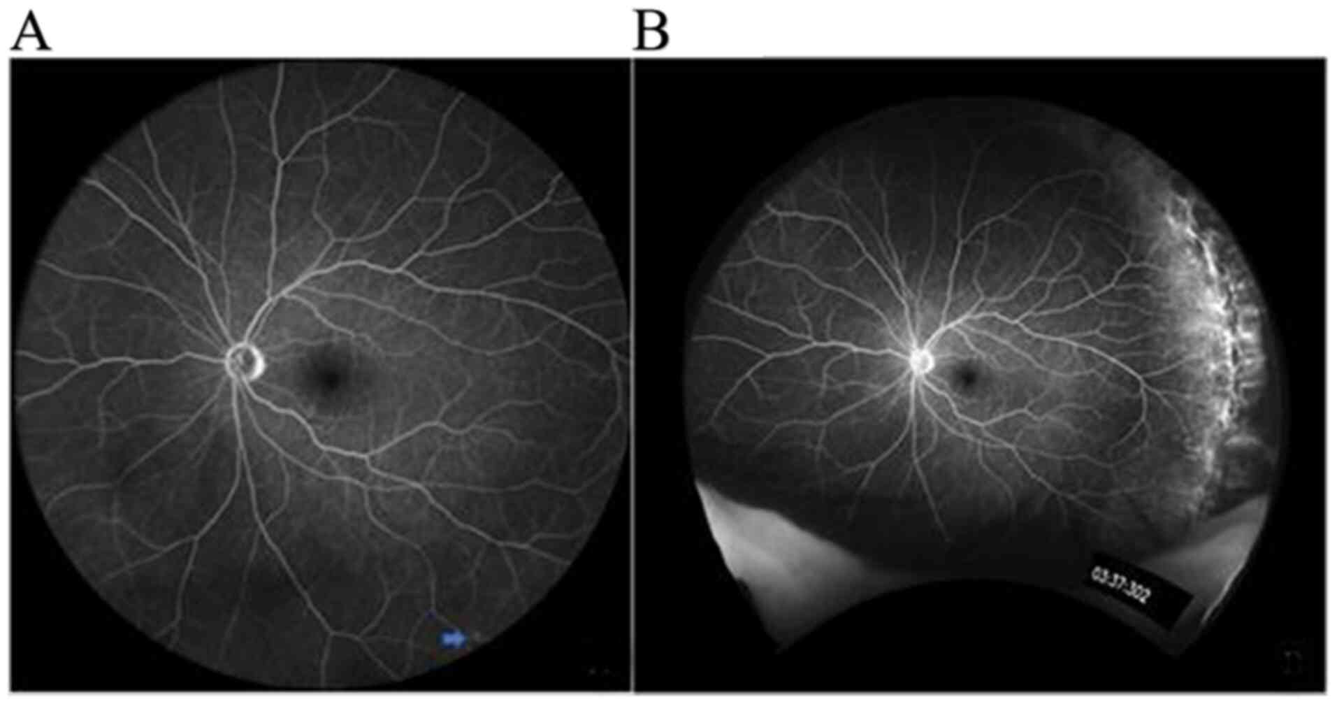
The present study aimed to compare the imaging performance of two ultra‑wide‑field fluorescein angiography imaging systems, namely the OptosOptomap 200Tx (Optos 200Tx) and the Heidelberg Spectralis (Spectralis). A total of 18 patients (36 eyes) underwent angiography using the two systems at the Department of Ophthalmology, Beijing Friendship Hospital (Beijing, China) between January and June 2017. The images were obtained as a single shot centered on the macula. The total area and area within each of four visualized quadrants were calculated and compared. The averages of the total and individual quadrant area captured by the Optos 200Tx were all larger than those obtained with the Spectralis (P<0.05). For pair‑wise comparison, the circular area centered on the macula (radius of 10 and 15 mm) was displayed: Optos 200Tx 10 mm (295.57 mm2) < Spectralis (520.11 mm2) < Optos 200Tx 15 mm (596.45 mm2) < Optos 200Tx (804.36 mm2) (P<0.01). The differences of each of the four quadrant areas were statistically significant between the two systems (P<0.05). The mean size of the areas was in the following order: Inferior < temporal < superior < nasal for the Optos 200Tx, and inferior < temporal < nasal < superior for the Spectralis. Further comparison of the four‑quadrant area indicated that the inferior quadrant of the Optos 200Tx was smaller than the other three quadrants (P<0.01) and the inferior quadrant of the Spectralis was smaller than the superior quadrant (P<0.01). The total retinal area and the retinal area of each quadrant captured by the Optos 200Tx were larger than those captured with the Spectralis. The total retinal area captured with the Optos 200Tx was able to cover the mid‑peripheral area and part of the far‑peripheral area of the retina, whereas the Spectralis only covered the mid‑peripheral area.
View Figures
View References

Related Articles

Journal Cover

January-2021
Volume 21 Issue 1

Print ISSN: 1792-0981
Online ISSN:1792-1015

Sign up for eToc alerts

Recommend to Library

Copy and paste a formatted citation
x
Spandidos Publications style
Li S, Wang J, Li H, Wang W, Tian M, Lang X and Wang K: Performance evaluation of two fundus oculi angiographic imaging system: Optos 200Tx and Heidelberg Spectralis. Exp Ther Med 21: 19, 2021
APA
Li, S., Wang, J., Li, H., Wang, W., Tian, M., Lang, X., & Wang, K. (2021). Performance evaluation of two fundus oculi angiographic imaging system: Optos 200Tx and Heidelberg Spectralis. Experimental and Therapeutic Medicine, 21, 19. https://doi.org/10.3892/etm.2020.9451
MLA
Li, S., Wang, J., Li, H., Wang, W., Tian, M., Lang, X., Wang, K."Performance evaluation of two fundus oculi angiographic imaging system: Optos 200Tx and Heidelberg Spectralis". Experimental and Therapeutic Medicine 21.1 (2021): 19.
Chicago
Li, S., Wang, J., Li, H., Wang, W., Tian, M., Lang, X., Wang, K."Performance evaluation of two fundus oculi angiographic imaging system: Optos 200Tx and Heidelberg Spectralis". Experimental and Therapeutic Medicine 21, no. 1 (2021): 19. https://doi.org/10.3892/etm.2020.9451