Introduction
The developmental potential of transferred embryos
is the major contributing factor to the outcomes of in vitro
fertilization (IVF) cycles (1),
while oocyte quality is an important factor affecting embryo
quality (2). Therefore, assessing
oocyte quality helps clinicians obtain the best oocytes and
subsequently improve the outcomes of IVF treatment. Oocyte quality
is influenced by patient-related factors, including age, body mass
index (BMI) and infertility type, and external factors, such as
ovarian stimulation protocols, laboratory procedures and culture
conditions, although age is the most important factor (3-5).
Cystic fibrosis transmembrane conductance regulator
(CFTR) is a cyclic adenosine monophosphate (cAMP)-dependent
chloride channel protein that is closely related to human
reproductive function and participates in multiple reproductive
events (6,7). CTFR is the only chloride channel in
the uterus and controls secretions of the cervix and uterus to
regulate a series of reproductive events such as sperm
capacitation, sperm migration, oocyte fertilization and embryo
implantation (8,9). When targeted to the plasma membrane,
CFTR functions as a small conductance Cl- channel
activated by protein kinase A-dependent phosphorylation (10).
To explore novel indices for evaluating oocyte
quality based on age, such as CFTR, the present study investigated
the relationship between CFTR in the follicular fluid (FF) or
cumulus cells (CCs) and age of the individuals.
Materials and methods
Patients
Between December 2017 and September 2018, 80 couples
undergoing assisted reproductive technology (ART) treatment at
Tongji Hospital of Tongji University (Shanghai, China) were
recruited. Couples had male factor infertility and underwent
intracytoplasmic sperm injection (ICSI) treatment were enrolled in
this study. Individuals with female infertility risk factors,
including fallopian tube obstruction, ovarian factor and underwent
in vitro fertilization treatment were excluded from this
study. The age of the females was between 25 and 42 years, their
BMI ranged from 18 to 27 kg/m2 and the duration of
infertility ranged from 0.6 to 10.2 years. The patients were
assigned to 3 groups according to female age (group A, ≤30 years;
group B, ≥31 and ≤35 years; and group C, ≥36 years) and underwent
controlled ovarian stimulation with the use of a standard
gonadotropin-releasing hormone agonist (GnRH-a) long protocol.
GnRH-a (0.1 mg/d; Diphereline; Ipsen Pharma Biotech) was
administered by intramuscular injection beginning on the 21st day
of the run-in cycle for 14 days. After the menstruation and when
estradiol (E2) <50 pg/ml, which indicated pituitary
block, 300 IU of recombinant human follitropin (GONAL-f; Merck
Serono) was administered until >2 follicles ≤18 mm in diameter
were observed by vaginal ultrasound. Subsequently, human chorionic
gonadotropin (HCG; LiZhu Co., Ltd.) was administered at a dose of
6,000 IU at 36 h prior to ultrasound-guided transvaginal oocyte
retrieval.
Sample collection Blood serum
sampling
Blood samples were obtained from all females during
their menstrual cycles and on the day of HCG administration. Blood
samples were kept at room temperature for 0.5 h and centrifuged at
1,000 x g for 10 min at 26˚C. Serum was isolated and stored at
-80˚C.
FF collection
FF samples were collected from all females during
ultrasound-guided transvaginal oocyte retrieval and FF was
collected only from the first aspirated follicle with a diameter
>15 mm. Oocyte retrieval needles were not flushed with any
culture medium. Once the oocyte-corona-cumulus complex (OCCC) was
found, the FF supernatant was preserved after centrifugation at
1,000 x g for 10 min at 26˚C and stored at -80˚C.
CC retrieval
CCs were collected from each of the 3 groups. Only
the oocyte that was aspirated without flushing in the first
aspiration was used to obtain cumulus cells in each patient. A
petri dish containing 100 µl hyaluronidase (80 IU/ml; FUJIFILM
Irvine Scientific, Inc.) in the middle and 4 20-µl droplets of
gamete handling medium (G-MOPS plus; Vitrolife) around the
hyaluronidase droplet was prepared. After incubation for 2 h at
37˚C in an atmosphere with 6% CO2 and 5% O2
in fertilization medium (G-IVF plus; Vitrolife), the OCCC was
digested in hyaluronidase for 1 min to remove ovarian mural
granulosa cells (GCs). Further mechanical denudation was performed
in enzyme-free MOPS-buffered medium droplets by repeated aspiration
through a Pasteur pipette with an inner diameter of 140 µm.
Finally, only the CCs remaining in the MOPS-buffered medium
droplets were collected for future study, while the GCs in enzyme
drops were discarded. The 80 µl of MOPS-buffered medium containing
the CC suspension was divided into two parts. The first 40 µl CC
suspension was seeded in a cell culture dish (353001; Falcon; BD
Biosciences), covered with 3 ml oil (OVOIL; FUJIFILM Irvine
Scientific, Inc.) and cultured at 37˚C with 6% CO2 and
5% O2 for 24 h for subsequent immunofluorescence. The
second 40-µl CC suspension was subjected to total RNA extraction
for reverse transcription-quantitative (RT-q)PCR analysis.
Morphologically normal metaphase II
(MII) oocyte rate calculation
After denudation, oocytes were determined to be MII
oocytes when an extruded first polar body (IPB) was evident and
then used for ICSI. In addition, based on the methods reported by
Ubaldi and Rienzi (11),
morphological assessment of the MII oocytes was performed and
included the zona pellucida (ZP), perivitelline space (PVS), IPB
and cytoplasm. The morphologically normal MII oocytes of the 3
groups were then calculated using the following formula: The number
of morphologically normal MII oocytes/the number of total MII
oocytes.
ICSI procedure and embryo culture
ICSI was performed on the heated plate (37˚C) of an
inverted microscope (IX73; Olympus Corp.) with a micromanipulator
system (Narishige). MII oocytes were selected for ICSI according to
the method described in the Textbook of Assisted Reproductive
Technologies, Third Edition (12).
The injected oocytes were cultured in embryo culture medium (G-1
plus; Vitrolife) and in tissue culture dishes (cat. no. 353001;
Falcon; BD Biosciences) with one oocyte per 30 µl medium.
Assessment of cleavage-stage
embryos
Morphological assessment of cleavage-stage embryos
was performed at 72 h after ICSI. Cleavage-stage embryo grading was
based on the criteria of the Society of ART (13).
Serum hormonal measurements and FF
CFTR assessment
Serum E2, LH, FSH and P levels were
measured by chemiluminescence immunoassay (I2000sr; Abbott
Laboratories). FF CFTR levels were determined using an ELISA kit
(cat. no. CSB-EL005292HU; Cusabio Technology LLC) according to the
manufacturer's protocol. The CFTR concentration was calculated
according to a standard curve.
Immunofluorescence
Immunofluorescence staining was performed on the cell
culture dishes containing CCs that had been retrieved from OCCC
digestion 24 h earlier. CCs were fixed with 4% paraformaldehyde
(Sinopharm Co., Ltd.), permeabilized with Triton X-100 (Sangon Co.,
Ltd.), blocked with normal goat serum (Solarbio Co., Ltd.) and then
incubated with mouse anti-CFTR primary antibody (1:100 dilution;
cat. no. ab2784; Abcam Co., Ltd.) at 4˚C overnight. Incubation with
polyclonal goat anti-mouse immunoglobulin/FITC secondary antibody
(1:50 dilution; cat. no. 33207ES60; Yeasen Co. Ltd.) was performed
for 30 min at room temperature. DAPI dihydrochloride (Sangon Co.,
Ltd.) staining was performed to visualize nuclei. Stained samples
were observed with a DM 4000 microscope (Leica Microsystems). Cell
immunofluorescence images were analyzed with MATLAB R2013a
(Mathworks).
RT-qPCR analysis of CFTR
CCs from each of the 3 groups were subjected to
RT-qPCR to measure CFTR levels. Total RNA from CCs in 40-µl cell
suspensions was extracted using RNA extraction kit (cat. no.
12183020; PureLink™ RNA Mini Kit; Invitrogen; Thermo Fisher
Scientific, Inc.) and subjected to RT according to the protocol
provided by the manufacturer of the BeScript One Step RT-PCR Kit
(cat. no. CV003; Shanghai Colegen biological technology Co., Ltd.;
http://www.colegen.com/products_16/28.html). RT-qPCR
for CFTR was performed with Perfect SYBR Green Master Mix (cat. no.
V001; Shanghai Colegen Biological Technology Co., Ltd.; http://www.colegen.com/products_16/31.html) and CFTR
levels were normalized to the β-actin endogenous control gene. A
7500 Sequence Detection System (SDS; Applied Biosystems; Thermo
Fisher Scientific, Inc.) was used to monitor the reaction
procedure. The primers were as follows: CFTR sense,
5'-TGTAAATGGAGGAGGAAATG-3' and antisense,
5'-GAGAAGATGATAAGTGGGAAA-3'; and β-actin sense,
5'-CTGGGACGACATGGAGAAAA-3' and antisense,
5'-AAGGAAGGCTGGAAGAGTGC-3'. The PCR conditions were as follows: 5
min at 95˚C, then 35 cycles of 15 sec at 95˚C and 15 sec at 60˚C,
followed by 10 min at 72˚C. Raw data were analyzed by ABI Prism
7500 SDS Software (Applied Biosystems; Thermo Fisher Scientific,
Inc.) The RT-qPCR results were quantified using the
2-ΔΔCq method (14).
Statistical analysis
SPSS 20.0 statistical software (IBM Corp.) was used
for data analysis. The measurement data are expressed as the mean ±
standard error of the mean. Statistical analysis for each variable
was performed using one-way ANOVA followed by Duncan's post-hoc
test. The multiple imputation method was used to manage
inconsistent numbers of cases between groups (15). The correlation between CFTR level in
FF and E2 on HCG injection day was analyzed by Pearson's
linear correlation analysis. P<0.05 was considered to indicate
statistical significance.
Results
Comparison of the general conditions
of the patients
Basal hormone levels during the menstrual cycles of
the 80 female patients in the 3 groups were measured and data on
the numbers of OCCCs retrieved were also recorded. No significant
differences in BMI and LH were observed among the three groups
(P>0.05). Significant differences in infertility duration,
E2, FSH, P and OCCC number were identified among the
groups (P<0.05; Table I).
 | Table IBasic demographic and laboratory data
of the patients. |
Table I
Basic demographic and laboratory data
of the patients.
| Parameter | Group A (n=32) | Group B (n=29) | Group C (n=19) | P-value |
|---|
| Age (years) | 25.88±2.01 | 32.25±1.19 | 37.62±1.47 | <0.001 |
| BMI
(kg/m2) | 20.9±2.1 | 21.7±2.2 | 23.0±4.8 | 0.468 |
| Duration of
infertility (years) | 3.6±1.9 | 2.8±1.9 | 7.1±4.4 | 0.002 |
| E2
(pmol/l) | 279.58±71.02 | 214±121.21 | 195.08±57.26 | 0.009 |
| LH (IU/l) | 5.39±3.19 | 3.95±1.58 | 5.13±3.27 | 0.400 |
| FSH (IU/l) | 4.19±1.4 | 5.81±1.59 | 6.73±1.6 | 0.004 |
| P (nmol/l) | 1.76±0.49 | 1.79±0.64 | 1.35±0.41 | 0.042 |
| OCCC number | 10.8±7.2 | 8.1±5.0 | 4.2±4.5 | 0.001 |
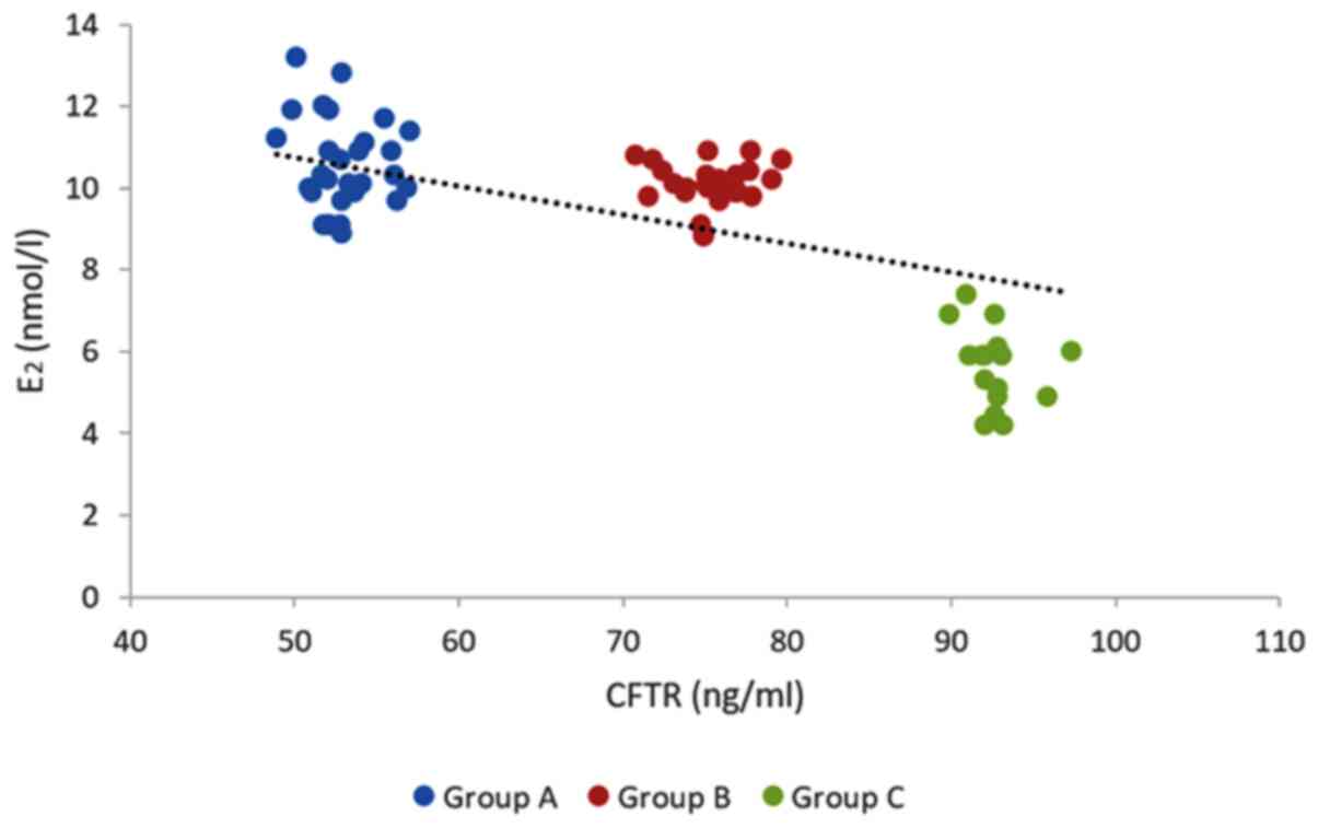
Association of FF CFTR levels with
indicators of oocyte quality
The level of FF CFTR on the day of oocyte retrieval
in group C was significantly higher than that in group A or group B
and it was higher in group B than that in group A (P<0.001;
Table II). The level of E2 on HCG
injection day decreased with increasing age, where the difference
of E2 among the three groups was statistically significant with
that in group C the lowest (P<0.05; Table II). A significant but weak negative
correlation was identified among the three groups of FF CFTR levels
and E2 levels on the HCG injection day (r=-0.131;
P=0.021; Fig. 1). There were no
statistical differences in the morphologically normal MII oocyte
rate and available embryo rate among the three groups, but the rate
of high-quality embryos declined with age, with that in group C the
lowest (P<0.05; Table II).
| Table IIIndicators of oocyte quality. |
Table II
Indicators of oocyte quality.
| Parameter | Group A (n=32) | Group B (n=29) | Group C (n=19) | P-value |
|---|
| CFTR in FF
(ng/ml) | 52.94±25.51 | 75.29±29.29 | 92.68±29.94 | <0.001 |
| E2 on HCG
injection day (nmol/l) | 10.70±6.84 | 10.65±8.07 | 5.81±5.18 | 0.021 |
| Morphologically
normal MII oocyte rate (%) | 80.34 | 78.91 | 73.56 | 0.066 |
| Available embryo
rate (%) | 63.69 | 80.43 | 60.61 | 0.089 |
| High-quality embryo
rate (%) | 58.33 | 40.58 | 33.33 | 0.019 |
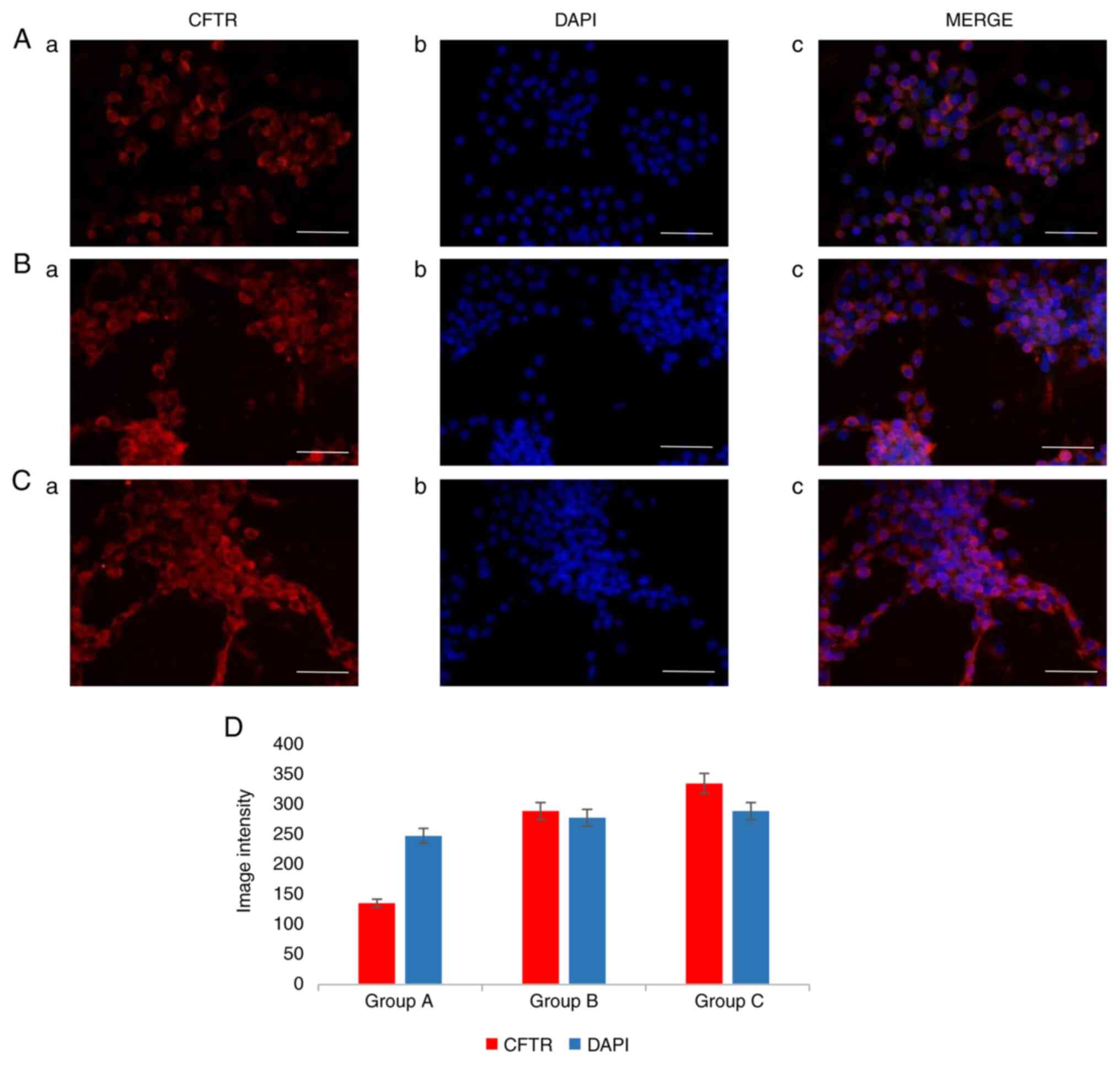
Localization and expression of CFTR in
cumulus GCs
CFTR expression in CCs (red expression) was detected
by immunofluorescence staining and was consistent with that denoted
in Gene Cards®. The fluorescence intensity of CFTR in
group C was higher than that in group B, while group A had the
lowest intensity (Fig. 2).
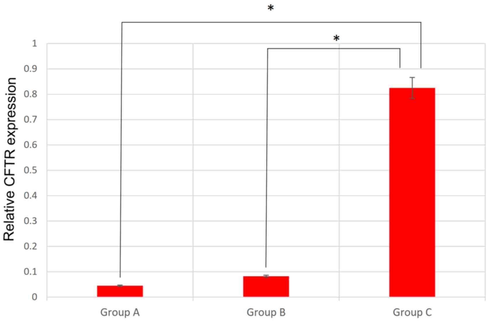
RT-qPCR analysis of CFTR
There are ~500 CCs around each oocyte and half of
the total CCs in each group were used for RT-qPCR. Due to the
limitation of sample size analysis in each group was performed
twice. The RT-qPCR results were analyzed using the
2-ΔΔCq method. Analysis of the three groups suggested
that the mean 2-ΔΔCq value of two experimental
replicates increased with age and there were significant
differences between groups A and C, and between groups B and C
(P<0.01), where group C had the highest value of
2-ΔΔCq (Fig. 3).
Discussion
Among the patient groups of the present study,
differences in proven age-associated indicators were identified,
suggesting that the selected patients were representative of the
general population (16). Numerous
indicators of oocyte quality exist, but each method has its own
shortcomings (17). Oocyte
morphological assessment is the most widely used method in clinical
practice; a normal human MII oocyte should have a round and clear
ZP, a small PVS containing a single IPB without fragmentation and
pale, moderately granular cytoplasm with no inclusions (18). However, oocyte morphological
assessment may be performed only after oocyte denudation in ICSI
cases and potential mechanisms that change the appearance of the
oocyte are multifactorial (19).
Age and the E2 level on the HCG day are indirect
indicators (20). Embryo quality
reflects oocyte quality only retrospectively and neglects
sperm-related factors (21).
Therefore, an index that is able to evaluate oocyte quality prior
to IVF is still required.
The present results indicated that with increasing
age, no significant changes in certain commonly used indicators of
oocyte quality occurred, although CFTR was more positively
correlated with age than these indicators. The CFTR is an anion
channel regulated by cAMP-dependent phosphorylation and is
expressed in epithelial cells of a wide variety of tissue types,
including the reproductive tracts (22). CFTR affects factors including sperm
transport, blastocyst implantation, sperm capacitation and function
by regulating fluid volume and bicarbonate secretion in the
reproductive tract (23).
The cumulus GC layer regulates oocyte maturation and
development through complex mechanisms such as gap junctions
(23). Detecting gene expression in
GCs may reflect changes in the follicular microenvironment and
further provide relevant information on oocyte development
(24). Evaluating egg quality and
embryonic development potential indirectly is helpful. In the
present study, CFTR expression in CCs was detected by indirect
immunofluorescence and the results suggested that CFTR was mainly
localized to the cumulus GC cytoplasm and membrane. The RT-qPCR
results also further demonstrated that CFTR gene expression in
cumulus GCs may have a positive correlation with age and may affect
the outcome of embryo development. In order to clarify the
relationship between the mature oocytes and the patient's age, only
the FF and CCs around mature oocytes obtained by the first
aspiration without rinsing were collected. FF or CCs were excluded
if there was no OCCC found or the oocytes were immature. Therefore,
the number of cases enrolled is limited, particularly in group C.
To verify this result, it is required to increase the sample sizes
of the different age groups.
In assisted reproductive technology, FF metabonomics
may be combined with other clinical factors such as age to predict
the quality of oocytes and other important information (25). Multiple studies have examined
correlations between lipid metabolism, amino acid metabolism or
glucose metabolism in FF and oocyte quality (26). Certain studies have indicated that
the levels of fatty acids in FF are related to oocyte maturity and
that certain changes in fatty acids may improve oocyte quality
(27). Regarding amino acid
metabolism, amino acids in FF have an important role in oocyte
maturation and fertilization and evaluating oocyte quality
according to amino acid levels in FF is a current research hotspot
(28). Regarding glycometabolism,
Robker et al (29) observed
that glucose levels in the FF of fertilized eggs with failed
cleavage are significantly higher compared with those in the FF of
fertilized eggs with normal cleavage, indicating that low oocyte
quality is related to high glucose levels in FF. In the present
study, CFTR levels were compared with several classical indexes
used to evaluate oocyte quality to identify novel correlations and
the correlation between age and the CFTR level in FF was confirmed
for the first time, to the best of our knowledge, i.e. that the
CFTR level in FF is positively correlated with age. Due to the
limited cases enrolled in this study, further research is needed.
Although the identification of specific predictive biochemical
markers is not yet possible, this method has potential application
value. FF metabonomics analysis is expected to open a new chapter
in the screening of high-quality oocytes and embryos.
Acknowledgements
Not applicable.
Funding
The present study was supported by grants from the
Tongji Hospital Clinical Research Project [grant nos. ITJ (QN) 1809
and ITJ (QN) 1813] and the Shanghai Key Laboratory of Embryo
Original Diseases (grant no. Shelab201903).
Availability of data and materials
The datasets used and/or analyzed during the current
study are available from the corresponding author on reasonable
request.
Authors' contributions
YW and HW performed the experiments. HW, FL, YJ and
ZS analyzed the data. XZ and YW conceived the study and wrote the
manuscript. All authors read and approved the final manuscript.
Ethics approval and consent to
participate
The present study was approved by the Ethics
Committee of Shanghai Tongji Hospital (Shanghai, China). Written
informed consent was provided by each patient prior to inclusion in
this study.
Patient consent for publication
Not applicable.
Competing interests
The authors declare that they have no competing
interests.
References
|
1
|
Moragianni D, Dryllis G, Andromidas P,
Kapeta-Korkouli R, Kouskouni E, Pessach I, Papalexis P, Kodonaki A,
Athanasiou N, Pouliakis A and Baka S: Genital tract infection and
associated factors affect the reproductive outcome in fertile
females and females undergoing in vitro fertilization.
Biomed Rep. 10:231–237. 2019.PubMed/NCBI View Article : Google Scholar
|
|
2
|
Wang X, Cai J, Liu L, Jiang X, Li P, Sha A
and Ren J: Association between outdoor air pollution during in
vitro culture and the outcomes of frozen-thawed embryo transfer.
Hum Reprod. 34:441–451. 2019.PubMed/NCBI View Article : Google Scholar
|
|
3
|
Smits MAJ, Wong KM, Mantikou E, Korver CM,
Jongejan A, Breit TM, Goddijn M, Mastenbroek S and Repping S:
Age-related gene expression profiles of immature human oocytes. Mol
Hum Reprod. 24:469–477. 2018.PubMed/NCBI View Article : Google Scholar
|
|
4
|
Aboulmakarim S, Benbella A, Hardizi H and
Bezad R: Retrospective assessment of an assisted reproductive
technology method. Ann Biol Clin (Paris). 76:11–21. 2018.PubMed/NCBI View Article : Google Scholar
|
|
5
|
Colaco S and Sakkas D: Paternal factors
contributing to embryo quality. J Assist Reprod Genet.
35:1953–1968. 2018.PubMed/NCBI View Article : Google Scholar
|
|
6
|
Chan LN, Tsang LL, Rowlands DK, Rochelle
LG, Boucher RC, Liu CQ and Chan HC: Distribution and regulation of
ENaC subunit and CFTR mRNA expression in murine female reproductive
tract. J Membr Biol. 185:165–176. 2002.PubMed/NCBI View Article : Google Scholar
|
|
7
|
Su M, Guo Y, Zhao Y, Korteweg C and Gu J:
Expression of cystic fibrosis transmembrane conductance regulator
in paracervical ganglia. Biochem Cell Biol. 88:747–55.
2010.PubMed/NCBI View
Article : Google Scholar
|
|
8
|
Zheng XY, Chen GA and Wang HY: Expression
of cystic fibrosis transmembrane conductance regulator in human
endometrium. Hum Reprod. 19:2933–2941. 2004.PubMed/NCBI View Article : Google Scholar
|
|
9
|
Field PD and Martin NJ: CFTR mutation
screening in an assisted reproductive clinic. Aust N Z J Obstet
Gynaecol. 51:536–539. 2011.PubMed/NCBI View Article : Google Scholar
|
|
10
|
Sheppard DN and Welsh MJ: Structure and
function of the CFTR chloride channel. Physiol Rev. 79 (Suppl
1):S23–S45. 1999.PubMed/NCBI View Article : Google Scholar
|
|
11
|
Ubaldi F and Rienzi L: Morphological
selection of gametes. Placenta. 29 (Suppl B):S115–S120.
2008.PubMed/NCBI View Article : Google Scholar
|
|
12
|
David KG, Ariel W, Colin MH and Zeev S:
Textbook of Assisted Reproductive Technologies. 3rd edition.
Informa Healthcare Corp., pp171-180, 2009.
|
|
13
|
Racowsky C, Vernon M, Mayer J, Ball GD,
Behr B, Pomeroy KO, Wininger D, Gibbons W, Conaghan J and Stern JE:
Standardization of grading embryo morphology. Fertil Steril.
94:1152–1153. 2010.PubMed/NCBI View Article : Google Scholar
|
|
14
|
Schmittgen TD and Livak KJ: Analyzing
real-time PCR data by the comparative C(T) method. Nat Protoc.
3:1101–1108. 2008.PubMed/NCBI View Article : Google Scholar
|
|
15
|
Kmetic A, Joseph L, Berger C and
Tenenhouse A: Multiple imputation to account for missing data in a
survey: Estimating the prevalence of osteoporosis. Epidemiology.
13:437–444. 2002.PubMed/NCBI View Article : Google Scholar
|
|
16
|
Tarin JJ, Ten J, Vendrell FJ and Cano A:
Dithiothreitol prevents age-associated decrease in oocyte/conceptus
viability in vitro. Hum Reprod. 13:381–386. 1998.PubMed/NCBI View Article : Google Scholar
|
|
17
|
Gosden LV: Oocyte retrieval and quality
evaluation. Methods Mol Biol. 1154:343–360. 2014.PubMed/NCBI View Article : Google Scholar
|
|
18
|
Camargos M, Rodrigues JK, Lobach VN, El
Cury-Silva T, Nunes MEG, Camargos AF and Reis FM: Human oocyte
morphometry before and after cryopreservation: A prospective cohort
study. Cryobiology. 88:81–86. 2019.PubMed/NCBI View Article : Google Scholar
|
|
19
|
Hajiyavand AM, Saadat M, Abena A, Sadak F
and Sun X: Effect of injection speed on oocyte deformation in ICSI.
Micromachines (Basel). 10(226)2019.PubMed/NCBI View Article : Google Scholar
|
|
20
|
Chen H, Zheng JB, Wang DM, Xing H and Wang
H: Association between vascular endothelial growth factor and
clinical outcomes of IVF-ET/ICSI. J Coll Physicians Surg Pak.
29:19–23. 2019.PubMed/NCBI View Article : Google Scholar
|
|
21
|
Devreker F, Pogonici E, De Maertelaer V,
Revelard P, Van Den Bergh M and Englert Y: Selection of good
embryos for transfer depends on embryo cohort size: Implications
for the ‘mild ovarian stimulation’ debate. Hum Reprod.
14:3002–3008. 1999.PubMed/NCBI View Article : Google Scholar
|
|
22
|
Chan HC, Ruan YC, He Q, Chen MH, Chen H,
Xu WM, Chen WY, Xie C, Zhang XH and Zhou Z: The cystic fibrosis
transmembrane conductance regulator in reproductive health and
disease. J Physiol. 587:2187–2195. 2009.PubMed/NCBI View Article : Google Scholar
|
|
23
|
Dehghan Z, Mohammadi-Yeganeh S and Salehi
M: MiRNA-155 regulates cumulus cells function, oocyte maturation,
and blastocyst formation. Biol Reprod. 103:548–559. 2020.PubMed/NCBI View Article : Google Scholar
|
|
24
|
Bonnet A, Cabau C, Bouchez O, Sarry J,
Marsaud N, Foissac S, Woloszyn F, Mulsant P and Mandon-Pepin B: An
overview of gene expression dynamics during early ovarian
folliculogenesis: Specificity of follicular compartments and
bi-directional dialog. BMC Genomics. 14(904)2013.PubMed/NCBI View Article : Google Scholar
|
|
25
|
Bracewell-Milnes T, Saso S, Abdalla H,
Nikolau D, Nornam-Taylor J, Johnson M, Holmes E and Thum MY:
Metabolomics as a tool to identify biomarkers to predict and
improve outcomes in reproductive medicine: A systematic review. Hum
Reprod Update. 23:723–736. 2017.PubMed/NCBI View Article : Google Scholar
|
|
26
|
Arya BK, Haq AU and Chaudhury K: Oocyte
quality reflected by follicular fluid analysis in poly cystic ovary
syndrome (PCOS): A hypothesis based on intermediates of energy
metabolism. Med Hypotheses. 78:475–478. 2012.PubMed/NCBI View Article : Google Scholar
|
|
27
|
Fayezi S, Leroy J, Ghaffari NM and Darabi
M: Oleic acid in the modulation of oocyte and preimplantation
embryo development. Zygote. 26:1–13. 2018.PubMed/NCBI View Article : Google Scholar
|
|
28
|
Anchordoquy JP, Lizarraga RM, Anchordoquy
JM, Nikoloff N, Rosa DE, Fabra MC, Peral-Garcia P and Furnus CC:
Effect of cysteine, glutamate and glycine supplementation to in
vitro fertilization medium during bovine early embryo development.
Reprod Biol. 19:349–355. 2019.PubMed/NCBI View Article : Google Scholar
|
|
29
|
Robker RL, Akison LK, Bennett BD, Thrupp
PN, Chura LR, Russell DL, Lane M and Norman RJ: Obese women exhibit
differences in ovarian metabolites, hormones, and gene expression
compared with moderate-weight women. J Clin Endocrinol Metab.
94:1533–1540. 2009.PubMed/NCBI View Article : Google Scholar
|