Introduction
Paracetamol, also known as acetyl-para-amino-phenol
or acetaminophen, is an analgesic and antipyretic that is referred
to as a N-(4-hydroxyphenyl) ethanamide, N-(4-hydroxyphenyl)
acetamide by the International Union of Pure and Applied Chemistry
and has the chemical formula
C8H9NO2 (1). It is suitable for intravenous
administration, and its injectable form has significant ease of
use. Accordingly, this drug is used as a moderate analgesic and
antipyretic following surgical intervention in almost all areas of
medicine, including neurosurgery (2).
Paracetamol is an analgesic drug and an
indispensable pharmaceutical preparation in emergency departments
that can be administered orally, rectally and intravenously
(3). This drug and its precursors
are believed to be among the most risk-prone pharmaceutical
preparations. It has been reported to cause hundreds of deaths each
year via acute liver failure even within industrialized countries,
and it has toxic effects on liver and kidney tissues (4).
Many pharmaceutical preparations are used to treat a
wide range of diseases. However, any medication may adversely
affect the tissues of regions surrounding the targeted tissues
(5,6); therefore, drugs may accumulate in
fluid compartments and tissue layers. The majority of analgesics
are composed of organic acids with high plasma protein binding
capacity. Paracetamol typically exhibits low levels of binding to
plasma proteins, but the binding level increases with higher
doses.
Increased binding to plasma proteins facilitates
drug accumulation in tissues. As a result, non-ionizing parts of
the drug increase, and the interaction level of the drug and the
lipid structure of the cell membranes also increase (7-9).
A notable number of studies have been performed to
identify an appropriate treatment procedure for damaged osseous
joints, facet joints and intervertebral disc structures, and
investigations on the toxicity of drugs in non-degenerative,
healthy tissues has gained momentum (10,11).
Certain side effects and adverse events associated
with paracetamol have been reported previously (12); however, no pharmaco-molecular
studies to date have investigated the damage to non-degenerate,
intact intervertebral disc tissue cells caused by paracetamol.
The present study investigated the toxicity of
paracetamol, an anilid derivate that is frequently prescribed alone
or in combination with other non-steroidal anti-inflammatory drugs
following major surgical procedures or minimally invasive surgeries
for numerous patients with pain or fever, using healthy human
intervertebral disc tissue cell cultures.
Proliferation analyses and chondroadherin (CHAD)
gene expression levels, as well as continuous expression of the
nucleus pulposus (NP) specific marker, were investigated. The level
of the cartilage oligo matrix protein (COMP) directly or indirectly
involved in the catabolic mechanisms of the disc structure was also
investigated.
In addition to these markers, the expression of
matrix metalloproteinase (MMP)-7, MMP-13 and MMP-19, known to be
responsible for the catabolic pathways (13,14),
were investigated. The changes in intervertebral disc and
extracellular matrix structures were assessed using a reverse
transcriptase-quantitative polymerase chain reaction (RT-qPCR). The
gene expression levels of IL-1β, a prominent proinflammatory
cytokine in disc metabolism (15,16),
were also investigated in the present study.
Materials and methods
Ethical approval
The present study was approved by the Namik Kemal
University School of Medicine's Local Ethics Committee (dated
28.06.2018 and numbered 2018/107/07/13). Written informed consent
was obtained from the patients undergoing surgery whose tissues
were used to obtain the primary cell cultures.
Materials
The cell dishes used for the preparation of cell
cultures were obtained from Zhejiang Sorfa Life Science Research
Co., Ltd. (cat. no. SCD-11-035), and a 96-well FG-Microplate (cat.
no. 4346906) was purchased from Thermo Fisher Scientific, Inc.
Fetal bovine serum (cat. no. 10500064), L-glutamine (cat. no.
25030024), penicillin-streptomycin (10.000 U/ml; cat. no. 15140122)
and amphotericin B (250 µg/ml; cat. no. 12183018) were obtained
from Thermo Fisher Scientific, Inc. The paracetamol-active
pharmacological agent administered to cell cultures was in the form
of a 100-ml vial (10 mg/ml; Perfalgan®) and was
purchased from Bristol-Myers Squibb Company. The commercial kit
used for the cell viability, toxicity and proliferation analyses
was Vybrant MTT Cell Proliferation assay (cat. no. V13154; Thermo
Fisher Scientific, Inc.). The ELISA with which spectrophotometric
analysis was performed was Mindray MR 96A (Georgia Institute of
Technology).
For RT-qPCR, a PureLink™ RNA Mini kit (cat. no.
15290018), High-Capacity cDNA Reverse Transcription kit (cat. no.
4368814), TaqMan® Gene Expression assay-Hs00154382_m1
(cat. no. 4331182), CHAD TaqMan® Gene Expression assay
(cat. no. 4331182), COMP TaqMan® Gene Expression assay
(cat. no. 4331182), MMP-7 TaqMan® Gene Expression assay
(cat. no. 4331182), MMP-13 TaqMan® Gene Expression assay
(cat. no. 4331182), MMP-19 TaqMan® Gene Expression assay
(cat. no. 4331182), IL-1β TaqMan® Gene Expression assay
(cat. no. 4331182), and internal control gene (housekeeping gene,
β-actin) Taqman Fast Advanced mix (5 ml; cat. no. 4444557) were
purchased from Thermo Fisher Scientific, Inc.
Inclusion and exclusion criteria
Tissues were obtained from the patients admitted to
the Department of Neurosurgery, Namik Kemal University, School of
Medicine between July 2018 and August 2019. The tissues of patients
with malignancy or pregnancy were not used. Patients who were
referred to the emergency department with a complaint of spinal
trauma and then diagnosed with spinal instability or traumatic disc
hernia following physical, neurological and imaging examinations
were enrolled (n=17). The tissues of the patients diagnosed with
degenerative intervertebral disc herniation following MRI scans
were excluded from the study (n=2).
Paracetamol may interact with beta-adrenergic
receptor blockers (n=3), including propranolol, ethyl alcohol (n=3)
and oral anticoagulants (n=2), and its efficacy may be inhibited by
certain drugs, including chlorpromazine (n=1). Therefore, the
tissues of the patients who had taken the aforementioned drugs were
excluded.
Intervertebral disc tissues obtained from 6
patients, included 3 males and 3 females (mean body mass index,
27.9 kg/m2; mean age, 28.62±8.18 years, range, 20-37)
were resected and transferred into sterile falcon tubes containing
penicillin streptomycin.
Resection of the tissues via surgery
and preparation of the primary cell cultures
Non-degenerate healthy disc material [intact NP and
annulus fibrosus (AF) tissue] was obtained as surplus surgical
material. Human primary intervertebral disc cell cultures were
established according to standard protocols (15,16).
Physical and neurological examinations of the
patients with spinal trauma who were referred to the emergency
department were performed. Subsequently, surgical intervention was
decided on. All patients were placed in the prone position under
endotracheal general anesthesia. Surgical field antisepsis was
provided with 10% povidone-iodine solution and covered in a sterile
manner. A median skin incision was performed on the skin and under
the skin. Paravertebral muscles were dissected subperiosteally from
osseous structures. The regions with neural compression were
decompressed via laminectomy and traumatic disc excision. Spinal
stabilization was performed through the transpedicular screw-rod
system (17). The resected disc
tissues were transferred to sterile Falcon tubes at 40˚C, and
letter coding was conducted.
Preparation of the primary cell
cultures and application of paracetamol to the cultures
The tissues placed in the flow cabinet were
transferred to Petri dishes in a sterile manner and were irrigated
with 0.9% isotonic sodium chloride solution to differentiate them
from the red blood cells. Subsequently, the tissues were
mechanically minced. The minced tissues were transferred to Falcon
tubes containing 50 ml Hank's Balanced Salt Solution and 0.375 µg
collagenase type II and then incubated overnight in an incubator
set to a temperature of 37˚C and 5% CO2. The samples
were centrifuged at 4˚C and 161 x g consecutively twice for 10 min.
Following centrifugation, the tubes were returned to the flow
cabinet. The tube caps were opened in a sterile environment. The
supernatant at the top of the tubes was discarded. The pellet
located at the bottom of the tube was resuspended with the freshly
prepared culture medium. This freshly prepared culture medium
consisted of Dulbecco's Modified Eagle's Medium (cat. no. D5796),
15% FBS (cat. no. 10500064), 1% L-glutamine (cat. no. 25030024),
penicillin-streptomycin (10,000 U/ml; cat. no. 15140122) and
amphotericin B (250 µg/ml; cat. no. 12183018) (all Gibco; Thermo
Fisher Scientific, Inc.). The cells, cultured in a T25 flask, were
incubated for 72 h in an incubator set to 37˚C and 5%
CO2. Standard human primary cell cultures were performed
(6,9,13,16).
Cells were trypsinized with trypsin-EDTA (0.25%) (cat. no. T3924;
Sigma-Aldrich; Merck KGaA), and viable cells were counted were
counted with Inverted light microscope (CKX41, Olympus Europa SE
& Co. KG) at a magnification of x10, using the Neubauer
chamber following trypan blue staining at 22.4˚C for 12 min. For
MTT analysis and acridine orange/propidium iodine (AO/PI) staining,
cells were cultivated with 15,000 cells/well in 96-well plates, and
for RNA isolation, cells were cultivated with 1.2x10⁶
cells/well in 6 well plates. At the end of the 24-h incubation
period, paracetamol content was applied to the cell cultures in the
experimental group. A 10 mg/m paracetamol stock solution was used
by diluting it with Dulbecco's modified Eagle's medium to a final
concentration of 15.22 µl/ml. The final concentration was
calculated based on the doses reported in previous studies
(18,19).
MTT analysis
MTT analyses were performed on the day of drug
applications and on the 10th and 20th days following the
applications, with at least three experimental and three technical
repetitions. The commercial Vybrant MTT cell proliferation assay
kit (V-13154) was used. The 12 mM MTT stock solution was prepared
by adding 1 ml sterile PBS to the vial containing 5 mg MTT. Next,
90 µl fresh aforementioned medium was added to the confluent cells
in 96-well plates. A total of 10 µl MTT stock solution was added,
protected from light, and incubated at 37˚C for 2 h. At the end of
this period, 25 µl of the samples were discarded and the same
amount of DMSO was added. This was incubated for an additional 10
min at 37˚C and absorbance measured for each well at 540 nm using
the ELISA reader (Mindray MR 96A; PRC; Georgia Institute of
Technology).
Inverted light microscopy analyses and
Acridine orange and propidium iodide (AO/PI) staining
Cell morphology and confluency were analyzed using
an inverted microscope. Microphotographs of the cell organizations
were obtained in the confocal/contrast phase prior to and following
the application of chemical agents at x10 and/or x20 magnifications
(Fig. 1; first lane). The cells
were stained with Giemsa (GS500; Sigma-Aldrich; Merck KgaA; 37˚C
for 10 min) stain to demonstrate the cells' organization more
clearly (Fig. 1; third lane). In
order to determine the presence of cell death and whether the death
was apoptotic, cells were stained (at 22.4˚C for 10 min) with AO/PI
and examined using a fluorescent microscope. Microphotographs of
cell structures were obtained (Fig.
1; second lane), and the images were evaluated using the
CytoVision® v 7.0 (DM6000B; Leica Microsystems GmbH)
capture station imaging program (6-16).
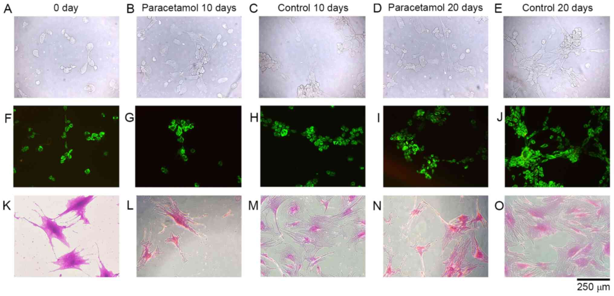 | Figure 1Microscopic evaluation of cultured
cells. Lane 1, inverted microscopy of (A) 0 day, (B) 10 days of
paracetamol application, (C) 10 day control, (D) 20 days of
paracetamol application and (E) 20 day control without staining.
Lane 2, Acridine orange and propidium iodide stained cell cultures
of (F) 0 day, (G) 10 days of paracetamol application, (H) 10 day
control, (I) 20 days of paracetamol application and (J) 20 day
control. Lane 3, inverted microscopy of Giemsa-stained cultures of
(K) 0 day, (L) 10 days of paracetamol application, (M) 10 day
control, (N) 20 days of paracetamol application and (O) 20 day
control. |
AO/PI stain is a cell viability dye that causes
viable nucleated cells to fluoresce green and non-viable nucleated
cells to fluoresce red. The AO/PI stain was prepared with 5 g
sodium-EDTA, 2 mg PI, 25 ml FBS and 2 mg AO dissolved in 1 ml 99%
ethanol; sterile distillated water was added to reach a final
volume of 100 ml. Cell cultures were stained with AO/PI at room
temperature for 10 min, and then cell death and cell viability were
monitored at a magnification of x10.
RT-qPCR analysis
Prior to performing the RNA isolation, a live cell
count was conducted by staining with trypan blue using the
Neubauer chamber. RNA isolation was performed on
5.3x10⁶ cells using the Pure Link, Ambion kit (Thermo
Fisher Scientific, Inc.). RNA samples obtained were converted into
cDNA copies by reverse transcription (Applied Biosystems; Thermo
Fisher Scientific, Inc.). The Applied Biosystems 7300/7500 RT-PCR
system was used with the following reaction protocol: Hold at
50˚C for 2 min, hold at 95˚C for 10 min, and
alternate between 15 sec at 95˚C and 1 min at 60˚C for 40 cycles.
RT-qPCR was performed using primers and probes specific to COMP,
MMP-7, MMP-13, MMP-19, IL-1β and CHAD genes with the obtained cDNA
samples. Commercial Taq-Man gene expression kits (cat. no. 4331182,
Thermo Fisher Scientific, Inc.) were used for each gene region.
Hs00164359_m1 was used for the COMP gene region, Hs01042796_m1 for
the MMP-7 gene region, Hs00419424_m1 for the MMP-13 gene region,
Hs00419424_m1 for the MMP-19 gene region, and Hs001541101-coded
products were used for the CHAD gene region (information regarding
primers used here: https://www.thermofisher.com/order/genome-database/).
Relative quantification (RQ) was performed according to the average
Ct values obtained using the b-actin (Hs01060665_g1) gene as an
endogenous control and the control group as a calibrator (7,8). Using
this method, the difference in gene expression between the control
group and experimental groups was determined as a fold-change.
Statistical analyses
The program Minitab R18 was used for the statistical
analysis. The results are expressed as the mean ± standard
deviation. The results were evaluated using an analysis of variance
(ANOVA; at a 95% confidence interval to assess whether there were
significant differences across groups. When differences across
groups were observed, Tukey's honest significant difference (HSD)
post hoc test was used for multiple pairwise comparisons. P<0.05
was considered to indicate a statistically significant
difference.
Results
Cell morphology, confluency and
viability
The surface morphologies of the samples were
preserved, and the cells were healthy, viable and had proliferated
in all microscopic analyses performed at all time periods in the
control group (Fig. 1A, C, E,
F, H, K,
M and O).
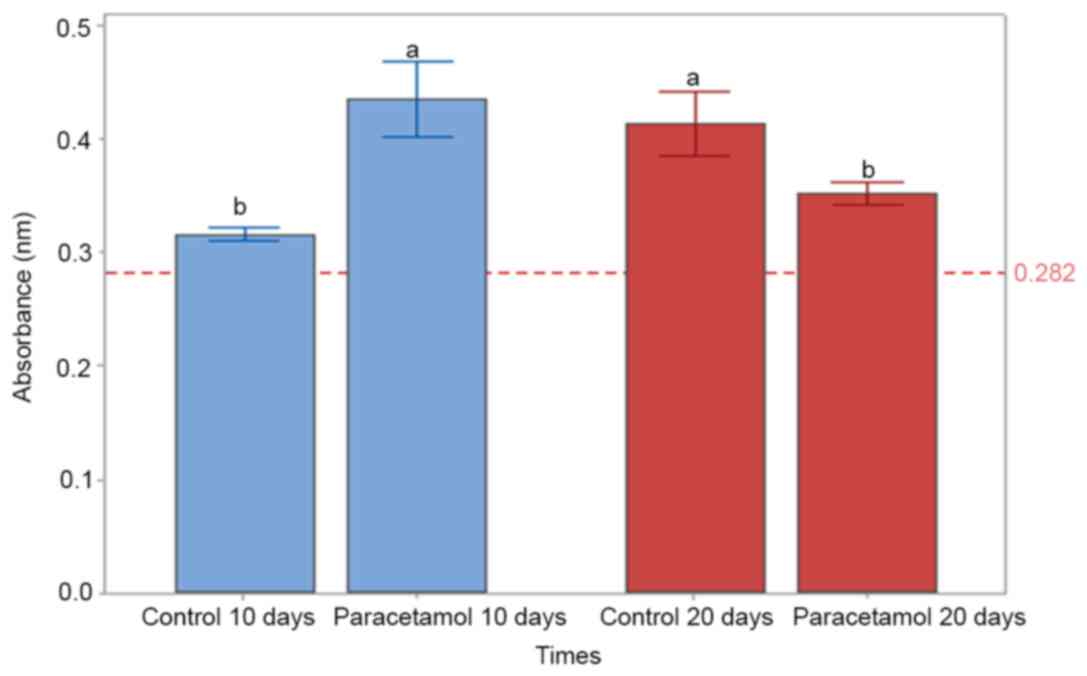
Cell proliferation based on MTT
analysis
Proliferation was suppressed on the 10th day
(P=0.05) and the 20th day (P<0.05) in the paracetamol
supplemented groups and the non-drug-administered control group
samples, respectively, and this difference is statistically
significant (P<0.05 Figs. 1B,
D, G, I,
L, N, and 2).
However, in addition to cell proliferation suppression, it is
notable that no round-shaped cells, indicating cytotoxicity, were
identified. Furthermore, the images obtained via fluorescent
microscopy also supported these results. The dead cells stained red
were not identified in the images from the 10th day (Fig. 1G) or the 20th day (Fig. 1I). Additionally, on the 10th day
(Fig. 1L) and the 20th day
(Fig. 1N), analyses of the
microscopic images with Giemsa staining, the numbers of dead cells
and the matrix structures around the cells deteriorated and broke
down in the experimental group, compared with the cell samples of
the control group (Fig. 1M and
O).
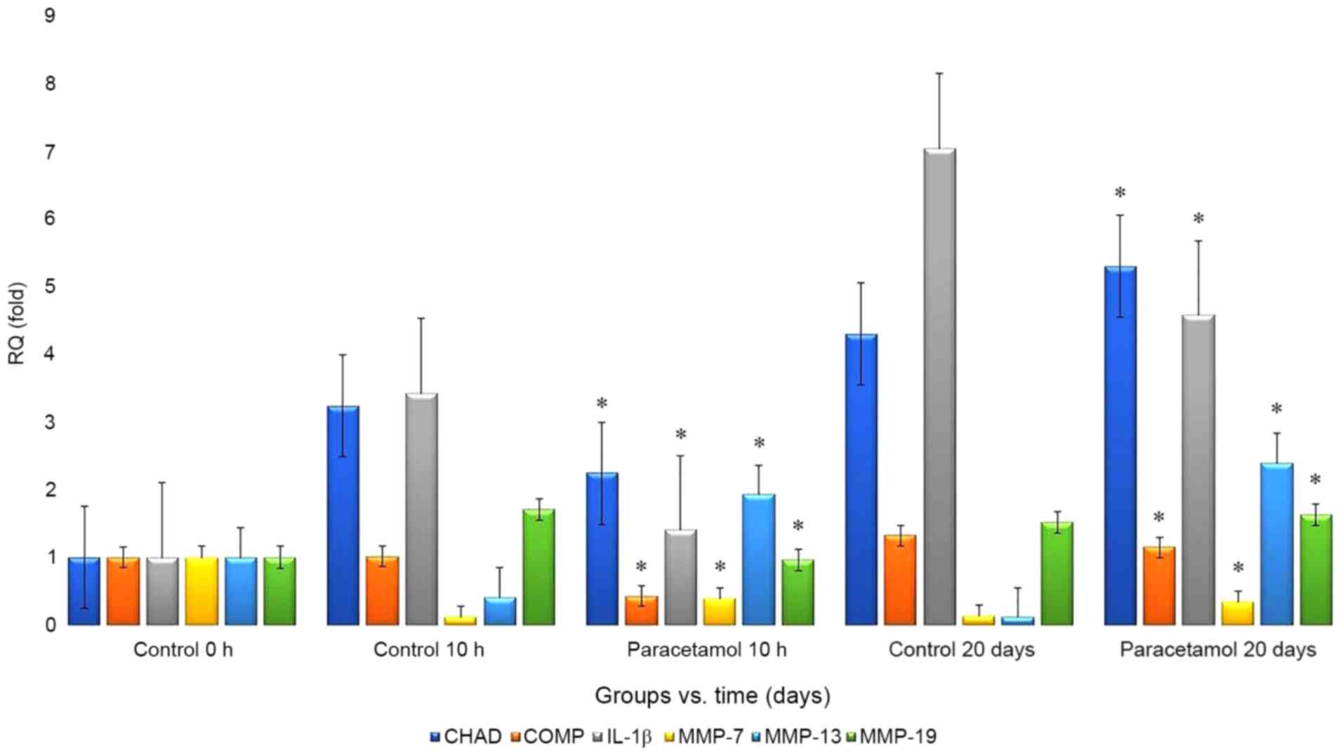
RT-qPCR analysis
RT-qPCR analysis revealed that the RQ values for
CHAD, MMP-7, MMP-13 and MMP-19 increased in the
paracetamol-administered samples compared with that in the control
group as follows: From 4.29-fold to 5.29-fold, from 0.13-fold to
0.34-fold, from 0.12-fold to 2.39-fold, and from 1.52-fold to
1.63-fold, respectively. However, the RQ values for the COMP and
IL-1β markers decreased from 1.32-fold to 0.34-fold and from
7.03-fold to 4.57-fold, respectively (Fig. 3).
Discussion
Paracetamol is a metabolite of phenacetin (20), and the mechanism of its analgesic
action has not yet been fully elucidated (20). However, it is known to be effective
in inhibiting prostaglandin synthesis in the central nervous system
(21). Paracetamol has been widely
used since 1950(22). The
increasing use of this drug has resulted in certain adverse events,
including an increased mortality rate due to liver toxicity
originating from excessive dose uptake (23).
Paracetamol is rapidly and uniformly distributed in
body tissues. It is frequently prescribed as an analgesic and
antipyretic in the treatment of headache, toothache, migraine,
dysmenorrhea, myalgia, neuralgia, musculoskeletal, tonsillectomy
pain, the common cold, influenza, and other bacterial and viral
infections. It can also be used as a pain controller for numerous
cases in neurosurgery and emergency departments, including
posterior lumbar laminectomy (24),
vertebral compression fractures (25), spinal primary and metastatic tumors
(26), post-craniotomy in
intracranial pathology surgery (27), traumatic fractures of bones
(28), urinary colic (29) and acute appendicitis (30).
In addition to liver and kidney toxicities caused by
paracetamol (31,32), certain studies have reported that it
may lead to maculopapular rashes (33) or nausea in very rare cases (34), and its long-term use may cause
hemolytic anemia (35),
thrombocytopenic purpura (36) and
agranulocytosis (37). However, no
studies have investigated the side effects and adverse events of
paracetamol on intervertebral disc tissue cells.
Ramachandran et al (38) reported that more recent
investigations into acetaminophen hepatotoxicity provided insights
into the critical role of mitochondrial dysfunction in mediating
liver injury. The authors stated that some studies clarified the
mechanisms of acetaminophen-induced hepatocyte cell death. They
suggested that a significant controversy on the role of innate
immunity in APAP-induced hepatotoxicity is ongoing, even though
mitochondrial oxidative and nitrosative stress was known to be a
key mechanistic feature involved in downstream signaling following
acetaminophen overdose (38).
An in vitro toxicity study reported that
differentiated HepaRG cells preserved liver-specific functions,
including drug-metabolizing enzymes (39). In that study, the feasibility of
HepaRG cells as a human hepatocyte was investigated using selected
hepatotoxic compounds. The possible adverse effects of
acetaminophen have also been investigated in another study
(40). It was reported that
acetaminophen, commonly used in clinics, had time- and
dose-dependent side effects; therefore, further animal studies are
required to accurately investigate possible adverse effects
(40). It was concluded that
acetaminophen-mediated specific cytotoxicity was associated with
the molecular mechanisms facilitating apoptosis and inflammatory
stress in the liver and kidney.
Knockaert et al (41) investigated the role of mitochondrial
CYP2E1 in the toxicity of acetaminophen and ethanol. The effects of
these two compounds were compared in cells expressing CYP2E1 in
either mitochondria only or both endoplasmic reticula and
mitochondria and in mock-transfected cells (41). It was reported that when
acetaminophen or ethanol was used as a CYP2E1 substrate, the
exclusive localization of CYP2E1 within mitochondria may cause
reactive oxygen species overproduction, depletion of reduced
glutathione, increased expression of mitochondrial Hsp70,
mitochondrial dysfunction and cytotoxicity (41). The study emphasized that these
deleterious events occurred despite the lower cellular level and
activity of CYP2E1 compared with cells expressing CYP2E1 in
endoplasmic reticula and mitochondria and that this was
particularly apparent in the case of acetaminophen (41). The authors proposed that
mitochondrial CYP2E1 may serve a significant role in oxidative
stress and cell death (41).
Patel et al (42) reported that acetaminophen
administered to rats during chronic exercise decreased skeletal
collagen and cross-linking. It was suggested that the effect of
acetaminophen on the muscle extracellular matrix (ECM) may be
mediated by the dysregulation of the balance between MMPs and their
tissue inhibitors (TIMPs). In a double-blind, placebo-controlled,
randomized cross-design study, two male volunteers performed two
trials of knee extension (42). A
placebo (PLA) and acetaminophen were administered (1,000 mg/6 h)
for 24 h prior to and following resistance exercise (RE). Vastus
lateralis biopsies were performed at 0, 1 and 3 h following RE
(42). They reported that mRNA
expression of MMP-2, type I collagen and type III collagen was not
changed by exercise or acetaminophen (P>0.05). They also stated
that, compared with the control group, the TIMP-1 expression was
lower at 1 h post-RE with acetaminophen but higher at 3 h, and this
difference was statistically significant (P<0.05). They
emphasized that MMP-9 expression and protein levels were increased
at 3 h post-RE independent from treatment and that lysyl oxidase
expression was significantly higher at 3 h post-RE with
acetaminophen consumption, compared with the control group
(P<0.05). It was suggested that short-term acetaminophen
consumption prior to RE has a small impact on the measured ECM
molecules in human skeletal muscle following acute RE (42).
Commercial cell lines or cell cultures established
with animal tissues have been commonly used in previous studies
(6-11,13,14).
The sensitivity of animal tissue is known to differ from that of
human tissue (6-11,13,14).
Therefore, the results obtained from assays using animal tissues
may diverge from those using human tissues, which may result in
misleading outcomes (6-11,13,14).
Commercial cell lines are known to comprise only a single type of
cell, and there are no complicated coordination mechanisms in the
microenvironment of the cells (6-11,13,14).
They do not have the same genotypic and/or phenotypic
characteristics as in the human body; therefore, the results of
studies using cell lines may also be misleading (6-11,13,14).
The surface morphologies of the samples were
maintained, and microscopic analyses of healthy and viable cells
were performed. Proliferation was suppressed on days 10 (P=0.05)
and 20 (P<0.05) in the paracetamol-treated groups, compared with
the untreated control group samples. However, no round-shaped cell
images were observed. The dead cells that were stained red were not
observed in images obtained through fluorescent microscopy. AO/PI
results revealed a decrease in the number of cells and a
deterioration of matrix structures around the cells in the
paracetamol-treated samples at days 10 and 20.
CHAD, as a constantly expressed NP-specific marker,
is secreted from NP/AF cells serving a key role in the organization
of ECM and contributes toward the formation of a healthy
microenvironment in the IVD tissue. ECM aid in keeping cells and
tissues together and supports the cell structure and function in
the continuity of cell viability and migration of cells (6-8).
Cell-matrix interactions are regulated by proteolytic enzymes,
including MMP-7, MMP-13 and MMP-19 (6-8).
Consequently, the present study considered the gene
expressions of MMP-7, MMP-13 and MMP-19 in addition to CHAD, as
they may be associated with the degenerated cell morphology and
decreased cell proliferation that may be indicative of IVD
degeneration. The RQ values of CHAD, MMP-7, MMP-13 and MMP-19 were
higher in the paracetamol-treated samples while the RQ values of
the COMP and IL-1β markers were decreased. These results were
statistically significant (P<0.05).
Paracetamol is metabolized to
N-acetyl-β-benzoquinoneimine, a toxic metabolite by the cytochrome
P450 enzyme system in the liver following oral ingestion. This
metabolite is detoxified by endogenous glutathione at a normal dose
of paracetamol. Glutathione is depleted at its overdose use; thus,
n-acetyl-β-benzoquinoneimine cannot be detoxified, which results in
liveroxicity (43). The present
study has a number of limitations. It is an in vitro
experimental study; therefore, there is no compensatory mechanism
that may be detoxified by endogenous glutathione against
N-acetyl-β-benzoquinonimine. Further limitations include the
consideration that all the patients were from the same ethnic group
and that the cell cultures were obtained from a small number of
patients.
Paracetamol is considered to be in the category of
innocuous drugs, and its side effect profile has not been fully
elucidated. As with every drug, this pharmacological agent may be a
highly toxic chemical. Therefore, clinicians should be aware of the
potential side effects and adverse events associated with this
drug.
Acknowledgements
The authors would like to thank the Scientific
Research Project Unit of Namik Kemal University for providing
support.
Funding
The present study was supported by Namik Kemal
University (grant no. NKU.BAP.02.GA.18.187).
Availability of data and materials
The datasets used and/or analyzed during the current
study are available from the corresponding author on reasonable
request.
Authors' contributions
NK and IY conceived and designed the present study.
NK selected the patients who met the inclusion criteria and
acquired subjects and data. NK also acquired subjects and data, and
analyzed/interpreted the data, diagnosed and operated on patients
and removed tissues. IY performed inverted light microscopy,
conducted/analyzed ELISA and prepared/stored culture drugs. IY,
prepared drugs and adapted clinical doses to cell cultures,
performed statistical analyses and wrote the manuscript. DYS
prepared the human primary intervertebral disc cells culture and
performed/analyzed PCR. NK, IY and DYS, contributed to the
preparation and critical revision of the manuscript. All authors
have read and approved the final version of the manuscript.
Ethics approval and consent to
participate
The present study was undertaken with the approval
of the Ethics Committee of the Namik Kemal University School of
Medicine, Tekirdag, Turkey (2018/107/07/13). Written informed
consent was obtained from all patients.
Patient consent for publication
Written informed consent for publication was
obtained from all enrolled patients.
Competing interests
The authors declare that they have no competing
interests.
References
|
1
|
Reiss CA, Mechelen JB, Goubitz K and
Peschar R: Reassessment of paracetamol orthorhombic Form III and
determination of a novel low-temperature monoclinic Form III-m from
powder diffraction data. Acta Crystallogr C Struct Chem.
74:392–399. 2018.PubMed/NCBI View Article : Google Scholar
|
|
2
|
Blough ER and Wu M: Acetaminophen: Beyond
pain and Fever-relieving. Front Pharmacol. 2(72)2011.PubMed/NCBI View Article : Google Scholar
|
|
3
|
Singla NK, Parulan C, Samson R, Hutchinson
J, Bushnell R, Beja EG, Ang R and Royal MA: Plasma and
cerebrospinal fluid pharmacokinetic parameters after single-dose
administration of intravenous, oral, or rectal acetaminophen. Pain
Pract. 12:523–532. 2012.PubMed/NCBI View Article : Google Scholar
|
|
4
|
Brune K, Renner B and Tiegs G:
Acetaminophen/paracetamol: A history of errors, failures and false
decisions. Eur J Pain. 19:953–965. 2015.PubMed/NCBI View
Article : Google Scholar
|
|
5
|
Dogan M, Isyar M, Yilmaz I, Bilir B, Sirin
DY, Cakmak S and Mahirogullari M: Are the leading drugs against
Staphylococcus aureus really toxic to cartilage? J Infect Public
Health. 9:251–258. 2016.PubMed/NCBI View Article : Google Scholar
|
|
6
|
Kaya YE, Karaarslan N, Sirin DY, Ozbek H,
Kaplan N and Yilmaz I: Investigation of the effects of
methylphenidate, an amphetamine derivative, on intervertebral disc
tissue cell cultures and matrix structures. Turk Neurosurg.
29:734–742. 2019.PubMed/NCBI View Article : Google Scholar
|
|
7
|
Akgun FS, Sirin DY, Yilmaz I, Karaarslan
N, Ozbek H, Simsek AT, Kaya YE, Kaplan N, Akyuva Y, Caliskan T and
Ates O: Investigation of the effect of dipyrone on cells isolated
from intervertebral disc tissue. Exp Ther Med. 18:216–224.
2019.PubMed/NCBI View Article : Google Scholar
|
|
8
|
Caliskan T, Sirin D, Karaarslan N, Yilmaz
I, Ozbek H, Akyuva Y, Kaplan N, Kaya YE, Simsek AT, Guzelant AY and
Ates O: Effects of etanercept, a tumor necrosis factor receptor
fusion protein, on primary cell cultures prepared from intact human
intervertebral disc tissue. Exp Ther Med. 18:69–76. 2019.PubMed/NCBI View Article : Google Scholar
|
|
9
|
Kaplan N, Karaarslan N, Yilmaz I, Sirin
DY, Akgun FS, Caliskan T, Simsek AT and Ozbek H: Are intervertebral
disc tissue cells damaged when attempting to prevent thrombus
formation using dabigatran, a new oral anticoagulant? Turk
Neurosurg. 29:470–477. 2019.PubMed/NCBI View Article : Google Scholar
|
|
10
|
Kaplan N, Yilmaz I, Karaarslan N, Kaya YE,
Sirin DY and Ozbek H: Does nimodipine, a selective calcium channel
blocker, impair chondrocyte proliferation or damage extracellular
matrix structures? Curr Pharm Biotechnol. 20:517–524.
2019.PubMed/NCBI View Article : Google Scholar
|
|
11
|
Kaplan N, Yilmaz I, Karaarslan N, Sirin
DY, Simsek AT, Caliskan T, Bircan R and Ozbek H: Evaluation of the
effect of daptomycin, a glycopeptide agent, on intact
intervertebral disc tissue. Turk Neurosurg. 29:522–529.
2019.PubMed/NCBI View Article : Google Scholar
|
|
12
|
Ohlsson A and Shah PS: Paracetamol
(acetaminophen) for patent ductus arteriosus in preterm or low
birth weight infants. Cochrane Database Syst Rev.
1(CD010061)2020.PubMed/NCBI View Article : Google Scholar
|
|
13
|
Karaarslan N, Yilmaz I, Sirin DY, Ozbek H,
Kaplan N, Kaya YE, Akyuva Y, Gurbuz MS, Oznam K and Ates O:
Pregabalin treatment for neuropathic pain may damage intervertebral
disc tissue. Exp Ther Med. 16:1259–1265. 2018.PubMed/NCBI View Article : Google Scholar
|
|
14
|
Karaarslan N, Yilmaz I, Ozbek H, Sirin DY,
Kaplan N, Caliskan T, Ozdemir C, Akyuva Y and Ates O: Are
radio-contrast agents commonly used in discography toxic to the
intact intervertebral disc tissue cells? Basic Clin Pharmacol
Toxicol. 124:181–189. 2019.PubMed/NCBI View Article : Google Scholar
|
|
15
|
Karaarslan N, Yilmaz I, Ozbek H, Sirin DY,
Kaplan N, Akyuva Y, Gonultas A and Ates O: Are Specific gene
expressions of extracellular matrix and nucleus pulposus affected
by primary cell cultures prepared from intact or degenerative
intervertebral disc tissues? Turk Neurosurg. 29:43–52.
2019.PubMed/NCBI View Article : Google Scholar
|
|
16
|
Kaya YE, Akalan H, Yilmaz I, Karaarslan N,
Sirin DY and Ozbek H: Evaluation of the expression and
proliferation of degenerative markers in primary cell cultures
obtained from human intervertebral disc tissue. Ann Med Res.
27:711–716. 2020.
|
|
17
|
Somay H and Karaarslan N: Sequestrectomy
or microdiscectomy in patients with lumbar disc herniation. Ann Med
Res. 26:753–758. 2019.
|
|
18
|
Nelson L, Navarro M, Treskes P, Samuel K,
Tura-Ceide O, Morley SD, Hayes PC and Plevris JN: Acetaminophen
cytotoxicity is ameliorated in a human liver organotypic co-culture
model. Sci Rep. 5(17455)2015.PubMed/NCBI View Article : Google Scholar
|
|
19
|
Holownia A, Menez JF and Braszko JJ: The
role of calcium in paracetamol (acetaminophen) cytotoxicity in PC12
cells transfected with CYP4502E1. Inflammopharmacol. 6:133–142.
1998.PubMed/NCBI View Article : Google Scholar
|
|
20
|
Clissold SP: Paracetamol and phenacetin.
Drugs. 32 (Suppl 4):S46–S59. 1986.PubMed/NCBI View Article : Google Scholar
|
|
21
|
Greco A, Ajmone-Cat MA, Nicolini A,
Sciulli MG and Minghetti L: Paracetamol effectively reduces
prostaglandin E2 synthesis in brain macrophages by inhibiting
enzymatic activity of cyclooxygenase but not phospholipase and
prostaglandin E synthase. J Neurosci Res. 71:844–852.
2003.PubMed/NCBI View Article : Google Scholar
|
|
22
|
Prescott LF: Paracetamol: Past, present,
and future. Am J Ther. 7:143–147. 2000.PubMed/NCBI
|
|
23
|
Rotundo L and Pyrsopoulos N: Liver injury
induced by paracetamol and challenges associated with intentional
and unintentional use. World J Hepatol. 12:125–136. 2020.PubMed/NCBI View Article : Google Scholar
|
|
24
|
Cakan T, Inan N, Culhaoglu S, Bakkal K and
Basar H: Intravenous paracetamol improves the quality of
postoperative analgesia but does not decrease narcotic
requirements. J Neurosurg Anesthesiol. 20:169–173. 2008.PubMed/NCBI View Article : Google Scholar
|
|
25
|
Megale RZ, Pollack A, Britt H, Latimer J,
Naganathan V, McLachlan AJ and Ferreira ML: Management of vertebral
compression fracture in general practice: BEACH program. PLoS One.
12(e0176351)2017.PubMed/NCBI View Article : Google Scholar
|
|
26
|
El Sayed SM, Mohamed WG, Seddik MA, Ahmed
AS, Mahmoud AG, Amer WH, Helmy Nabo MM, Hamed AR, Ahmed NS and
Abd-Allah AA: Safety and outcome of treatment of metastatic
melanoma using 3-bromopyruvate: A concise literature review and
case study. Chin J Cancer. 33:356–364. 2014.PubMed/NCBI View Article : Google Scholar
|
|
27
|
Dunn LK, Naik BI, Nemergut EC and Durieux
ME: Post-craniotomy pain management: Beyond opioids. Curr Neurol
Neurosci Rep. 16(93)2016.PubMed/NCBI View Article : Google Scholar
|
|
28
|
Casey SD, Stevenson DE, Mumma BE, Slee C,
Wolinsky PR, Hirsch CH and Tyler K: Emergency department pain
management following implementation of a geriatric Hip Fracture
Program. West J Emerg Med. 18:585–591. 2017.PubMed/NCBI View Article : Google Scholar
|
|
29
|
García-Perdomo HA, Echeverría-García F,
López H, Fernández N and Manzano-Nunez R: Pharmacologic
interventions to treat renal colic pain in acute stone episodes:
Systematic review and meta-analysis. Prog Urol. 27:654–665.
2017.PubMed/NCBI View Article : Google Scholar
|
|
30
|
Robb AL, Ali S, Poonai N and Thompson GC:
Pediatric Emergency Research Canada (PERC) Appendicitis Study
Group. Pain management of acute appendicitis in Canadian pediatric
emergency departments. CJEM. 19:417–423. 2017.PubMed/NCBI View Article : Google Scholar
|
|
31
|
Kennon-McGill S and McGill MR:
Extrahepatıc toxicity of acetaminophen: Critical evaluation of the
evidence and proposed mechanisms. J Clin Transl Res. 3:297–310.
2017.PubMed/NCBI View Article : Google Scholar
|
|
32
|
Jetten MJ, Gaj S, Ruiz-Aracama A, de Kok
TM, van Delft JH, Lommen A, van Someren EP, Jennen DG, Claessen SM,
Peijnenburg AA, et al: Omics analysis of low dose acetaminophen
intake demonstrates novel response pathways in humans. Toxicol Appl
Pharmacol. 259:320–328. 2012.PubMed/NCBI View Article : Google Scholar
|
|
33
|
Pena MÁ, Pérez S, Zazo MC, Alcalá PJ,
Cuello JD, Zapater P and Reig R: A case of toxic epidermal
necrolysis secondary to acetaminophen in a child. Curr Drug Saf.
11:99–101. 2016.PubMed/NCBI View Article : Google Scholar
|
|
34
|
Hartling L, Ali S, Dryden DM, Chordiya P,
Johnson DW, Plint AC, Stang A, McGrath PJ and Drendel AL: How safe
are common analgesics for the treatment of acute pain for children?
A systematic review. Pain Res Manag. 2016(5346819)2016.PubMed/NCBI View Article : Google Scholar
|
|
35
|
Rickner SS, Cao D and Simpson SE:
Hemolytic crisis following acetaminophen overdose in a patient with
G6PD deficiency. Clin Toxicol (Phila). 55:74–75. 2017.PubMed/NCBI View Article : Google Scholar
|
|
36
|
Moulis G, Sommet A, Sailler L,
Lapeyre-Mestre M and Montastruc JL: French Association of Regional
Pharmacovigilance Centers. Drug-induced immune thrombocytopenia: A
descriptive survey in the French PharmacoVigilance database.
Platelets. 23:490–494. 2012.PubMed/NCBI View Article : Google Scholar
|
|
37
|
Humphreys BD, Forman JP, Zandi-Nejad K,
Bazari H, Seifter J and Magee CC: Acetaminophen-induced anion gap
metabolic acidosis and 5-oxoprolinuria (pyroglutamic aciduria)
acquired in hospital. Am J Kidney Dis. 46:143–146. 2005.PubMed/NCBI View Article : Google Scholar
|
|
38
|
Ramachandran A and Jaeschke H:
Acetaminophen toxicity: Novel insights into mechanisms and future
perspectives. Gene Expr. 18:19–30. 2018.PubMed/NCBI View Article : Google Scholar
|
|
39
|
Yokoyama Y, Sasaki Y, Terasaki N, Kawataki
T, Takekawa K, Iwase Y, Shimizu T, Sanoh S and Ohta S: Comparison
of drug metabolism and its related hepatotoxic effects in HepaRG,
Cryopreserved human hepatocytes, and HepG2 cell cultures. Biol
Pharm Bull. 41:722–732. 2018.PubMed/NCBI View Article : Google Scholar
|
|
40
|
Guo C, Xie G, Su M, Wu X, Lu X, Wu K and
Wei C: Characterization of acetaminophen-induced cytotoxicity in
target tissues. Am J Transl Res. 8:4440–4445. 2016.PubMed/NCBI
|
|
41
|
Knockaert L, Descatoire V, Vadrot N,
Fromenty B and Robin MA: Mitochondrial CYP2E1 is sufficient to
mediate oxidative stress and cytotoxicity induced by ethanol and
acetaminophen. Toxicol In Vitro. 25:475–484. 2011.PubMed/NCBI View Article : Google Scholar
|
|
42
|
Patel SH, D'Lugos AC, Eldon ER, Curtis D,
Dickinson JM and Carroll CC: Impact of acetaminophen consumption
and resistance exercise on extracellular matrix gene expression in
human skeletal muscle. Am J Physiol Regul Integr Comp Physiol.
313:44–50. 2017.PubMed/NCBI View Article : Google Scholar
|
|
43
|
Mant TG, Tempowski JH, Volans GN and
Talbot JC: Adverse reactions to acetylcysteine and effects of
overdose. Br Med J (Clin Res Ed). 289:217–219. 1984.PubMed/NCBI View Article : Google Scholar
|