Introduction
As the development of laparoscopic technology and
the update of laparoscopic instruments, laparoscopic hepatectomy is
considered to be a more favorable option for the treatment of
hepatolithiasis compared with open hepatectomy (1). Compared to conventional open
approaches, the laparoscopic approach has been shown to cause
reduced intraoperative blood loss, fewer post-operative
complications, a shorter hospital stay and a shorter intestinal
function recovery time, even in elderly patients (2). However, for surgeons, a high infective
complication rate in elderly patients is still a substantial
challenge, even when using minimally invasive techniques such as
laparoscopic hepatectomy, which results in mild trauma to patients
(3).
The immune system is activated by surgical stress
and trauma and plays important roles in resisting injury and
facilitating wound healing (4).
However, in the case of immune dysfunction, the temporary
post-traumatic or post-operative immune suppression predisposes the
patient for septic complications, especially for elderly patients
as a result of immunosenescence (5). Studies have shown that circular RNA
(circRNA) plays an important role in immune regulation (6,7).
circRNAs show tissue-specific expression profiles and are
abnormally expressed in various diseases. They also participate in
post-transcriptional cutting, editing, translocation, translation
and degradation (8). Studies have
shown that circRNAs are involved in the development of various
disease, including liver cancer, cholangiocarcinoma, leukemia,
acute kidney transplantation, Alzheimer's disease and rheumatoid
arthritis (6,7). However, the role of (circRNA)_102911
(102911) in T lymphocytes immune function in elderly patients with
laparoscopic left hepatectomy (LLH) for hepatolithiasis has not
been elucidated. MicroRNAs (miRNAs), a subgroup of non-coding RNA
molecules which are ~17-24 nucleotides in length, can negatively
regulate gene expression by base pairing with the 3'-untranslated
regions (3'-UTRs) of their target mRNAs, resulting in transcript
inhibition and/or mRNA degradation (9). Currently, 474 miRNAs have been
verified in the human genome and they are estimated to regulate
about ~30% of protein-coding genes (10). The proposed competing endogenous RNA
theory, which suggests that circRNAs may work as miRNA sponges,
preventing their binding to mRNAs, has received increasing
attention (11). circRNAs exhibit a
network-like regulatory pattern in age-related diseases and various
circRNAs play important roles in immune regulation (12). Abnormally expressed miRNAs and
circRNAs may prove to be a significant target for the prevention
and effective management of post-operative complications for
patients with post-operative atrial fibrillation (13). In the present study, it was
confirmed that 102911 targets miR-129-5p and that miR-129-5p can
target SOX6, which together play important roles in elderly-related
diseases. In the present report, the difference of T lymphocyte
immune changes between elderly and young patients after LLH for
hepatolithiasis and the essential roles of 102911 on the
proliferation and apoptosis of CD3+/CD4+ T
lymphocytes were evaluated, which may provide a novel insight for
the treatment of elderly patients with hepatolithiasis.
Patients and methods
Patients
The present study was approved by the ethics
committee of the Eighth Medical Center of the Chinese PLA General
Hospital (Beijing, China). All experiments were performed in
accordance with the guidelines set by the medical laboratory
management committee of the Eighth Medical Center of the Chinese
PLA General Hospital. Informed consent was obtained from all
participants. All of the operations were performed by the same
surgical team. The inclusion criteria were as follows: i)
Left-sided hepatolithiasis with irreversible diseases, such as
biliary strictures, severe parenchymal fibrosis or atrophy; and ii)
left-sided hepatolithiasis with or without common bile duct stones.
The exclusion criteria included: i) Patients with immunological
diseases or those who were on steroids or other immunosuppressive
drugs; ii) Child-Pugh class C liver function, cardiopulmonary or
hepato-renal impairment and associated tumors; iii) previous upper
abdominal surgery, but not for cholecystectomy; and iv) necessity
of bilio-enteric anastomosis or left caudate lobectomy. A total of
164 patients who underwent LLH for hepatolithiasis from Jan
2010-Dec 2017 were enrolled in the present study. Among these
prospectively enrolled patients, 88 were elderly patients, >65
years (elderly group) and 76 were young patients (<65 years old,
younger group). The age range of all patients was 18-85 years (37
males and 127 females). All patients received follow-up (5.2±2.9
months) after the operation. Any post-operative complication was
diagnosed using either physical examination or standard diagnostic
testing and treated based on surgeon discretion using accepted
standard methods of treatments.
Operative procedures
LLH was performed under general anesthesia with
patients in the supine position. After set of carbon dioxide
pneumoperitoneum at 12-15 mmHg accordingly, four ports were
established: One 10 mm port below the umbilicus, one 12 mm port
under the left costal margin, and two 5 mm ports in the right
midabdomen. Ligaments around the left liver lobe were firstly
divided, then the hepatoduodenal ligament was dissected bluntly,
the left hepatic artery and the left branch of the portal vein were
freed and clamped. The liver parenchyma was transected using
laparoscopic hepatectomy by curettage and aspiration as previously
described (11). Laparoscopic
common bile duct exploration and T-tube drainage were performed
routinely in patients with stones in the dilated common bile ducts
(>8 mm) and the stones were removed using forceps or by
choledochoscopy, according to their location. Endoscopic
sphincterotomy was performed to remove stones in the nondilated
common bile ducts.
Data collection
The patients' demographic data, operation duration
and post-operative stay were recorded. Complications such as wound
infection, pneumonia and peritonitis were assessed at follow-up
(mean 3 months, by telephone). T lymphocyte proliferative
abilities, levels of IL-2 in peripheral blood T lymphocytes, %
CD3+/CD4+ T lymphocytes and the apoptotic
rate of CD3+/CD4+T lymphocytes in peripheral
blood were compared between elderly group and the middle-aged group
during the preoperative period and post-operative day 1, 3, 7 and
14.
Blood samples
Blood samples were collected from peripheral veins
of all patients at five time points, namely prior to the operation
and on post-operative day 1, 3, 7 and 14. A non-pyrogenic test tube
containing Dipotassium EDTA was used to collect 3 ml blood samples
and mixed with the same volume of aseptic isotonic saline. After
slowly placing 6 ml blood solution on the surface of sterile,
pyrogen-free Ficoll-Hypaque Solution (Inno-Train Diagnostik GmbH),
the liquid was centrifuged at 18-20˚C and 1,500 x g for 20 min to
get the mononuclear cells in the second layer. The property of
monocytes and polymorphonuclear leukocytes adhering to plastic and
capillary glass tubes were used to further separate and purify
lymphocytes (14). Trypan blue was
used to detect cell activity >97%. The cells were counted, and
the concentration was adjusted to 5x10⁶ cells/ml.
Isolation and detection of
CD3+/CD4+ T lymphocytes
The cell concentration of lymphocytes was adjusted
to 1x10⁶ cells/ml using PBS (Beijing Solarbio Science
& Technology Co., Ltd.) and 100 µl suspension was added to a
flow tube. After incubating with CD3-FITC (BD Pharmingen; BD
Biosciences) 20 µl and CD4-allophycocyanin (APC; BD Pharmingen; BD
Biosciences) 20 µl for 15 min in the dark, 400 µl PBS was added.
The % of CD3+/CD4+ T lymphocytes was isolated
using a flow cytometer (BD Pharmingen; BD Biosciences) and then
analyzed using FlowJo version 7.6 software (FlowJo LLC).
Cell culture and transfection
Cells were maintained in RPMI-1640 medium (HyClone;
GE Healthcare) supplemented with 10% FBS (HyClone; GE Healthcare),
100 U/ml penicillin and 100 µg/ml streptomycin (both from HyClone;
GE Healthcare) at 37˚C in a humidified incubator containing 5%
CO2. Small interfering RNA (siRNA) sequences targeting
102911 (si-102911, 5'-CUUCUAUUAAGUAAUUGUGUA-3'), negative control
(si-NC, 5'-GAGCAAGAAGUAGAUGCCU-3'), miR-129-5p mimic
(5'-CUUUUUGCGGUCUGGGCUUGC-3'), miR-129-5p inhibitor (the sequence
was 5'-GCAAGCCCAGACCGCAAAAAG-3') and the corresponding negative
controls (miR-NC; miR mimic control, 5'-ACAUACUUCUUUAUAUGCCCAU-3'
or miR inhibitor control, 5'-UUCUCCGAACGUGUCACGUTT-3') were
obtained from Shanghai Genepharm Co., Ltd. Wild-type (WT) 102911
(LV-102911), SOX6 (LV-SOX6) or mutant (LV-NC;
5'-TGCTTCTATTAAGTAATTGTG-3') fragments were amplified using PCR
from 293T cDNA using Taq polymerase (Thermo Fisher Scientific,
Inc.) and subsequently inserted into a pcDNA3.1 vector (Shanghai
Genepharm Co., Ltd.). The primer sequences used were as follows:
102911 forward, 5'-GCCTCCATAGGTGTGGAAGAT-3' and reverse
5'-TTCGCCTCCATAGGTGTGGAA-3'; SOX6 forward,
5'-CTTCGCCTCCATAGGTGTGGA-3' and reverse
5'-TCGCCTCCATAGGTGTGGAAG-3'. The following thermocycling conditions
were used: 95˚C for 30 sec, followed by 30 cycles at 95˚C for 15
sec, 60˚C for 20 sec, 68˚C for 10 sec and a final extension at 68˚C
for 5 min. All the transfections were performed using
Lipofectamine® 2000 (Invitrogen; Thermo Fisher
Scientific, Inc.) at room temperature for 24 h according to the
manufacturer's protocols. All transfections and further western
blotting at 8 h following transfection were performed on
lymphocytes isolated from elderly patients.
Reverse transcription-quantitative PCR
(RT-qPCR)
Total RNA of CD3+/CD4+ T
lymphocyte were extracted using TRIzol® (Invitrogen;
Thermo Fisher Scientific, Inc.) according to the manufacturer's
protocol. Reverse transcription was performed using
PrimeScript™ RT Reagent kit (Takara Biotechnology Co.,
Ltd.) according to the manufacturer's protocols. PCR primers were
designed and synthesized by Shanghai Genepharm Co., Ltd. and gene
expressions were detected using SYBR Premix Ex Taq II (Takara
Biotechnology Co., Ltd.). The expression levels of mRNAs and miRNA
were normalized to β-actin and U6, respectively. The sequences of
forward and reverse primers were as follows: 102911 forward
5'-ATGCCACAACGCAGATTGAT-3' and reverse 5'-CGAGAAACGCACAAGAAGG-3';
miR-129-5p forward 5'-GCGACTGACGTCTTTTTGCGGTCTGG-3' and reverse
5'-CAGAACAGTGTCGTGACAGTGACGAT-3'; SOX6 forward,
5'-CCCCTCTGAACATGGTGGTGGC-3' and reverse,
5'-TGAGACTGCCCCTGCCGAGT-3'; β-actin forward,
5'-GCACCACACCTTCTACAAG-3' and reverse, 5'-TGCTGCTGATCCACATCTG-3';
and U6 forward 5'-GCTTCGGCAGCACATATACTAAAAT-3' and reverse,
5'-CGCTTCACGAATTTGCGTGTCAT-3'. The following thermocycling
conditions were used for the qPCR: Initial denaturation at 95˚C for
5 min, followed by 45 cycles of 95˚C for 15 sec, 60˚C for 20 sec
and 72˚C for 10 sec. Relative expression was calculated using the
2-∆∆Cq method (15).
Western blotting
Total protein from cells was isolated using protein
extraction reagent RIPA buffer (Beyotime Institute of
Biotechnology). The protein concentration was measured using the
BCA protein assay kit (Beyotime Institute of Biotechnology). A
total of 40 µg protein was added to 10% SDS-PAGE and separated
proteins were transferred onto polyvinylidene fluoride membranes,
which were then blocked with 5% non-fat milk in TBS at room
temperature for 1 h. Subsequently, these membranes were washed with
TBS-Tween-20 (1X TBS, 0.1% Tween-20 and 5% nonfat milk powder;
Wuhan Boster Biological Technology, Ltd.) three times and incubated
with antibodies targeting caspase-3 (1:500; cat. no. ab49822;
Abcam), SOX6 (1:1,000; cat. no. ab64946; Abcam) or β-actin
(1:5,000; cat. no. ab179467; Abcam) overnight at 4˚C. Subsequently,
the membranes were incubated with horseradish peroxidase
(HRP)-conjugated secondary antibody targeting anti-rabbit IgG
(1:5,000; cat. no. ab6721; Abcam) for 1 h. The protein signals were
determined using an ECL detection kit (Thermo Fisher Scientific,
Inc.).
Luciferase assay
WT fragments of the 3'UTR of 102911/SOX6 with
potential binding sites of miR-129-5p were synthesized into
pmiR-GLO (Promega Corporation) according to the manufacturer's
protocols. 102911/SOX6-3'UTR-mutant reporter containing the mutant
binding sites of miR-129-5p was established using the QuikChange
Multi Site-Directed Mutagenesis kit (Stratagene; Agilent
Technologies GmbH). The constructed vectors were then
co-transfected with miR-NC or miR-129-5p mimics into DH5α competent
cells using Lipofectamine® 2000 (Invitrogen; Thermo
Fisher Scientific, Inc.). Luciferase activity was determined 48 h
post-transfection using the Promega luciferase assay (Promega
Corporation) according to the manufacturer's instructions. Firefly
luciferase activity was normalized to that of Renilla
luciferase.
T cell proliferation assay
Cells (2x10⁶ cells/ml) were inoculated
into 96-well plates and incubated at 37˚C in 5% CO2 for
4 h. Subsequently, cells were treated with 5 µg/ml concanavalin A
(Sigma-Aldrich; Merck KGaA) at 37˚C for 24 h. A total of 20 µl
Thiazolyl blue (MTT; Sigma-Aldrich; Merck KGaA) was then added to
each well. After culturing for 4 h, 100 µl Triton-ISOP (10% Triton
X-100, 50% isopropanol and 0.01 M HCl) solution was added to
dissolve the MTT crystals. When all the crystals had been dissolved
through repeated blowing with a pipette, the optical density was
measured using a microplate reader (Spectra MR, Dynex) at a
wavelength of 540 nm.
Enzyme-linked immunosorbent assay
IL-2 and IFN-γ levels in culture supernatant were
measured using commercially available ELISA kits for human proteins
(human IL-2 Quantikine ELISA kit; cat. no. PD2050 and human IFN-γ
Quantikine ELISA kit; cat. no. PDIF50C; R&D Systems, Inc.).
ELISAs were performed strictly following the protocols provided by
the manufacturer. Plates were read using a microplate reader
(Spectra MR, Dynex).
Detection of the apoptotic rate of
CD3+/CD4+ T lymphocytes
Using PBS, the concentration of isolated peripheral
blood mononuclear cells was adjusted to 1x10⁶ cells/ml.
The supernatant was discarded by centrifugation and the cell pellet
was suspended in flow with 0.1 ml of PBS. A total of 20 µl of
CD3-FITC, 20 µl of CD4-APC, 5 µl of Annexin V and 5 µl of 7-amino
actinomycin D were then added and incubated for 15 min in the dark.
Finally, 0.4 ml of binding buffer was added, and the cells were
examined by a flow cytometer (BD Pharmingen; BD Biosciences) within
1 h and analyzed using FlowJo version 7.6 software (FlowJo
LLC).
Statistical analysis
SPSS 15.0 (SPSS Inc.) for Windows was used for data
analysis and the normality assumption of parametric tests was
checked. The χ2 test and Student's t-tests were
used to compare categorical, parametric and nonparametric data
between two groups. A paired t-test was performed when analyzing
the preoperative and post-operative changes whining the groups.
Statistical significance was analyzed using one-way ANOVAs with
Tukey's post hoc tests among multiple groups. Two-way ANOVAs,
followed by Bonferroni's corrections were used to analyze the main
effect of time, main effect of age and the interaction between age
and time. Pearson's correlation analysis was performed to determine
the correlations between RNA expression levels. A P-value <0.05
was considered statistically significant for all tests.
Results
Patient demographics and clinical
outcomes
The patient demographic data are presented in
Table I. A total of 164 patients
were included in the study. There were no significant differences
in the ratio of male to female subjects, body mass index (BMI),
Child-Pugh class or preoperative white blood count. However, the
basic quantity of peripheral lymphocyte in the elderly group was
significantly lower than that in the younger group, preoperatively.
Table II shows the clinical
outcomes of LLH categorized by the patients' age grouping. These 2
groups had similar operation duration, blood loss and types of
operative procedures. The elder patients required a longer time to
resume to a normal diet and a longer stay in hospital. In terms of
infective complications, no noteworthy differences were found in
wound infection rates, but the elder patients group had higher
rates of pneumonia and peritonitis. The probability of peritonitis
in elderly patients may be closely related to bile leakage.
 | Table IDemographic data. |
Table I
Demographic data.
| Parameter | Age ≤65 years | Age >65
years | P-value |
|---|
| Patients
numbers | 76 | 88 | 0.437 |
| Sex ratio,
male/female | 17/59 | 20/68 | 0.353 |
| BMI
(kg/m2), | 23.1±2.6 | 22.7±3.1 | 0.447 |
| Child-Pugh
class | | | |
|
A | 70 | 79 | - |
|
B | 6 | 9 | |
| Preoperative WBC
(x109/l) | 6.47±1.73 | 7.34±1.8 | 0.254 |
| Preoperative LYM
(x109/l) | 2.64±0.65 | 1.81±0.78 | 0.005 |
| Table IILLH outcome according to age. |
Table II
LLH outcome according to age.
| Parameter | Age ≤65 years | Age >65
years | P-value |
|---|
| Operation duration,
min | 231±75 | 229±59 | 0.762 |
| Blood loss, ml | 337±184 | 329±172 | 0.583 |
| Operative
procedures, n | | | - |
|
LLH | 32 | 37 | |
|
LLH with
CBDE | 25 | 28 | |
|
LLH with
intraoperative endoscopic sphincterotomy | 3 | 5 | |
|
LLH with
preoperative endoscopic sphincterotomy | 16 | 18 | |
| Resumption to diet,
days | 1.9±0.7 | 2.8±1.1 | 0.056 |
| Hospital stays | 4.28±1.2 | 5.07±2.2 | 0.006 |
| Infective
complications | | | |
|
Wound
infection | 2 | 5 | 0.212 |
|
Pneumonia | 2 | 9 | 0.000 |
|
Peritonitis | 1 | 7 | 0.000 |
T lymphocyte proliferative ability and
IL-2 levels
Data from the preoperative period showed that
compared with the younger group, the proliferative ability of
peripheral T lymphocytes in elderly patients was significantly
attenuated (1.17±0.18 vs. 0.98±0.22, respectively). After
operation, T lymphocyte proliferative ability of both groups was
significantly inhibited; however, in the elderly patient group,
this trend lasted for a longer period (Table III). IL-2 expression levels in the
elderly patient group were significantly lower than that in
middle-age group in both the pre- and post-operative period
(Table IV). After operation, T
lymphocytes secreted reduced IL-2 in both groups. In the elderly
patient group, expression levels of IL-2 at POD14 were still
significantly lower than the preoperative levels, but in the
younger group, T lymphocyte secreted a normal level of IL-2
(Table IV).
| Table IIIComparison of the T lymphocyte
proliferative ability in the two age groups. |
Table III
Comparison of the T lymphocyte
proliferative ability in the two age groups.
| | Time-points for
detection |
|---|
| Group | Pre-operation | POD1 | POD3 | POD7 | POD14 |
|---|
| >65 years | 0.98±0.22 |
0.68±0.15a |
0.53±0.14a |
0.59±0.25a |
0.63±0.21a |
| ≤65 years |
1.17±0.18b |
0.93±0.27a,b |
0.63±0.17a,c |
0.81±0.27a,b |
1.02±0.17a,b |
| Table IVComparison of levels of IL-2 in
peripheral blood T lymphocytes (pg/ml). |
Table IV
Comparison of levels of IL-2 in
peripheral blood T lymphocytes (pg/ml).
| | Time-points for
detection |
|---|
| Group | Pre-operation | POD1 | POD3 | POD7 | POD14 |
|---|
| >65 years | 21.7±7.8 |
11.3±4.8a |
10.8±3.3a |
11.1±3.6a |
13.1±4.5a |
| ≤65 years |
27.8±5.9b |
22.7±6.7a,b |
15.8±5.6a,b |
17.3±4.8a,b |
25.7±3.6b |
Apoptotic rate of
CD3+/CD4+T lymphocytes in the peripheral
blood
As described in Table
I, the basic quantity of peripheral lymphocytes in the elderly
group was significantly lower than that in the younger group
pre-operatively (2.64±0.65 vs. 1.81±0.78). However, data from
Table V showed that there was no
difference in the percentage of CD3+/CD4+ T
lymphocytes in the peripheral blood from these two groups
pre-operatively. After operation, the % of
CD3+/CD4+ T lymphocytes in the elderly
patient group levels rapidly fell by POD1, which remained low
during the entire monitoring period. For the younger-age group, the
% CD3+/CD4+ T lymphocytes dropped in POD3 and
POD7, but in POD14 the percentage of
CD3+/CD4+ T lymphocytes in the peripheral
blood raised to levels similar to the pre-operative levels
(Table V). After the operation, the
apoptotic rate of CD3+/CD4+ T lymphocytes in
the elderly patients group rose significantly by POD1 and still
significantly increased until POD14. For the younger-age group, the
apoptotic rate of CD3+/CD4+ T increased in
POD3 and POD7, but in POD14, the apoptotic rate of
CD3+/CD4+ T in the peripheral blood declined
to levels similar to the pre-operative levels (Table VI).
| Table VComparison of the %
CD3+/CD4+T lymphocytes in the peripheral
blood. |
Table V
Comparison of the %
CD3+/CD4+T lymphocytes in the peripheral
blood.
| | Time-points for
detection |
|---|
| Group | Pre-operation | POD1 | POD3 | POD7 | POD14 |
|---|
| >65 years | 42±7 | 25±6a | 23±7a | 27±5a | 26±5a |
| ≤65 years | 44±9 | 40±5b,c | 30±7a,c | 38±5a,c | 42±7c |
| Table VIComparison of the % apoptotic rate of
CD3+/CD4+T lymphocytes in peripheral
blood. |
Table VI
Comparison of the % apoptotic rate of
CD3+/CD4+T lymphocytes in peripheral
blood.
| | Time-points for
detection |
|---|
| Group | Pre-operation | POD1 | POD3 | POD7 | POD14 |
|---|
| >65 years | 12.5±4.9 |
17.5±5.4a |
19.3±6.8a |
18.4±7.3a |
16.3±6.7a |
| ≤65 years | 10.7±5.8 |
12.5±3.1b |
18.9±5.7a |
15.3±4.7a,c |
11.7±4.5b |
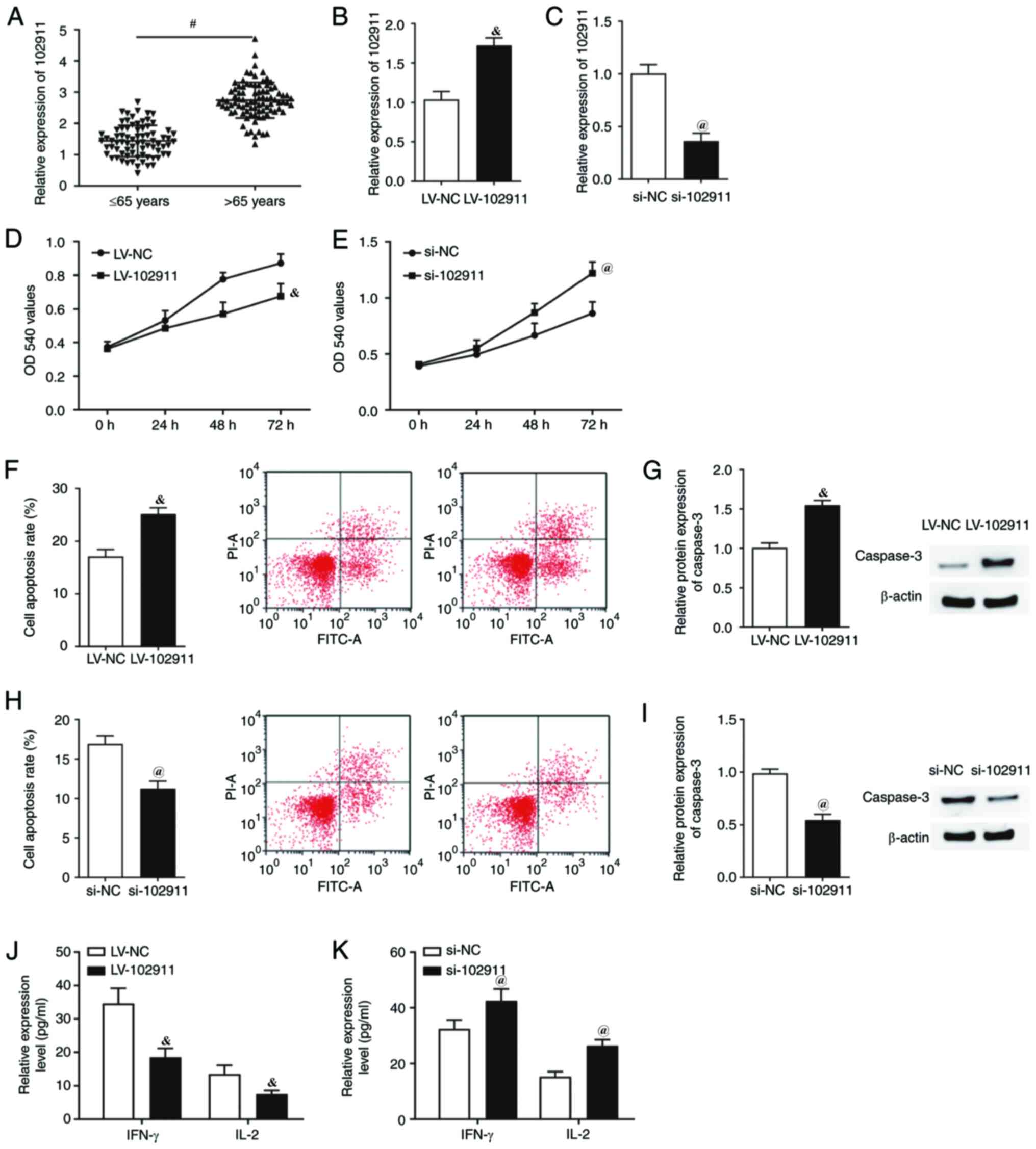
102911 is involved in the
proliferation and apoptosis of CD3+/CD4+ T
lymphocytes
The expression levels of 102911 were significantly
elevated in the CD3+CD4+ T lymphocytes
obtained from the elderly patients (Fig. 1A). However, there was no significant
difference in the pre-operative and post-operative (POD1-14)
expression levels of 102911 in the CD3+CD4+ T
lymphocytes of the two groups of patients, and the expression
levels of 102911 were significantly increased in the
CD3+CD4+ T lymphocytes obtained from the
elderly patients in the pre-operative and post-operative groups
(POD1-14) (Table VII). After
CD3+CD4+ T lymphocytes were transfected with
LV-102911, the expression levels of 102911 were significantly
increased (Fig. 1B). The expression
of 102911 was significantly decreased following transfection of
CD3+CD4+ T lymphocytes with si-102911
(Fig. 1C). Overexpression of 102911
significantly inhibited CD3+CD4+ T
lymphocytes proliferation (Fig.
1D), promoted CD3+CD4+ T lymphocytes
apoptosis (Fig. 1F and G) and decreased the expression levels of
IL-2 and IFN (Fig. 1J). Similarly,
reduced expression of 102911 significantly promoted T cell
proliferation (Fig. 1E), inhibited
T cell apoptosis (Fig. 1H and
I) and increased IL-2 and IFN
expression levels (Fig. 1K). These
results suggested that 102911 may be involved in the regulation of
CD3+CD4+ T lymphocytes in elderly
patients.
| Table VIIComparison of pre-operative and
post-operative mRNA expression of 102911 in
CD3+/CD4+T lymphocytes using RT-qPCR. |
Table VII
Comparison of pre-operative and
post-operative mRNA expression of 102911 in
CD3+/CD4+T lymphocytes using RT-qPCR.
| | Time-points for
detection |
|---|
| Group | Pre-operation | POD1 | POD3 | POD7 | POD14 |
|---|
| >65 years | 2.7±0.5 | 2.8±0.7 | 2.9±0.6 | 2.7±0.8 | 2.6±0.5 |
| ≤65 years |
1.4±0.5a |
1.4±0.4a |
1.5±0.7a |
1.6±0.6a |
1.5±0.6a |
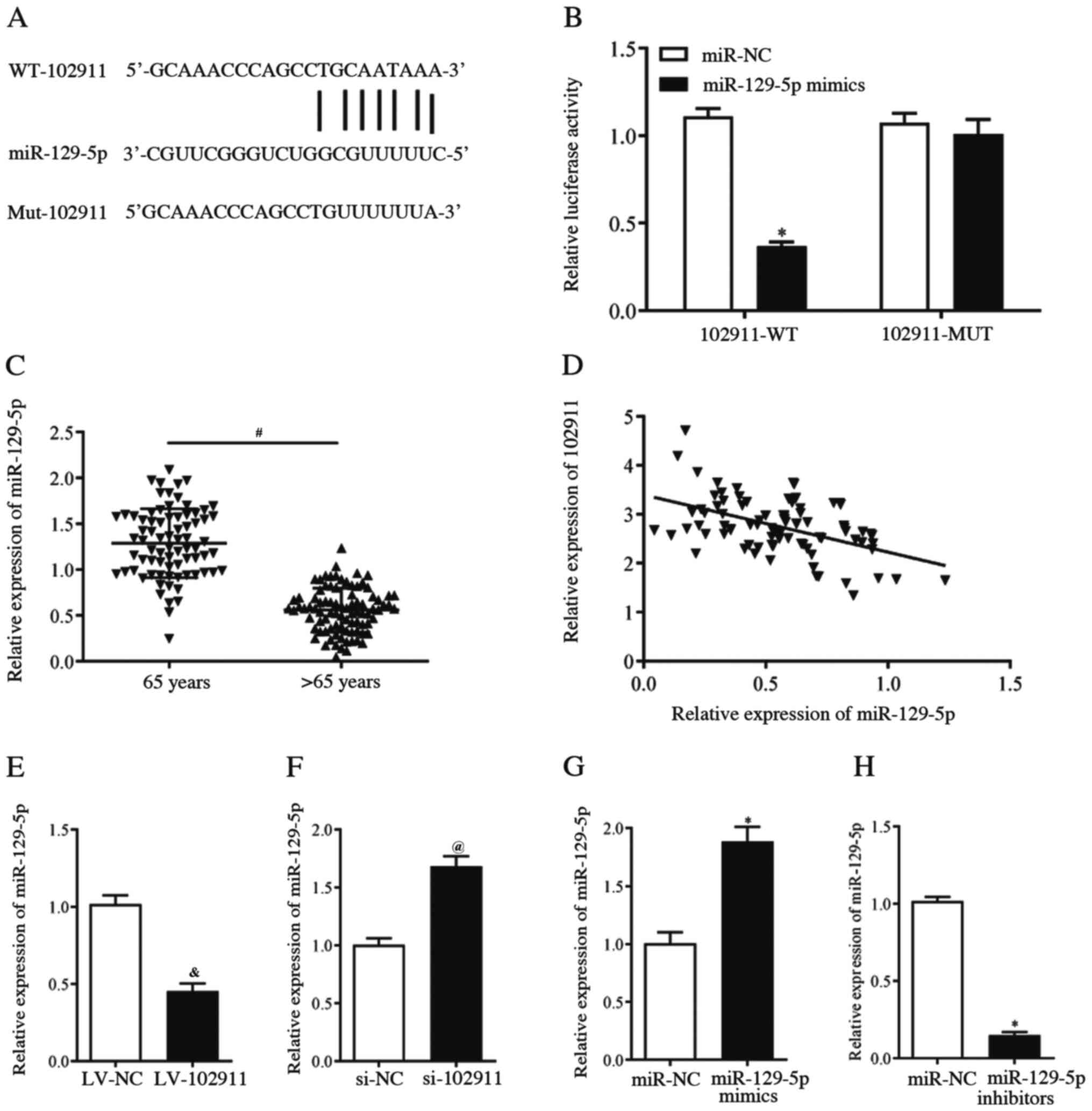
102911 directly binds with miR-129-5p
in CD3+CD4+ T lymphocytes
The target binding site of 102911 and miR-129-5p is
shown in Fig. 2A. Dual luciferase
reporter assays showed that the activity of double fluorescein was
significantly decreased after transfection with miR-129-5p mimics
in 102911-WT (Fig. 2B).
Additionally, RT-qPCR was used to detect the expression level of
miR-129-5p in the CD3+CD4+ T lymphocytes of
the two groups. The results showed that the expression levels of
miR-129-5p in CD3+CD4+ T lymphocytes of the
elderly patients was significantly decreased (Fig. 2C). There was a significant negative
correlation between the expression levels of 102911 and miR-129-5p
in CD3+CD4+ T lymphocytes of elderly patients
(Fig. 2D). Furthermore, the
expression levels of miR-129-5p were significantly reduced
following the overexpression of 102911 (Fig. 2E) and the expression levels of
miR-129-5p were significantly increased following the reduction of
102911 expression (Fig. 2F). After
CD3+CD4+ T lymphocytes were transfected with
miR-129-5p mimics, the expression levels of miR-129-5p were
significantly increased (Fig. 2G).
The expression levels of miR-129-5p were significantly decreased
following transfection of CD3+CD4+ T
lymphocytes with miR-129-5p inhibitors (Fig. 2H), demonstrating the successful
transfection of these agents. These results suggested that 102911
may play a role in CD3+CD4+ T lymphocytes
through targeting miR-129-5p.
 | Figure 2102911 directly binds with miR-129-5p
in CD3+/CD4+ T lymphocytes. (A) The putative
binding sites between the transcripts of 102911 and miR-129-5p. (B)
Luciferase gene reporter assays demonstrated that 102911 directly
bound with miR-129-5p (Student's t-test). (C) The expression levels
of miR-129-5p in CD3+/CD4+ T lymphocytes of
two groups were determined using RT-qPCR (Student's t-test). (D)
Spearman's correlation analysis indicated a negative correlation
between 102911 and miR-129-5p expression levels in
CD3+/CD4+ T lymphocytes of elderly patients.
miR-129-5p expression levels of CD3+/CD4+ T
lymphocytes transfected with (E) LV-102911, (F) si-102911, (G)
miR-129-5p mimics or (H) miR-129-5p inhibitors, were determined
using RT-qPCR (Student's t-tests). #P<0.05 vs. <65
years; *P<0.05 vs. miR-NC; &P<0.05
vs. LV-NC; @P<0.05 vs. si-NC. All assays were
repeated three times in duplicates. 102911, circular RNA_102911;
miR, microRNA; Mut, mutant; NC, negative control; RT-qPCR, reverse
transcription-quantitative PCR; si, small interfering RNA; WT,
wild-type. |
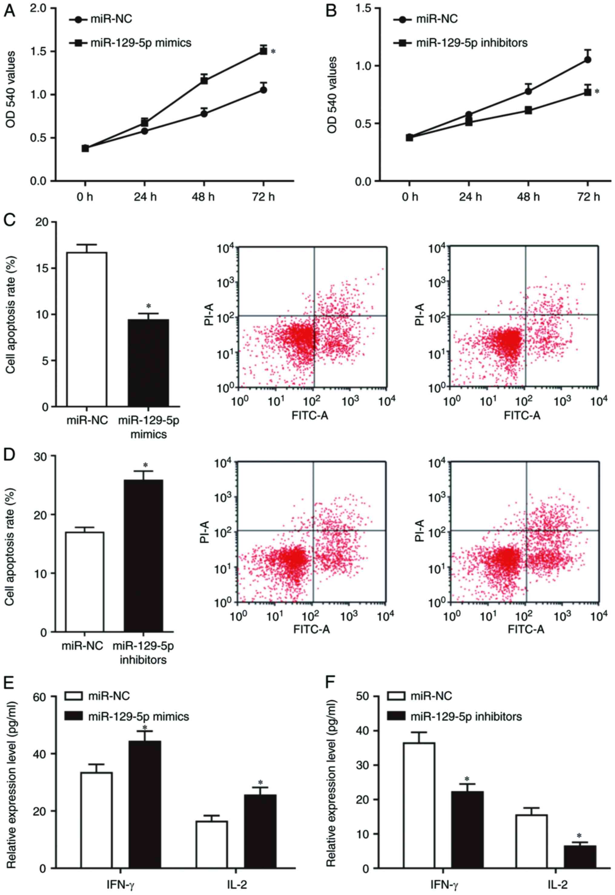
miR-129-5p is involved in the
proliferation and apoptosis of CD3+CD4+ T
lymphocytes
To further explore the role of miR-129-5p in
CD3+CD4+ T lymphocytes, miR-129-5p mimics and
miR-129-5p inhibitors were transfected into
CD3+CD4+ T lymphocytes. Overexpression of
miR-129-5p significantly promoted T cell proliferation (Fig. 3A), inhibited T cell apoptosis
(Fig. 3C) and increased IL-2 and
IFN expression levels (Fig. 3E). In
contrast, low expression of miR-129-5p inhibited
CD3+CD4+ T lymphocyte proliferation (Fig. 3B), promoted
CD3+CD4+ T lymphocyte apoptosis (Fig. 3D) and decreased IL-2 and IFN
expression levels (Fig. 3F). These
results showed that miR-129-5p was also involved in the
proliferation and apoptosis of CD3+CD4+ T
lymphocytes.
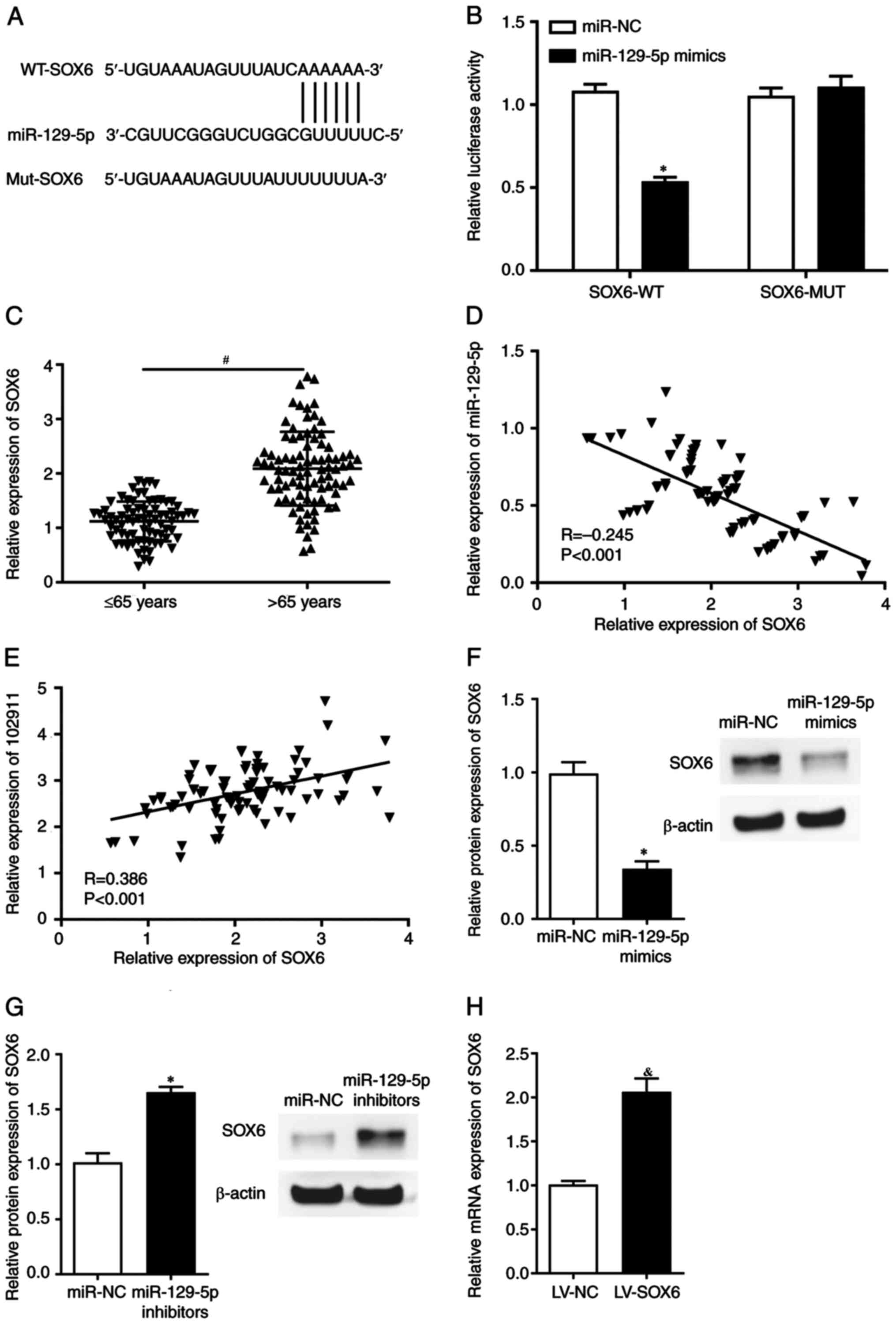
miR-129-5p directly binds with SOX6 in
CD3+CD4+ T lymphocytes
To further study the downstream targets of
miR-129-5p, the target binding site between miR-129-5p and SOX6 was
found (Fig. 4A). Dual luciferase
reporter assays showed that the activity of double fluorescein was
significantly decreased after transfection with miR-129-5p mimics
in SOX6-WT (Fig. 4B). Furthermore,
RT-qPCR was used to detect the expression level of SOX6 in
CD3+CD4+ T lymphocytes of the two groups. The
results showed that the mRNA expression levels of SOX6 in
CD3+CD4+ T lymphocytes of elderly patients
was significantly increased (Fig.
4C). Meanwhile, there was a significant negative correlation
between the expression levels of miR-129-5p and SOX6 in the
CD3+CD4+ T lymphocytes of the elderly
patients (Fig. 4D) and a positive
correlation between expression levels of 102911 and SOX6 (Fig. 4E). Furthermore, the expression level
of SOX6 were significantly reduced after
CD3+CD4+ T lymphocytes were transfected with
miR-129-5p mimics (Fig. 4F) and the
expression levels of SOX6 were significantly raised after the
reduction of miR-129-5p expression levels (Fig. 4G). In order to investigate the
effects of SOX6 on the biological behavior of
CD3+CD4+ T lymphocytes, the cells were
transfected with LV-NC or LV-SOX6, and the transfection efficiency
were determined using RT-qPCR, with the results showing that a
successful transfection, leading to an increased expression level
of SOX-6 (Fig. 4H). These results
showed that miR-129-5p may play a role in
CD3+CD4+ T lymphocytes through targeting
SOX6.
The 102911/miR-129-5p/SOX6 axis is
involved in the proliferation and apoptosis of
CD3+CD4+ T lymphocytes
In order to further study the regulatory roles that
102911 has on CD3+CD4+ T lymphocytes through
regulating the miR-129-5p/SOX6 axis, CD3+CD4+
T lymphocytes were co-transfected with LV-102911 and miR-129-5p
mimics, or miR-129-5p and LV-SOX6 respectively. The effects of
LV-102911 on the proliferation and apoptosis of
CD3+CD4+ T lymphocytes were abolished
following co-transfection with miR-129-5p mimics (Fig. 5A, C
and E). Meanwhile, the effects of
miR-129-5p mimics on the proliferation and apoptosis of
CD3+CD4+ T lymphocytes were abolished
following co-transfection with SOX6 (Fig. 5B, D
and F). Taken together, these data
show that the 102911/miR-129-5p/SOX6 axis was involved in T
lymphocytes immune function, which provided a novel insight for the
treatment of elderly patients with hepatolithiasis.
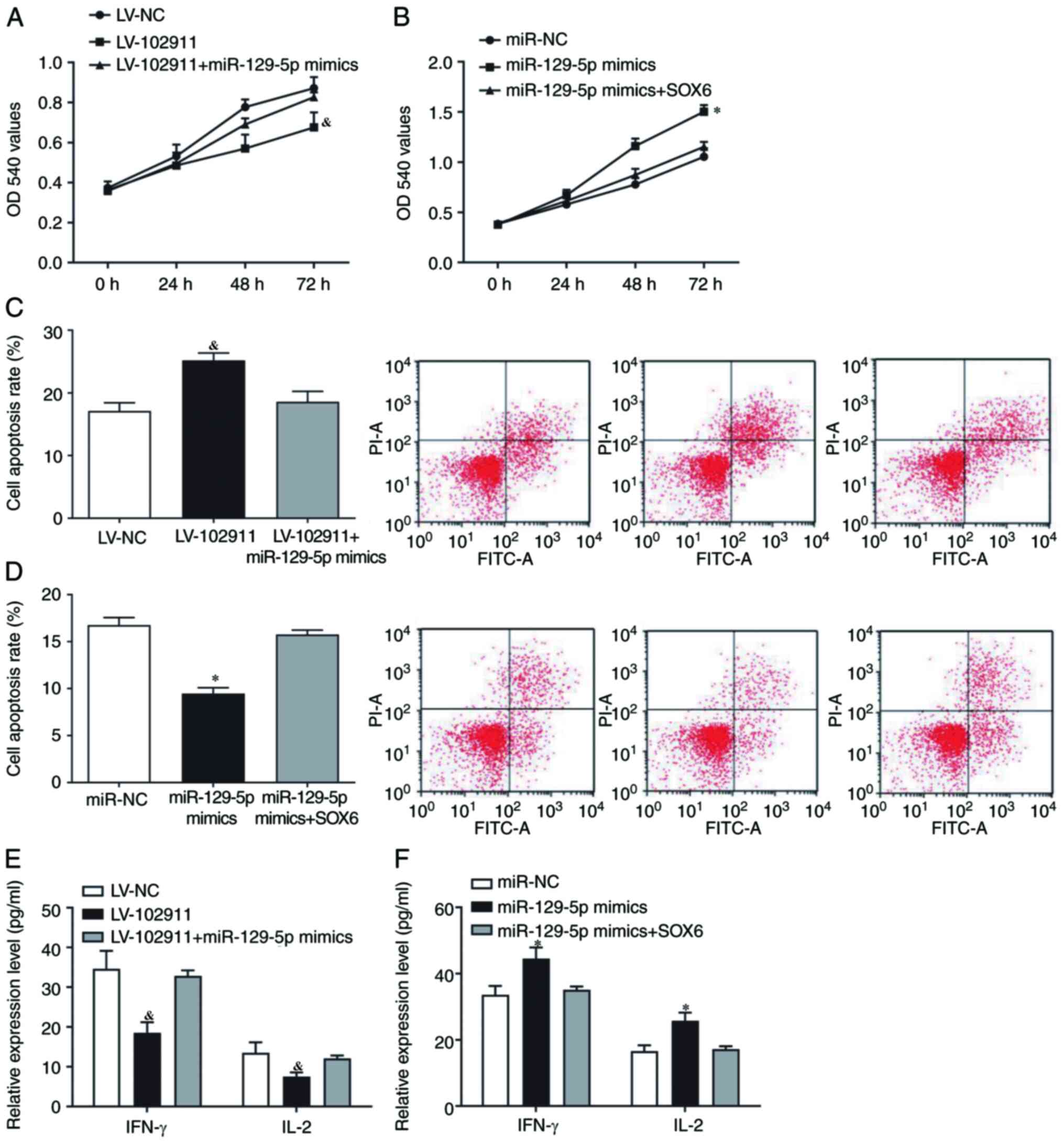 | Figure 5The 102911/miR-129-5p/SOX6 axis is
involved with the proliferation and apoptosis of
CD3+/CD4+ T lymphocytes. The proliferation of
CD3+/CD4+ T lymphocytes co-transfected with
(A) LV-102911 and miR-129-5p mimics, or (B) miR-129-5p and SOX6,
was determined using MTT assays (Two-way ANOVA). The apoptosis of
CD3+/CD4+ T lymphocytes co-transfected with
(C) LV-102911 and miR-129-5p mimics, or (D) miR-129-5p and SOX6,
was determined using flow cytometry (one-way ANOVA). IL-2 and IFN-γ
protein expression levels of CD3+/CD4+ T
lymphocytes co-transfected with (E) LV-102911 and miR-129-5p
mimics, (F) or miR-129-5p and SOX6, were detected using ELISAs
(one-way ANOVAs). *P<0.05 vs. miR-NC;
&P<0.05 vs. LV-NC. All assays were repeated three
times in duplicates. FITC-A, anti-fluorescein isothiocyanate; miR,
microRNA; NC, negative control; OD, optical density. |
Discussion
Increases in the average age of populations is a
worldwide phenomenon (16). In
recent years, as life expectancies increase, the aged population is
expanding dramatically in China (17). Due to the increasing aged
population, there is increased demand on surgical services to treat
diseases and improve patient quality of life (18). Hepatolithiasis is a common disease
in China and a risk factor for cholangiocarcinoma (19). Usually, open partial hepatectomy is
used as an effective treatment for hepatolithiasis (20). The most frequent location of
intrahepatic ductal stones in hepatolithiasis is in segments I-IV
of Couinaud (21). With the
development of minimally invasive concepts, technologies and
anatomical features, laparoscopic partial left LLH has become
widely used and established as a more effective treatment for
hepatolithiasis (22).
The improved treatment protocol has become
especially significant for aged patients. As reported by Chen et
al (23) and Jin et al
(24), LLH is associated with a
shorter hospital stay, decreased morbidity and more importantly,
expedited post-operative recovery compared with an open procedure.
The present study found that post-operative infective complication
was still a problem affecting the patients. Data from the present
report showed that although the immediate clinical outcome was fair
for both groups of patients in this study, the elderly patients
were more likely to develop infective complications, even after
minimally invasive surgery such as LLH. The probability of
peritonitis in elderly patients may be closely related to bile
leakage from bile duct stones (25).
Several factors have been revealed to be related to
elderly patient's vulnerability to infective complications after
trauma, of which immune dysfunction after trauma is of importance
(26). This effect is most
pronounced in elderly patients due to immunosenescence (27). Immunosenescence is the age-related
decline in immune functions and affects various cell types such as
neutrophils, monocytes, macrophages, natural killer cells and
dendritic cells from the innate arm, as well as B and T cells from
the adaptive arm of the immune system (26). Although aging alters both arms of
the immune system, more severe detrimental changes occur in the
adaptive immune system (28). The
data from the present study showed that the basic quantity of
peripheral lymphocytes in the elderly group was significantly lower
than that in the younger group pre-operatively. Secondly, after
surgical trauma, the immune function of T lymphocytes went through
a significant decline and lasted longer (no less than 2 weeks) when
compared with that of the younger patients, which was reflected by
perioperative changes to the T lymphocyte proliferative ability,
levels of IL-2 secreted by T lymphocytes and the % of
CD3+CD4+T lymphocytes in the peripheral
blood. Finally, surgical trauma was found to further affect the
number of T lymphocytes through altered levels of apoptosis. The
changes in T lymphocytes may be why elderly patients are more prone
to post-operative complications.
It has been revealed that the
immunosenescence-mediated decline in cell-mediated immune functions
results in a chronic inflammatory state (29). The age-related increases in
cytokines and other components of inflammation are partially
responsible for the increased prevalence and severity of infections
(30). This pro-inflammatory
response of aged patients is also believed to be responsible for
the increased risk for developing complications following injury
(31). The present study revealed
that after trauma, the number and function of T lymphocytes in aged
patients altered more drastically than that in the younger patients
and took longer to return to the original levels. This
theoretically worsens the immune function and increases the risk
that elderly patients will suffer from infectious diseases.
However, the exact mechanism of action has not yet been fully
elucidated.
In the present study, 102911 was upregulated and
miR-129-5p was downregulated in the CD3+CD4+
T lymphocytes from elderly patients with LLH for hepatolithiasis.
102911 overexpression inhibited CD3+CD4+ T
lymphocyte proliferation and promoted cell apoptosis, with the
opposite observations occurring following knockdown of 102911.
miRNAs are a class of non-coding small RNAs that are ubiquitous in
the genome of an organism, which are ~20-24 nt (32). circRNA has the ability to recognize
complementary sequences and bind to miRNAs (33). With the in-depth study of miRNAs,
there is increasing evidence that they are closely related to the
progression of various immune-related and age-related diseases
(34,35).
The present study found that 102911 directly binds
with miR-129-5p in CD3+CD4+ T lymphocytes and
there was a significant negative correlation between expression
levels of 102911 and miR-129-5p in CD3+CD4+ T
lymphocytes from elderly patients. Overexpression of miR-129-5p
significantly promoted T cell proliferation, inhibited T cell
apoptosis and increased IL-2 and IFN expression levels. In
contrast, reduced expression of miR-129-5p inhibited
CD3+CD4+ T lymphocyte proliferation, promoted
CD3+CD4+ T lymphocyte apoptosis and decreased
IL-2 and IFN expression levels. These results showed that
miR-129-5p could be a promising target of 102911 and that
miR-129-5p is involved in the proliferation and apoptosis of
CD3+CD4+ T lymphocytes.
SOX6 is a multifaceted transcription factor that
participates in cell proliferation, apoptosis, differentiation and
release of inflammatory factors (36). SOX6 also plays an important role in
embryonic development (37). SOX6
has multiple cytotoxic T-lymphocyte and helper epitopes to induce
antitumor activity and the effectiveness of SOX6-DNA vaccine for
the prevention and treatment of glioma (38). In the present study, the expression
levels of SOX6 in CD3+CD4+ T lymphocytes from
elderly patients was significantly increased and SOX6 was found to
be a downstream molecule of miR-129-5p.
In conclusion, the present study has demonstrated
that the immune function and the number of T lymphocytes went
through a significant decline and this decline lasted longer in
elderly patients with LLH for hepatolithiasis after surgical
trauma, compared with the younger patients. However, there were
limitations to the present study. For example, in vivo
studies should be performed to confirm the existing findings.
Furthermore, other assays such as immunocytostaining should be
carried out to evaluate the expression profile of the associated
proteins. Despite the limitations, the present results suggested
that the 102911/miR-129-5p/SOX6 axis was involved in T lymphocytes
immune function, which provided a novel insight for potential
treatments for elderly patients with hepatolithiasis.
Acknowledgements
Not applicable.
Funding
This work is supported by National Natural Science
Foundation of China (grant no. 81771717).
Availability of data and materials
The datasets used and/or analyzed during the
current study are available from the corresponding author on
reasonable request.
Authors' contributions
WL designed the study. HZ, XS, HL, QL and JL
performed the experiments and analyzed the data. HZ and XS drafted
the manuscript. All authors read and approved the final
manuscript.
Ethics approval and consent to
participate
This study was approved by the Ethics Committee of
The Eighth Medical Center of the Chinese PLA General Hospital
(Beijing, China). Written informed consents were obtained from all
the patients.
Patient consent for publication
Not applicable.
Competing interests
The authors declare that they have no competing
interests.
References
|
1
|
Cai X: Laparoscopic liver resection: The
current status and the future. Hepatobil Surg Nutr. 7:98–104.
2018.PubMed/NCBI View Article : Google Scholar
|
|
2
|
Chen K, Pan Y, Maher H, Zhang B and Zheng
XY: Laparoscopic hepatectomy for elderly patients: Major findings
based on a systematic review and meta-analysis. Medicine
(Baltimore). 97(e11703)2018.PubMed/NCBI View Article : Google Scholar
|
|
3
|
Chong KC, Leung CC, Yew WW, Zee BCY, Tam
GCH, Wang MH, Jia KM, Chung PH, Lau SYF, Han X and Yeoh EK:
Mathematical modelling of the impact of treating latent
tuberculosis infection in the elderly in a city with intermediate
tuberculosis burden. Sci Rep. 9(4869)2019.PubMed/NCBI View Article : Google Scholar
|
|
4
|
Islam MN, Bradley BA and Ceredig R:
Sterile post-traumatic immunosuppression. Clin Transl Immunology.
5(e77)2016.PubMed/NCBI View Article : Google Scholar
|
|
5
|
Nikolich-Žugich J: The twilight of
immunity: Emerging concepts in aging of the immune system. Nat
Immunol. 19:10–19. 2018.PubMed/NCBI View Article : Google Scholar
|
|
6
|
Gaffo E, Boldrin E, Dal Molin A, Bresolin
S, Bonizzato A, Trentin L, Frasson C, Debatin KM, Meyer LH, Te
Kronnie G and Bortoluzzi S: Circular RNA differential expression in
blood cell populations and exploration of circRNA deregulation in
pediatric acute lymphoblastic leukemia. Sci Rep.
9(14670)2019.PubMed/NCBI View Article : Google Scholar
|
|
7
|
Lei X, Fang Z and Guo L: Predicting
circRNA-disease associations based on improved collaboration
filtering recommendation system with multiple data. Front Genet.
10(897)2019.PubMed/NCBI View Article : Google Scholar
|
|
8
|
Deng W, Peng W, Wang T, Chen J, Qiu X, Fu
L and Zhu S: Microarray profile of circular RNAs identifies
hsa_circRNA_102459 and hsa_circRNA_043621 as important regulators
in oral squamous cell carcinoma. Oncol Rep. 42:2738–2749.
2019.PubMed/NCBI View Article : Google Scholar
|
|
9
|
Chang W, Wang J, Tao D, Zhang Y, He J and
Shi C: Identification of a novel miRNA from the ovine ovary by a
combinatorial approach of bioinformatics and experiments. J Vet Med
Sci. 77:1617–1624. 2016.PubMed/NCBI View Article : Google Scholar
|
|
10
|
Agrawal M, Pandey N, Rastogi M, Dogra S
and Singh SK: Chikungunya virus modulates the miRNA expression
patterns in human synovial fibroblasts. J Med Virol. 92:139–148.
2020.PubMed/NCBI View Article : Google Scholar
|
|
11
|
Ji Q, Zhang C, Sun X and Li Q: Circular
RNAs function as competing endogenous RNAs in multiple types of
cancer. Oncol Lett. 15:23–30. 2018.PubMed/NCBI View Article : Google Scholar
|
|
12
|
Zhang Y, Yu F, Bao S and Sun J: Systematic
characterization of circular RNA-associated CeRNA network
identified novel circRNA biomarkers in Alzheimer's disease. Front
Bioeng Biotechnol. 7(222)2019.PubMed/NCBI View Article : Google Scholar
|
|
13
|
Khan MS, Yamashita K, Sharma V, Ranjan R
and Dosdall DJ: RNAs and gene expression predicting postoperative
atrial fibrillation in cardiac surgery patients undergoing coronary
artery bypass grafting. J Clin Med. 9(1139)2020.PubMed/NCBI View Article : Google Scholar
|
|
14
|
Bjarnsholt T, Jensen PØ and Alhede M:
Revival of krebs-ringer balanced salt solution for the
investigation of polymorphonuclear leukocytes and pseudomonas
aeruginosa biofilm interaction. Pathog Dis.
77(ftz052)2019.PubMed/NCBI View Article : Google Scholar
|
|
15
|
Livak KJ and Schmittgen TD: Analysis of
relative gene expression data using real-time quantitative PCR and
the 2(-Delta Delta C(T)) method. Methods. 25:402–408.
2001.PubMed/NCBI View Article : Google Scholar
|
|
16
|
Harper S: Economic and social implications
of aging societies. Science. 346:587–591. 2014.PubMed/NCBI View Article : Google Scholar
|
|
17
|
Chen LK: Population ageing is a global
phenomenon, which affects both Taiwan and China. Preface. Ageing
Res Rev. 9 (Suppl 1)(S1)2010.PubMed/NCBI View Article : Google Scholar
|
|
18
|
Franco MR and Fernandes NM: Dialysis in
the elderly patient: A challenge of the XXI century-narrative
review. J Bras Nefrol. 35:132–141. 2013.PubMed/NCBI View Article : Google Scholar : (In English,
Portuguese).
|
|
19
|
Zhang N, Li Y, Zhao M, Chang X, Tian F, Qu
Q and He X: Sarcomatous intrahepatic cholangiocarcinoma: Case
report and literature review. Medicine (Baltimore).
97(e12549)2018.PubMed/NCBI View Article : Google Scholar
|
|
20
|
Jarufe N, Figueroa E, Muñoz C, Moisan F,
Varas J, Valbuena JR, Bambs C, Martínez J and Pimentel F: Anatomic
hepatectomy as a definitive treatment for hepatolithiasis: A cohort
study. HPB (Oxford). 14:604–610. 2012.PubMed/NCBI View Article : Google Scholar
|
|
21
|
Li EL, Yuan RF, Liao WJ, Feng Q, Lei J,
Yin XB, Wu LQ and Shao JH: Intrahepatic bile duct exploration
lithotomy is a useful adjunctive hepatectomy method for bilateral
primary hepatolithiasis: An eight-year experience at a single
centre. BMC Surg. 19(16)2019.PubMed/NCBI View Article : Google Scholar
|
|
22
|
Zizzo M, Ugoletti L, Castro Ruiz C,
Zanelli M, De Marco L, Pedrazzoli C and Annessi V: Laparoscopic
liver resection for malignancies confined to Couinaud's segment VII
in the robotic surgery era. Hepatobil Surg Nutr. 8:439–441.
2019.PubMed/NCBI View Article : Google Scholar
|
|
23
|
Chen S, Huang L, Qiu FN, Zhou SQ, Yan ML,
Bai YN, Lai ZD, Tian YF and Wang YD: Total laparoscopic partial
hepatectomy versus open partial hepatectomy for primary left-sided
hepatolithiasis: A propensity, long-term follow-up analysis at a
single center. Surgery. 163:714–720. 2018.PubMed/NCBI View Article : Google Scholar
|
|
24
|
Jin RA, Wang Y, Yu H, Liang X and Cai XJ:
Total laparoscopic left hepatectomy for primary hepatolithiasis:
Eight-year experience in a single center. Surgery. 159:834–841.
2016.PubMed/NCBI View Article : Google Scholar
|
|
25
|
Parra-Membrives P, Martínez-Baena D,
Lorente-Herce JM and Jiménez-Vega J: Laparoscopic common bile duct
exploration in elderly patients: Is there still a difference? Surg
Laparosc Endosc Percutan Tech. 24:e118–e122. 2014.PubMed/NCBI View Article : Google Scholar
|
|
26
|
Salminen A, Kaarniranta K and Kauppinen A:
Immunosenescence: The potential role of myeloid-derived suppressor
cells (MDSC) in age-related immune deficiency. Cell Mol Life Sci.
76:1901–1918. 2019.PubMed/NCBI View Article : Google Scholar
|
|
27
|
Behrens MI, Silva M, Schmied A, Salech F,
Manzur H, Rebolledo R, Bull R, Torres V, Henriquez M and Quest AF:
Age-dependent increases in apoptosis/necrosis ratios in human
lymphocytes exposed to oxidative stress. J Gerontol A Biol Sci Med
Sci. 66:732–740. 2011.PubMed/NCBI View Article : Google Scholar
|
|
28
|
López-Otín C, Blasco MA, Partridge L,
Serrano M and Kroemer G: The hallmarks of aging. Cell.
153:1194–1217. 2013.PubMed/NCBI View Article : Google Scholar
|
|
29
|
McElhaney JE and Effros RB:
Immunosenescence: What does it mean to health outcomes in older
adults? Curr Opin Immunol. 21:418–424. 2009.PubMed/NCBI View Article : Google Scholar
|
|
30
|
Maeve RI, Gibson DS, Victoria MG, Mcnerlan
SE, Denis AH and Ross OA: Age and age-related diseases: Role of
inflammation triggers and cytokines. Front Immunol.
9(586)2018.PubMed/NCBI View Article : Google Scholar
|
|
31
|
Kishimoto K, Hiraguri M, Koide N, Hanazaki
K and Adachi W: Postoperative suppression of inflammatory cytokines
after distal gastrectomy in elderly patients. Surg Today.
39:487–492. 2009.PubMed/NCBI View Article : Google Scholar
|
|
32
|
Li G, Liu B, Jiang Q, Zhang J, Xin S and
Xu K: The association of two common polymorphisms in miRNAs with
diabetes mellitus: A meta-analysis. Medicine (Baltimore).
98(e17414)2019.PubMed/NCBI View Article : Google Scholar
|
|
33
|
Jia N, Tong H, Zhang Y, Katayama H, Wang
Y, Lu W, Zhang S and Wang J: CeRNA expression profiling identifies
KIT-related circRNA-miRNA-mRNA networks in gastrointestinal stromal
tumour. Front Genet. 10(825)2019.PubMed/NCBI View Article : Google Scholar
|
|
34
|
Hong BS, Ryu HS, Kim N, Kim J, Lee E, Moon
H, Kim KH, Jin MS, Kwon NH, Kim S, et al: Tumor suppressor
microRNA-204-5p regulates growth, metastasis, and immune
microenvironment remodeling in breast cancer. Cancer Res.
79:1520–1534. 2019.PubMed/NCBI View Article : Google Scholar
|
|
35
|
Kang H, Liang QJ, Hu R, Li ZH, Liu Y and
Wang WN: Integrative mRNA-miRNA interaction analysis associated
with the immune response of epinephelus coioddes to vibrio
alginolyticus infection. Fish Shellfish Immunol. 90:404–412.
2019.PubMed/NCBI View Article : Google Scholar
|
|
36
|
Qi H, Yao L and Liu Q: MicroRNA-96
regulates pancreatic β cell function under the pathological
condition of diabetes mellitus through targeting Foxo1 and Sox6.
Biochem Biophys Res Commun. 519:294–301. 2019.PubMed/NCBI View Article : Google Scholar
|
|
37
|
Li Y, Deng S, Peng J, Wang X, Essandoh K,
Mu X, Peng T, Meng ZX and Fan GC: MicroRNA-223 is essential for
maintaining functional β-cell mass during diabetes through
inhibiting both FOXO1 and SOX6 pathways. J Biol Chem.
294:10438–10448. 2019.PubMed/NCBI View Article : Google Scholar
|
|
38
|
Ueda R, Kinoshita E, Ito R, Kawase T,
Kawakami Y and Toda M: Induction of protective and therapeutic
antitumor immunity by a DNA vaccine with a glioma antigen, SOX6.
Int J Cancer. 122:2274–2279. 2008.PubMed/NCBI View Article : Google Scholar
|