Introduction
Heidelbach and Gilbricht were among the first to
report the use of A-mode sonography for the analysis of maxillary
sinusitis cases, which was later used broadly due to the
enhancement of probes and because it represented a viable
alternative to standard radiology (1).
Evidently, the gold standard in sinus imaging are
computed tomography (CT) and magnetic resonance imaging and because
of their development the use of sonography was abandoned until
recently, when it was used in emergency departments in trauma cases
for rapid detection of hemosinus (2).
Promising results were obtained in the management of
maxillary acute sinusitis in pediatric cases given the advantage of
reducing child exposure to radiation and enabling the serial exam
during treatment (3). Therefore,
ultrasonography can be used in pregnant patients to exclude acute
sinusitis requiring active medical treatment precarious to
pregnancy.
After 2005, the increased accessibility to high
resolution ultrasound machines that were implemented aimed to
analyze the specificity and sensitivity of this method for the
detection of sinus fractures compared to standard radiology and CT
scans. According to Adeyemo and Akadiri, the sensitivity and
specificity for detecting orbital fractures was between 56-100 and
85-100%, respectively; for the detection of nasal fractures, they
recorded 90-100% sensitivity and 98-100% specificity compared to CT
scans (4).
For the present study, we implemented the ultrasound
exam of patients with inflammatory sinus pathology before they
received CT scans in order to prove the utility of sonography in
the primary management of these cases and in deciding for further
referral to CT scan imaging.
Materials and methods
General
A total of 81 cases (48 males and 33 females), with
a median age of 56 years, with sinus symptoms presented to a
private outpatient clinic (Galenus Medical Center, Targu Mures,
Romania) and were enrolled in the present study. Inclusion signs
and symptoms: Pus secretion in the nasal cavity, mild facial pain,
pressure sensation on sinus points, nasal obstruction, loss of
smell, fever, headache.
All cases received standard ENT exam with nasal
endoscopy, followed by sonography with an Acuson x300 machine.
Following these steps, some of the cases were referred to CT scan
imaging.
The study followed the international regulations in
accordance with the Declaration of Helsinki. The study was approved
by the Ethics Committee of Galenus Medical Center. Patient informed
consent for publication of the data/images associated with the
manuscript was obtained.
Method
The technique for ultrasound exam of sinuses is
based on two important aspects: Precise delineation of bony
structures in sonography window through the back-shadow effect, and
the fact that, in pneumatic cavities, the ultrasound trajectory is
reduced without a contiguous medium.
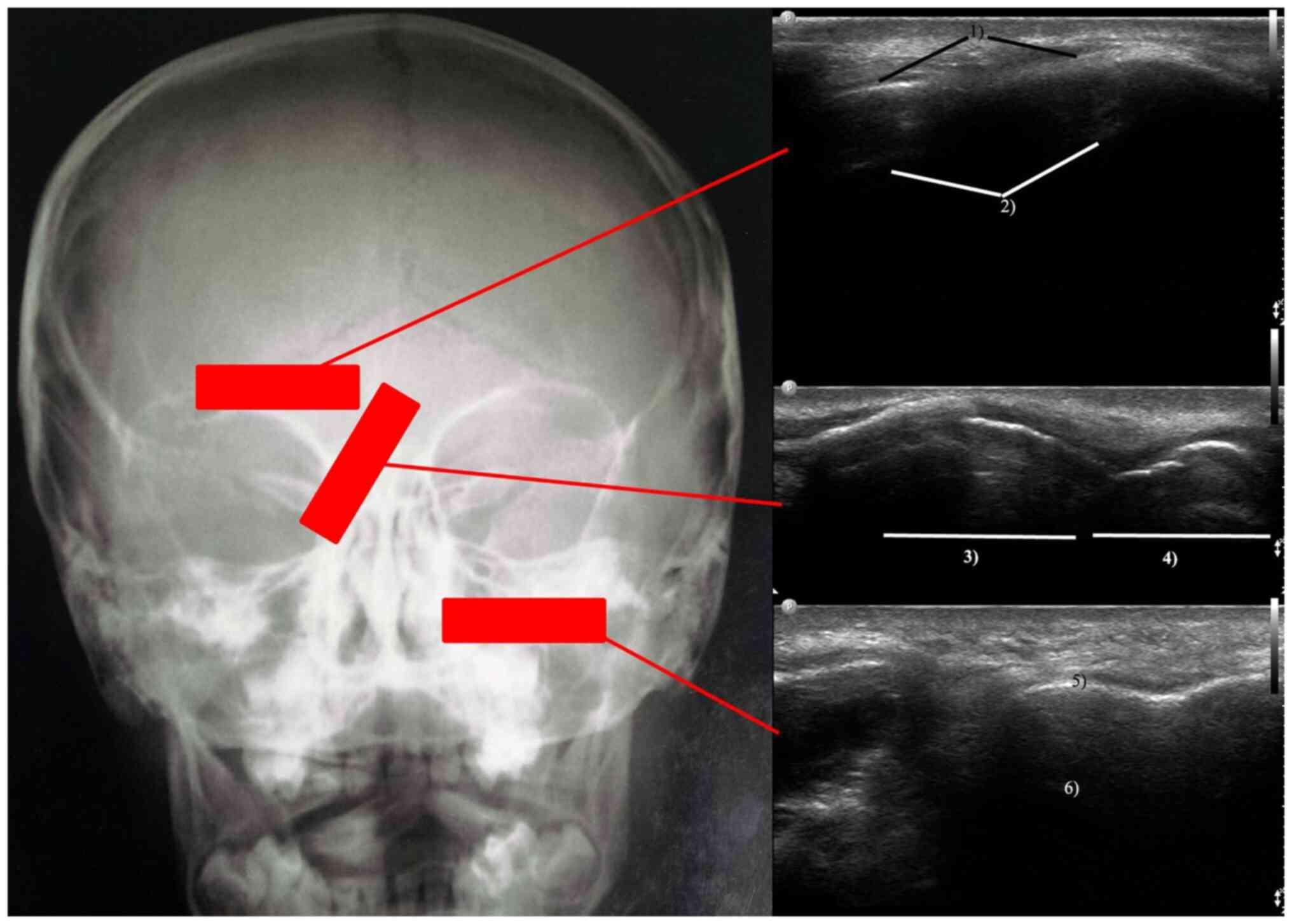
At the level of the frontal sinus, the transducer is
placed over the upper rim of the orbit centered on the median line.
Therefore, one can visualize the anterior wall of the sinus and the
posterior echo generated through the encounter of ultrasound rays
with the posterior sinus wall. In the case of acute sinusitis, the
fluid content filling the sinus will enable the contiguous
propagation of the ultrasound rays between the two walls. Sinus
sonography is performed with the patient sitting upright and facing
downwards. Therefore, the content of the sinus is moved downwards
on the anterior wall and can be measured in quantity. Subsequently,
the patient is examined with the head upright and if the content
moves inferiorly, it will confirm the suspicion of a frontal
purulent sinusitis. The absence of this movement will raise the
suspicion of a cyst and even a sinus tumor. These steps are shown
in Fig. 1.
Moreover, we examined the integrity of the nasal
bone pyramid while angling the transducer over the lateral aspect
obliquely to the inner angle of the eye. The structures visualized
are nasal bones with posterior shadow effect and alar
cartilages.
Imaging of the maxillary sinus is similar to that of
the frontal sinus following examination of the integrity of the
anterior sinus wall and the posterior back echo generated at the
level of the posterior wall of the sinus. There are limits in the
fact that cystic pathology on the posterior wall is hard to
visualize.
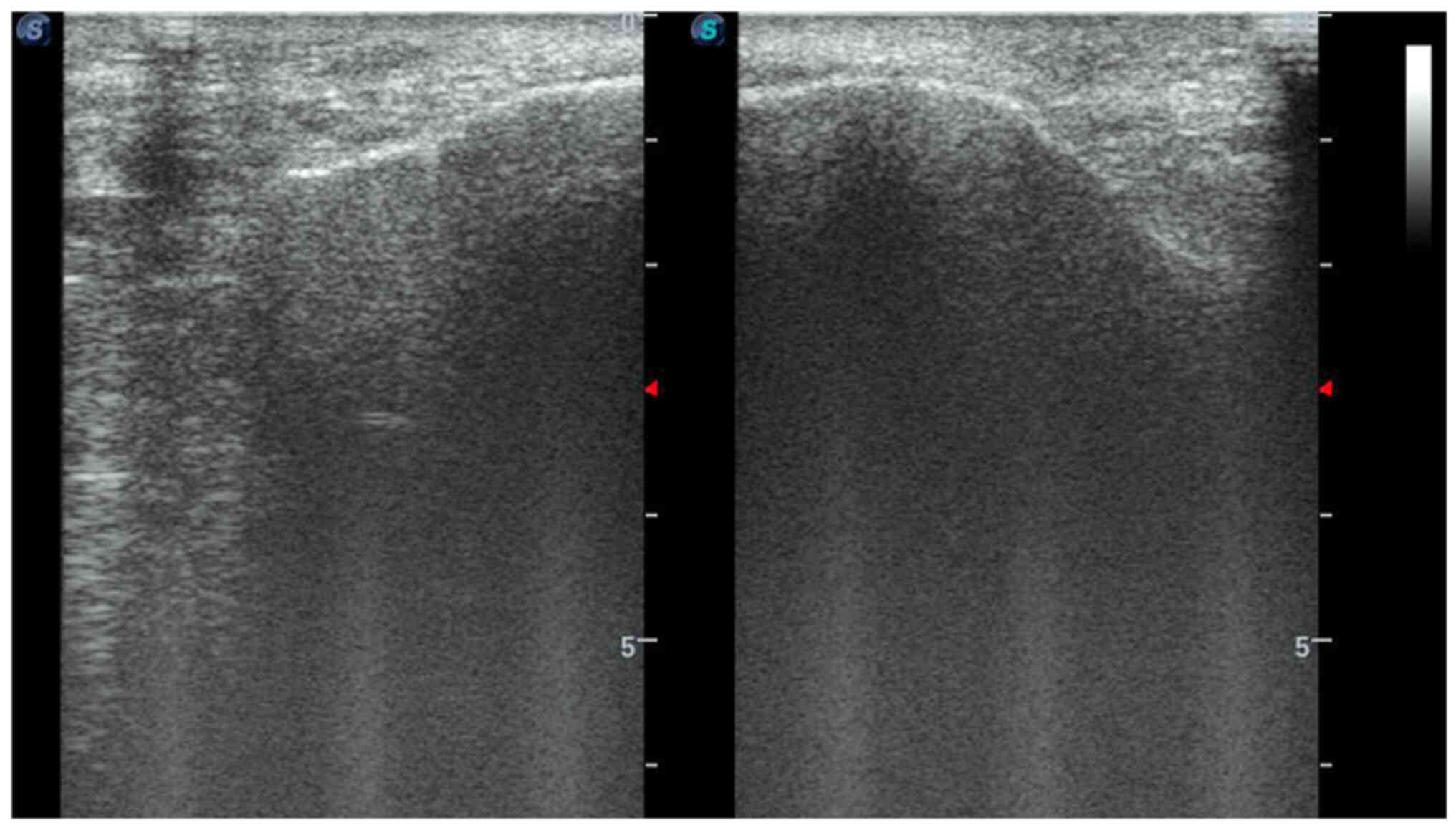
The case of a patient with polypoid maxillary mucosa
on the right side is presented. We enhanced the ultrasound exam
protocol by visualizing both sinuses in duplex comparative window
(Fig. 2).
In the duplex ultrasound window, the difference in
thickness of the sinus mucosa on the anterior right maxillary wall
compared to the thinner one on the anterior left maxillary wall is
underlined. Due to the presence of the content, we obtained an
important posterior echo on the right side, but on the left side,
the normal sinus content had an absent posterior echo.
Statistical analysis
Statistical analysis was performed with Microsoft
Excel Program using the Chi square test, and Spearman's correlation
for P-values <0.05. In order to ascertain the performance of
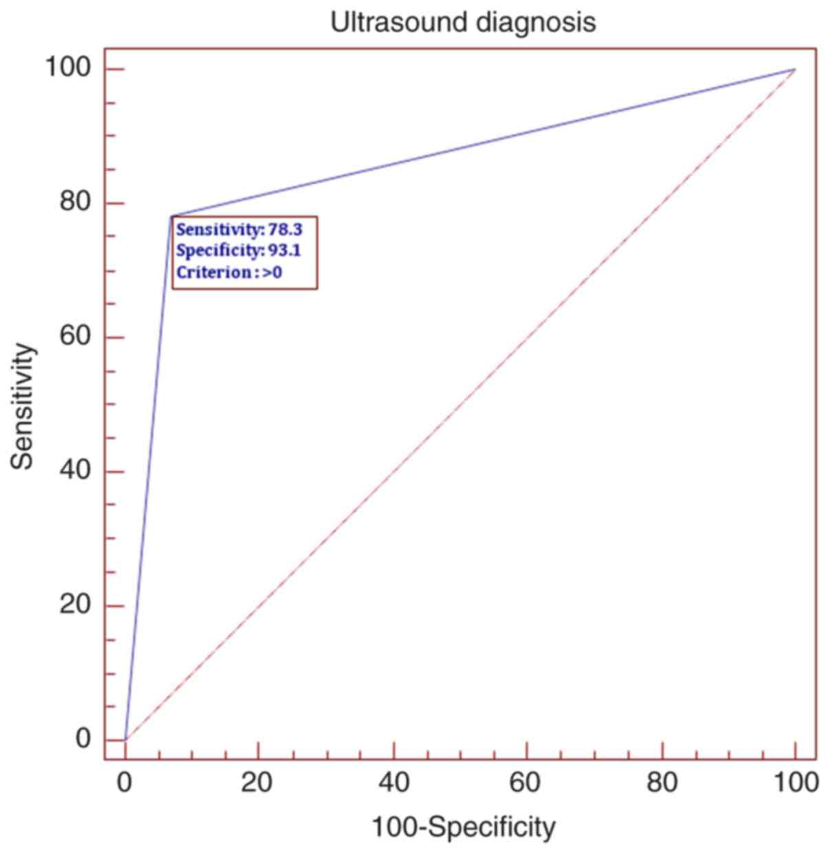
sonography as a screening test, we used the ROC curve therefore
calculating the sensitivity and specificity.
Results
Patients
The median age of the study subjects was 56 years.
The male predominance was of 59.25%. The ultrasound exam compared
to nasal endoscopy presented 78.3% sensitivity and 93.1%
specificity. Only 28.39% of the patients were further referred to
CT scan imaging. Two of the 81 patients proved to be suffering from
mucocele and sinus tumor.
Spearman's correlation
The age group distribution revealed a greater
incidence of sinus pathology of 27.16% between 41 and 50 years of
age.
In addition, the ultrasound exam suggested the
diagnosis of rhinosinusitis in 28.39% of the cases. We obtained a
statistical P-value of 0.002 for the correlation between sex and
age groups, with a lower incidence of male patients <40 years
and a female predominance between 41 and 50 years. Regarding the
correlation between clinical diagnosis and age groups, we did not
obtain a positive statistical result. However, we obtained a
statistical P-value of 0.04 for correlating ultrasound diagnosis
with age groups. Furthermore, there was an increased incidence of
ultrasound diagnosis of rhinosinusitis is in the age groups 41-50
and 71-80 years. Moreover, in the age group of 51 to 60 years, the
ultrasound confirmed the clinical suspicion of chronic
rhinosinusitis. While correlating the sex of the patients with the
ultrasound diagnosis we obtained a P-value of 0.01; in 40 male
cases, the ultrasound confirmed the clinical diagnosis of
rhinosinusitis. Further correlation between ultrasound imaging and
further referral to CT scan retrieved a significant P-value of
0.0001.
By plotting the ROC curve, we established that
sonography may be a screening method in sinus pathology with a high
sensitivity of 78.3 and 93.1% specificity, (Fig. 3).
Discussion
Data obtained thus far prove the utility in
screening pediatric population or in emergency departments. There
were also studies that analyzed the utility of sonography in
imaging the bone sections performed during rhinoplasty procedures
and for correct visualization of the repositioned bone fragments in
trauma cases (5,6).
The increased incidence of cranial and facial trauma
requires the development of new screening protocols in the
emergency departments to expedite the access of major cases to
complex imaging studies (7). This
is also the case for patients with sinusitis complications in which
the treatment progression could be actively followed using
ultrasound (8). Ultrasonography
could be the answer to these problems, as it attracts more ENT
specialists towards its use in outpatient settings (9,10).
Moreover, there is a current trend in
surgeon-performed sonography, as in the case of gastrointestinal
pathology, and this could also be applied in sinus pathology
lowering the burden in already crowded imaging departments
(11). Our sensitivity and
specificity data are consistent with the ones published on 74
patients by Hsu et al (12).
Correlating allergic rhinitis cases with nasal sinus
ultrasound could be a future step in the management of this
pathology (13). Thus, we hope that
our data will help shift the use of sonography from experimental to
clinical practice in ENT and other allied specialties, such as
ophthalmology, plastic surgery, and trauma surgery (14,15).
However, there is the drawback of receiving
incomplete data regarding nasal anatomy via this method, data that
are necessary for proper planning of functional endoscopic sinus
surgery.
In summary, sonography is an imaging method that has
many advantages: Lack of irradiation, it is fast and cheap, and it
permits serial dynamic examinations to ascertain the treatment
efficacy. In the present study, we have shown that, in outpatient
settings, sinus ultrasound has high sensitivity and specificity in
ruling out cases without rhinosinusitis.
Acknowledgements
Professional editing, linguistic and technical
assistance performed by Irina Radu, Individual Service Provider,
certified translator in Medicine and Pharmacy (certificate
credentials: Series E no. 0048).
Funding
Funding: No funding was received.
Availability of data and material
All data generated or analyzed during this study are
included in this published article.
Authors' contributions
AN and MD contributed substantially to the
conception and design of the study, the acquisition, analysis, and
interpretation of the data, and were involved in the drafting of
the manuscript. DV and AC contributed substantially to the analysis
and interpretation of the data and were involved in the drafting of
the manuscript. ANM and RC contributed substantially to the
interpretation of the data and were involved in the critical
revisions of the manuscript for important intellectual content. All
authors agreed to be accountable for all aspects of the work in
ensuring that questions related to the accuracy or integrity of any
part of the work are appropriately investigated and resolved. All
authors read and approved the final version of the manuscript.
Ethics approval and consent to
participate
The study followed the international regulations in
accordance with the Declaration of Helsinki. The study was approved
by the Ethics Committee of Galenus Medical Center. Patient informed
consent for publication of the data/images associated with the
manuscript was obtained.
Patient consent for publication
Not applicable.
Competing interests
The authors declare that they have no competing
interests.
References
|
1
|
Heidelbach JG and Gilbricht E: Practical
experience in ultrasonic diagnosis of the great paranasal sinuses
and their bases. Z Arztl Fortbild (Jena). 66:208–210.
1972.PubMed/NCBI(In German).
|
|
2
|
Bektas F, Soyuncu S and Yigit O: Acute
maxillary sinusitis detected by bedside emergency department
ultrasonography. Int J Emerg Med. 3:497–498. 2010.PubMed/NCBI View Article : Google Scholar
|
|
3
|
Puhakka T, Haikkinen T, Makela MJ, Alanen
A, Kallio T, Korsoff L, Suonpää J and Ruuskanen O: Validity of
ultrasonography in diagnosis of acute maxillary sinusitis. Arch
Otolaryngol Head Neck Surg. 126:1482–1486. 2000.PubMed/NCBI View Article : Google Scholar
|
|
4
|
Adeyemo WL and Akadiri OA: A systematic
review of the diagnostic role of ultrasonography in maxillofacial
fractures. Int J Oral Maxillo Surg. 40:655–661. 2011.PubMed/NCBI View Article : Google Scholar
|
|
5
|
Abu-Samra M, Selmi G, Mansy H and Agha M:
Role of intra-operative ultrasound-guided reduction of nasal bone
fracture in patient satisfaction and patient nasal profile (a
randomized clinical trial). Eur Arch Otorhinolaryngol. 268:541–546.
2011.PubMed/NCBI View Article : Google Scholar
|
|
6
|
Anghel I, Anghel AG, Soreanu CC and
Dumitru M: Craniofacial trauma produced by a violent mechanism.
Coltea ENT Clinic experience. Rom J Leg Med. 20:215–218. 2012.
|
|
7
|
Enache G, Rusu E, Ilinca A, Rusu F,
Costache A, Jinga M, Pănuş C and Radulian G: Prevalence of
overweight and obesity in a Roma Population from southern
Romania-Calarasi county. Acta Endocrinol (Buchar). 14:122–130.
2018.PubMed/NCBI View Article : Google Scholar
|
|
8
|
Rusu E, Jinga M, Rusu F, Ciurtin C, Enache
G, Dragomir A, Cristescu V, Stoica V, Costache A, Cheta D, et al:
Statin therapy in patients with diabetes and hepatitis C. Farmacia.
61:1204–1215. 2013.
|
|
9
|
Costache A, Dumitru M, Anghel I, Cergan R,
Anghel AG and Sarafoleanu C: Ultrasonographic anatomy of head and
neck-a pictorial for the ENT specialist. Med Ultrason. 17:104–108.
2015.PubMed/NCBI View Article : Google Scholar
|
|
10
|
Costache A, Dumitru M, Tweedie D,
Sarafoleanu C and Anghel I: Adult cervical
lymphangioma-ultrasonography, surgical removal, and pathology
results. Case report. Med Ultrason. 17:411–413. 2015.PubMed/NCBI View Article : Google Scholar
|
|
11
|
Georgescu EF, Mogoantă SŞ, Costache A,
Pârvănescu V, Totolici BD, Pătraşcu Ş and Stănescu C: The
assessment of matrix metalloproteinase-9 expression and
angiogenesis in colorectal cancer. Rom J Morphol Embryol.
56:1137–1144. 2015.PubMed/NCBI
|
|
12
|
Hsu CC, Sheng C and Ho CY: Efficacy of
sinus ultrasound in diagnosis of acute and subacute maxillary
sinusitis. J Chin Med Assoc. 81:898–904. 2018.PubMed/NCBI View Article : Google Scholar
|
|
13
|
Berghi NO, Dumitru M, Vrinceanu D,
Ciuluvica RC, Simioniuc-Petrescu A, Caragheorgheopol R, Tucureanu
C, Cornateanu SR and Giurcaneanu C: Relationship between chemokines
and T lymphocytes in the context of respiratory allergies (review).
Exp Ther Med. 20:2352–2360. 2020.PubMed/NCBI View Article : Google Scholar
|
|
14
|
Gradinaru S, Popescu LM, Piticescu RM,
Zurac S, Ciuluvica R, Burlacu A, Tutuianu R, Valsan SN, Motoc AM
and Voinea LM: Repair of the orbital wall fractures in rabbit
animal model using nanostructured hydroxyapatite-based implant.
Nanomaterials (Basel). 6(11)2016.PubMed/NCBI View Article : Google Scholar
|
|
15
|
Cherecheanu PA, Istrate S, Iancu R,
Popescu M, Bastian A and Ciuluvica R: Nanostructured hydroxyapatite
used as an augmenting material to expand the orbit. Acta
Ophthalmol: 95, 2017.
|