Introduction
Acute myocardial infarction (AMI) is a
life-threatening disease characterized by high morbidity and
mortality (1). Ischemia/reperfusion
(I/R) injury is often accompanied by the restoration of occluded
arteries, leading to excessive inflammation, oxidative stress and
cardiomyocyte apoptosis (2,3). Furthermore, I/R-mediated myocardial
damage increases the mortality of patients with AMI (4). Therefore, it is urgent to develop
effective therapeutic strategies for the prevention of I/R
injury.
Naringin (NRG), a citrus flavonoid, was initially
extracted from pomelo peel (5). In
traditional Chinese medicine, NRG is reported to exert protective
effects against multiple cardiovascular diseases. Chen et al
(6) demonstrated that NRG could
alleviate anoxia/reoxygenation induced cardiomyocyte apoptosis by
promoting nuclear factor erythroid 2-related factor 2 (Nrf2)
nuclear translocation in vitro. Jian et al (5) indicated that NRG was able to relieve
doxorubicin-induced cardiotoxicity by suppressing myocardial
apoptosis via the p38MAPK pathway. Additionally, NRG was observed
to attenuate sepsis-associated cardiac dysfunction by inhibiting
the inflammatory response and oxidative stress (7). Though these studies indicate that NRG
may be useful as a therapeutic agent in the treatment of
cardiovascular diseases, its effects on myocardial I/R injury
require further investigation.
The PI3K/Akt signaling pathway is a conserved family
of signal transduction pathway that participates in the modulation
of multiple biological processes, including cell proliferation,
oxidative response and cardiac apoptosis (8). Numerous studies have demonstrated that
pharmacological activation of the PI3K/Akt pathway could
effectively enhance the defensive ability of cardiomyocytes against
I/R injury, as manifested by an increase in anti-inflammatory,
anti-apoptotic and anti-oxidative capacity (9,10). In
addition, the PI3K/Akt signaling pathway was reported to depress
I/R-induced excessive autophagy thus attenuating I/R injury
(11,12). A study by Rani et al
(13) also suggested that the
cardioprotective effects of NRG were closely related to the
PI3K/Akt pathway.
In the present study whether NRG could alleviate
myocardial I/R injury in vivo was investigated. In addition,
the in-depth modulatory effect of NRG on the PI3K/Akt signaling
cascade was examined.
Materials and methods
Animals
All procedures involving animals were conducted
according to the Guide for the Care and Use of Laboratory Animals
(NIH Publication no. 85-23, revised 1996) (14) and were approved by the Institutional
Animal Care and Use Committee of Hubei University of Medicine. A
total of 80 male Sprague-Dawley rats (weight 210-250 g; age, 7-8
weeks) were purchased from Hubei University of Medicine. The rats
were acclimatized to the specific pathogen free conditions at a
room temperature 24±2˚C, humidity of 55±5% on a 12-h light/dark
cycle and were allowed free accesses to food and water.
I/R model and protocol for in vivo
experiments
An I/R model was established as previously described
(12). Briefly, rats were
anesthetized by intraperitoneal injection of sodium thiopentone (70
mg/kg) and then mechanically ventilated with tracheal intubation
(70 strokes/min). Subsequently, thoracotomy and pericardiotomy were
performed at the left side in the three to four intercostal space
to expose the heart. The left anterior descending (LAD) coronary
artery was occluded using a 6-0 silk suture 2-3 mm below the left
auricle. An obvious elevation in the ST segment was considered as
successful establishment of ischemia. After 30 min of LAD
occlusion, the artery was reperfused for 180 min. NRG (HPLC≥99%;
Shanghai Winherb Medical S&T Development Co., Ltd.) was
dissolved in normal saline (15)
and continuously administrated by intraperitoneal injection for 1
week before I/R induction. LY294002 (PI3K/Akt inhibitor, 0.3 mg/kg;
MedChemExpress) was given 30 min before I/R induction via caudal
vein injection.
Rats were first randomly assigned to the following
groups to determine the optimal dose of NRG administration: i) Sham
group; ii) I/R group; iii) NRG (25 mg/kg/day) + I/R group (NRG25 +
I/R); iv) NRG (50 mg/kg/day) + I/R group (NRG50 + I/R); and v) NRG
(100 mg/kg/day) + I/R group (NRG100 + I/R). The 100 mg/kg dose of
NRG was proven to exert the best cardioprotective effects and
subsequently the rats were randomly assigned into the following
groups to evaluate the functions of NRG in myocardial I/R injury:
i) Sham group; ii) I/R group; iii) NRG100 + I/R group; and iv)
NRG100 + I/R + LY294002 group (NRG100 + I/R + LY).
Evaluation of myocardial injury
After 180 min of reperfusion, blood specimens were
obtained and centrifuged 1,000 x g for 15 min at a room temperature
in preparation for the measurement of creatine kinase (CK), lactate
dehydrogenase (LDH) and cardiac troponin I (cTnI). Creatine kinase
isoenzyme Assay kit (cat. no. E006; Nanjing Jiancheng
Bioengineering Institute), lactate dehydrogenase assay kit (cat.
no. A020-1; Nanjing Jiancheng Bioengineering Institute) and
Troponin Assay kit (no. E019-1; Nanjing Jiancheng Bioengineering
Institute) were used for the analyses according to the
manufacturer's instructions.
Assessment of infarct size
2,3,5-triphenyltetrazolium chloride (TTC;
Sigma-Aldrich; Merck KGaA) staining was applied to evaluate the
infarcted size as previously described (8). Hearts were harvested and frozen at
-80˚C after 180 min of reperfusion. Subsequently, hearts were cut
from the apex to the base, these slices were then incubated with 1%
TTC for 20 min at 37˚C . The infarct area (white) were evaluated
using Image Pro Plus 6.0 (Media Cybernetics, Inc.).
Assessment of oxidative stress
Blood specimens were obtained for the measurement of
malondialdehyde (MDA) and superoxide dismutase (SOD) using
Malondialdehyde assay kit (cat. no. A003-1; Nanjing Jiancheng
Bioengineering Institute) and Superoxide Dismutase assay kit (cat.
no. A001-3; Nanjing Jiancheng Bioengineering Institute) according
to the manufacturer's instructions.
Detection of myocardial apoptosis
The degree of myocardial apoptosis was assessed by
terminal deoxynucleotidyl transferase dUTP nick end labeling
(TUNEL) staining using the In Situ Cell Death Detection kit (cat.
no. 11684817910; Roche Diagnostics GmBH) in accordance with the
manufacturer's instructions. Cardiomyocytes with marked nuclear
labeling were considered TUNEL-positive using a fluorescence
microscope. A total of 3 fields (magnification, x200) were randomly
selected per heart to determine apoptosis index (AI) using
image-pro 5.0 (Media Cybernetics, Inc.). The formula for
calculating AI was as follows, AI=number of apoptotic cells/total
number of cells counted.
Analysis of the inflammatory
response
The levels of interleukin-1β (IL-1β), interleukin-6
(IL-6) and tumor necrosis factor-α (TNF-α) released from myocardium
were evaluated using Interleukin-1β Assay kit (cat. no. H002;
Nanjing Jiancheng Bioengineering Institute), Interleukin-6 Assay
kit (cat. no. H007; Nanjing Jiancheng Bioengineering Institute) and
Tumor Necrosis Factor-α Assay kit (cat. no. H052; Nanjing Jiancheng
Bioengineering Institute) in accordance with the manufacturer's
instructions.
Histopathological examination
Heart samples were fixed in paraffin and cut into
transverse sections (5-µm thick) before staining with hematoxylin
and eosin (H&E) (16). The
stained slices were then analyzed under a light microscope (model
no. BX51; Olympus Corporation). A total of 5 fields (magnification,
x200) were randomly chosen from each group and scored in a blinded
manner by 2 individuals (Department of Cardiology, Suizhou
Hospital) according to the following criteria as previously
described (17): 0, no abnormality
was observed; 0.5, with minor lesions; 1.0, with sparse lesions;
1.5, with intermediate lesions; and 2.0, extensive hemorrhage and
leukocytic infiltration.
Echocardiography
Cardiac performance was analyzed by non-invasive
echocardiography after a 180 min period of reperfusion using an
ultrasound Doppler imaging system [Vinno Technology (Suzhou) Co.,
Ltd.]. The heart rate (HR), left ventricular internal dimension
systole (LVIDs), left ventricular internal dimension diastole
(LVIDd), ejection fraction (EF) and fractional shortening (FS) were
obtained as previously described (18).
Western blotting
Myocardial tissue was immediately harvested and
stored in liquid nitrogen 180 min after reperfusion. Western
blotting was carried out using a previously described method
(19). Primary antibodies applied
for western blotting were as follows: Bcl-2 (1:2,000; cat. no.
ab59348; Abcam); p-Akt (1:1,000, cat. no. 4060, Cell Signaling
Technology, USA); Akt (1:3000, cat. no. 4691, Cell Signaling
Technology, Inc.); GADH (1:10,000; cat. no. ab37168; Abcam);
cleaved caspase3 (1: 1,000; cat. no. AF7022, Affinity Biosciences
Ltd.); microtubule-associated protein 1B-light chain 3 (LC3BI;
1:1,000, ; cat. no. 4599; Cell Signaling Technology, Inc.); LC3BII
(1:1,000; cat. no. 3868; Cell Signaling Technology, Inc.); beclin-1
(1:2,000; cat. no. 3495; Cell Signaling Technology, Inc.) and
caspase 3 (1:1,000; cat. no. ab44976, Abcam).
Secondary antibody was horseradish peroxidase (HRP)
goat anti-Rabbit (1:10,000; cat. no. AS1107; Wuhan Aspen
Biotechnology Co. Ltd.)
Statistical analysis
Continuous variables are presented as the mean ±
standard deviation (SD) and were analyzed using SPSS 18.0 (SPSS,
Inc.). One-way analysis of variance (ANOVA) followed by Tukey's
post hoc test was used for multiple comparisons among groups.
P<0.05 was defined as statistically significant.
Results
NRG administration alleviates
myocardial I/R injury
The level of myocardial enzymes can reflect the
degree of I/R-induced myocardial damage (20). As demonstrated in Fig. 1A-C, I/R significantly promoted the
release of the cardiac enzymes CK-MB, cTnI and LDH in comparison
with sham surgery, while pretreatment with NRG decreased the levels
of these myocardial enzymes released following I/R. NRG
pretreatment at a dose of 100 mg/kg displayed the highest
cardioprotective effect against I/R injury, as indicated by the
significant reduction in the levels of creatine kinase myocardial
band (CK-MB), cTnI and LDH as compared to I/R. Therefore, the
optimal dose of NRG was considered to be 100 mg/kg. The results
also indicated that pretreatment with LY294002, a PI3K/Akt
inhibitor, reversed the cardioprotective effects of NRG (Fig. 1D-F). The myocardial structure in the
I/R group was altered in comparison with that in the sham animals
with a disorderly arrangement of cardiomyocytes, necrosis and
hemorrhage and relatively high damage scores (Fig. 1G-H). However, I/R-induced myocardial
structural changes were significantly reduced by NRG treatment with
a lower damage score, while LY294002 abolished the effects of
NRG.
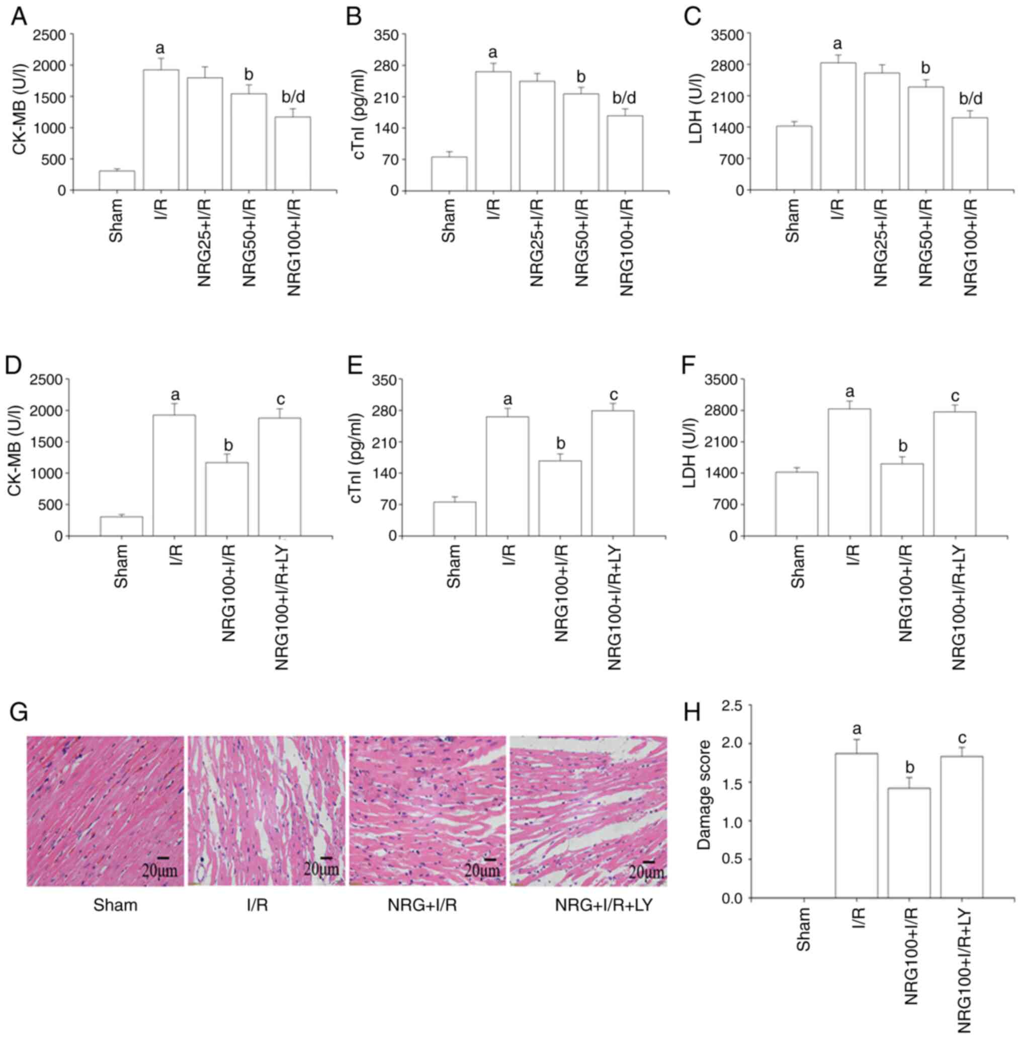 | Figure 1NRG pretreatment attenuates cardiac
I/R injury. Serum levels of (A) CK-MB, (B) cTnI and (C) LDH (n=5)
after I/R and following pretreatment with different NRG doses. The
expression levels of (D) CK-MB, (E) cTnI and (F) LDH (n=5) after
I/R with use of a PI3K/Akt inhibitor. (G) Representative images of
heart tissues following hematoxylin and eosin staining. (H) Damage
score (n=5). NRG, naringin; I/R, ischemia reperfusion; CK-MB,
creatine kinase myocardial band; cTn1, cardiac troponin 1; LDH,
lactate dehydrogenase; LY, PI3K/Akt inhibitor LY294002; NRG25, 25
mg/kg naringin; NRG50, 50 mg/kg naringin; NRG100, 100 mg/kg
naringin. aP<0.05 vs. sham group;
bP<0.05 vs. I/R group; cP<0.05 vs. I/R
group; dP<0.05 vs. NRG50 + I/R group. |
Anti-inflammatory and anti-oxidative
effects of NRG
The results of the present study suggested that I/R
triggered an inflammatory response by increasing the release of
inflammatory factors TNF-α, IL-1β and IL-6 (Fig. 2A-C). NRG pretreatment significantly
inhibited the release of inflammatory factors accelerated by I/R.
However, LY294002 partially abrogated the effects of NRG.
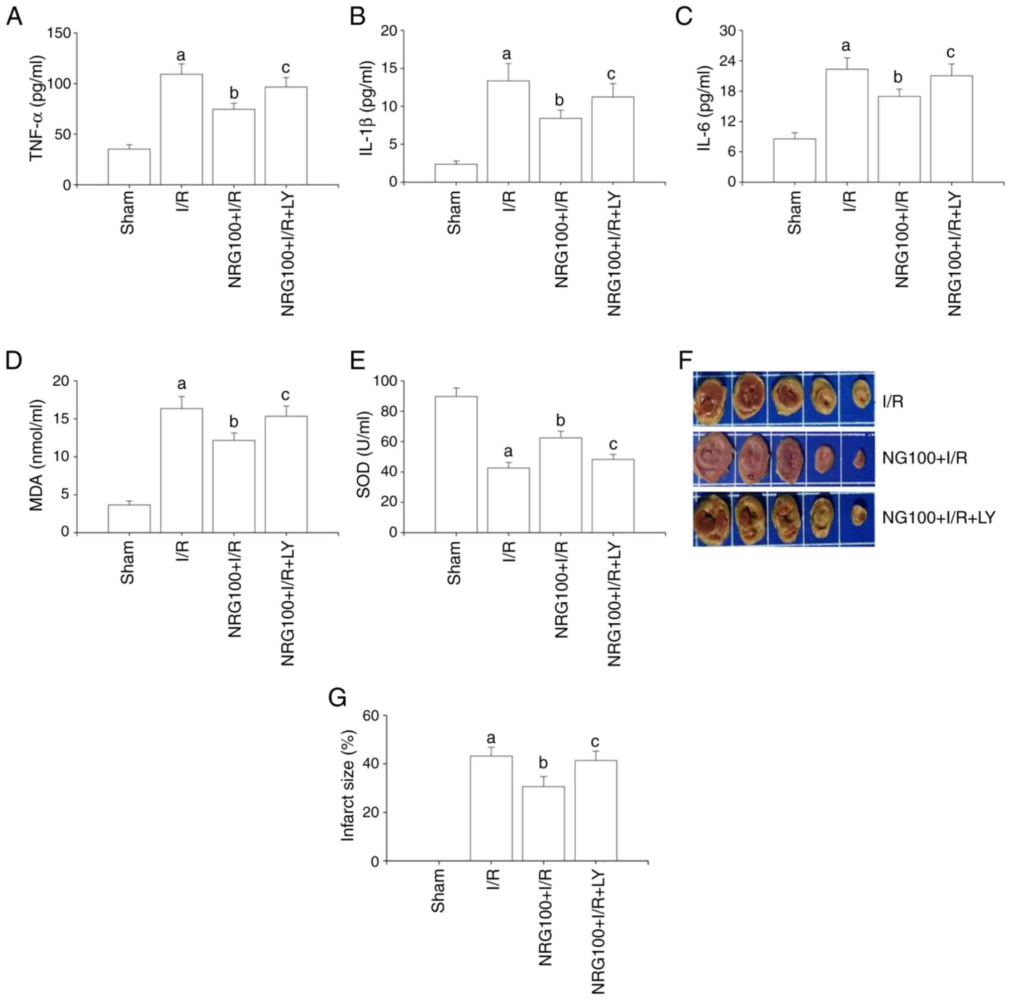 | Figure 2NRG pretreatment alleviates
I/R-accelerated inflammatory reactions, oxidative stress and
infarct size. The expression levels of (A) TNF-α, (B) IL-1β and (C)
IL-6 (n=5); (D) The level of MDA (n=5). (E) The level of SOD (n=5).
(F) Representative images of 2,3,5-triphenyltetrazolium chloride
stain. (G) Quantitative analysis of infarct size (n=3). NRG,
naringin; I/R, ischemia reperfusion; TNF-α, tumor necrosis
factor-α; IL, interleukin; MDA, malondialdehyde; SOD, superoxide
dismutase; NRG100, 100 mg/kg naringin; LY, PI3K/Akt inhibitor
LY294002. aP<0.05 vs. sham group;
bP<0.05 vs. I/R group; cP<0.05 vs.
NRG100 + I/R group. |
A significant reduction in SOD activity and MDA
concentration was observed in the NRG + I/R group in comparison
with I/R (Fig. 2D-E). However, the
effects of NRG were abolished by LY294002. These finding
demonstrated that NRG could mitigate I/R-induced oxidative stress
and inflammatory signaling.
Assessment of infarct size
As demonstrated in Fig.
2F and G, the infarct size in
the I/R group was significantly increased in comparison with the
sham group. However, NRG pretreatment notably reduced the infarct
size and these effects were abrogated by LY294002.
Effects of NRG on heart function
As displayed in Table
I, I/R significantly aggravated cardiac dysfunction by
enlarging LVIDs and impairing EF and FS. However, NRG pretreatment
markedly improved cardiac performance by narrowing LVIDs while
promoting FS. The administration of the PI3K/Akt inhibitor
LY294002, abrogated the effects of NRG.
 | Table IEffects of NRG on cardiac
performance. |
Table I
Effects of NRG on cardiac
performance.
| Group | HR (beats/min) | LVIDs (mm) | LVIDd (mm) | EF (%) | FS (%) |
|---|
| Sham | 442.32±16.32 | 2.41±0.31 | 4.98±0.18 | 84.62±3.21 | 59.48±2.69 |
| I/R | 429.85±15.75 |
3.24±0.36a | 5.02±0.21 |
55.36±4.23a |
32.25±1.38a |
| NRG100 + I/R | 432.48±17.63 |
2.95±0.25b | 5.07±0.23 |
66.31±2.41a |
37.62±1.57a |
| NRG100 + I/R
+LY | 425.17±19.32 |
3.19±0.32c | 5.04±0.20 |
53.36±4.06a |
31.24±1.45a |
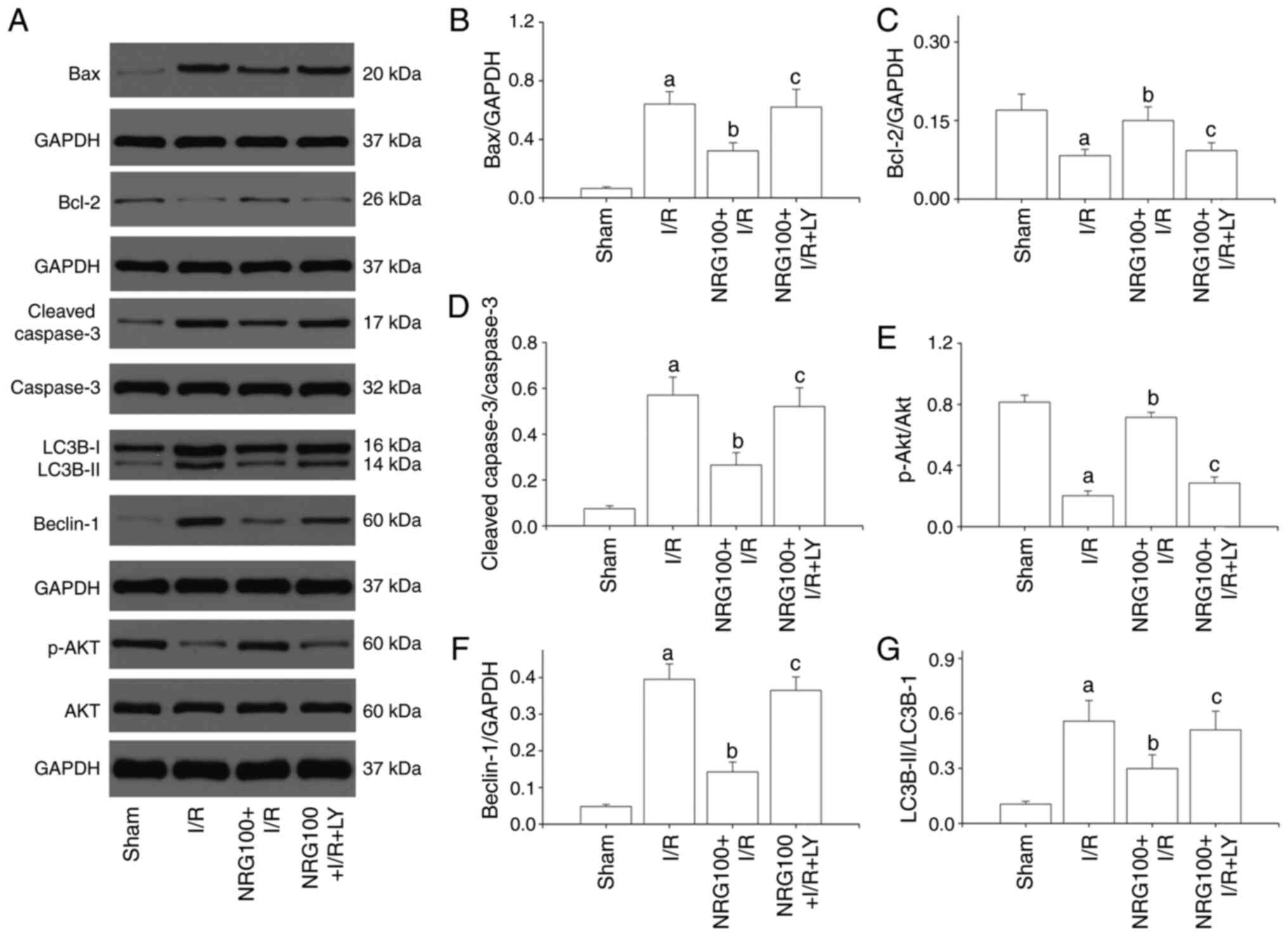
NRG pretreatment reduces myocardial
apoptosis
The present results showed that the AI (Fig. 3A-B) and the expression of cleaved
caspase3 were dramatically increased in the I/R group, while the
level of Bcl-2 was notably decreased in comparison with the sham
group (Fig. 4A-D). NRG pretreatment
markedly reduced AI and cleaved caspase3 expression and
significantly increased the level of Bcl-2 in comparison with the
I/R group. These results suggested that NRG could suppress I/R
induced myocardial apoptosis. However, LY294002 partially abolished
the cardioprotective effects of NRG.
NRG pretreatment activates the
PI3K/Akt signaling pathway
As shown in Fig. 4A
and E, I/R injury significantly
reduced the phosphorylation of Akt as compared to sham group.
However, NRG pretreatment remarkably promoted the phosphorylation
of Akt. Administration of LY294002 abrogated the effect of NRG,
indicating that NRG could activate the PI3K/Akt signaling
pathway.
NRG pretreatment inhibits myocardial
autophagy
Compared with the sham group, I/R significantly
increased the expression of Beclin-1 and the LC3BII/LC3BI ratio
(Fig. 4A, F and G).
However, I/R-induced increases in Benlin-1 expression and
LC3BII/LC3BI ratio were suppressed by NRG pretreatment, suggesting
that NRG could inhibit I/R-promoted myocardial autophagy. LY294002
partially abolished NRG-mediated anti-autophagy effect against
I/R.
Discussion
The results of the present study demonstrated that
NRG pretreatment could alleviate I/R-induced myocardial injury and
cardiac dysfunction by suppressing apoptosis, the inflammatory
response and oxidative stress. I/R-promoted myocardial autophagy
was also inhibited by NRG pretreatment. Furthermore, the
cardioprotective effects of NRG may be closely related to the
activation of the PI3K/Akt signaling cascades.
NRG is a pharmacologically active constituent of
tomentose pummelo peel, chemically known as
4,5,7-trihydroxy-flavonone-7-rhamnogglucoside (5). NRG has been reported to have multiple
cardioprotective effects. Rajadurai and Prince (21) demonstrated that NRG protected
against isoproterenol-induced myocardial infarction by increasing
the anti-oxidative capacity of the myocardium, as evidenced by the
enhanced levels of SOD, glutathione s-transferase and glutathione
peroxidase. Similar results were reported by Li et al
(22) who revealed that NRG could
suppress TNF-α-induced oxidative stress and the inflammatory
response via the PI3K/Akt pathway. I/R injury could significantly
promote oxidative stress thus reduces the activity of SOD (23,24).
It is known that Akt signaling plays a crucial role in the
modulation of multiple biological processes in cardiovascular
diseases. Numerous studies have demonstrated that pharmacological
activation of PI3K/Akt could effectively enhance the activities of
SOD against I/R injury (20,25).
Yang et al (26)
demonstrated that PI3K/Akt cascades could facilitate Nrf2 nuclear
translocation and therefore promote SOD activity in cardiomyocytes
after hypoxia-reoxygenation procedures. Sun et al (27) found that PI3K/Akt signaling could
notably activates the endothelial nitric oxide synthase pathway to
enhance the anti-oxidative capacity of cardiomyocytes against I/R
injury.
Myocardial I/R injury occurs with the restoration of
blood flow to the occluded coronary artery (7). Myocardial apoptosis, the inflammatory
response, oxidative stress and autophagy constitute crucial parts
in the pathophysiological procedure of I/R injury (7). Rani et al (13). demonstrated that NRG significantly
attenuated myocardial I/R injury by diminishing cardiac apoptosis
as well as oxidative stress. A study by Chen et al (6) reported that NRG pretreatment mitigated
anoxia/reoxygenation-induced apoptosis via the Nrf2 pathway in
vitro. Consistent with these findings, the results of the
present study also showed that NRG exerted cardioprotective effects
against I/R injury via a reduction in the levels of pro-apoptotic
proteins and oxidative stress, while promoting the expression of
anti-apoptotic protein Bcl-2.
Myocardial I/R injury is also characterized by an
excessive level of inflammation response (28). Inflammatory mediators such as IL-1β,
IL-6 and TNF-α could significantly contribute to the myocardial
dysfunction and structural alterations in the failing heart
(29). Additionally, TNF-α could
further exert deleterious effects on myocardial I/R injury by
promoting oxidative stress and the inflammatory response (30). Tong et al (20) demonstrated that I/R markedly
increased the release of inflammatory cytokines, while the
activation of the PI3K/Akt pathway could significantly repress
I/R-mediated inflammation via down-regulation of high mobility
group box protein. In sepsis-associated myocarditis, NRG also
displayed anti-inflammatory effects (7). The above evidence supported the
hypothesis that NRG could attenuate I/R injury via effectively
inhibiting inflammatory response.
Autophagy is a dynamic process in which cellular
waste is encapsulated into double membrane vesicles and then
transported to lysosomes for degradation (31). A normal state of autophagy is
necessary for the renewal of organelles (32). The signaling pathways involved in
autophagy have been extensively studied and the PI3K/Akt pathway is
widely recognized as a crucial mediator in the regulation of
autophagy (33,34). Li et al (35) demonstrated that pharmacological
activation of PI3K/Akt could effectively suppress
hypoxia/reoxygenation-induced autophagy in neonatal rats by
targeting rapamycin. Ye et al (12) also confirmed that phosphorylated Akt
could attenuate I/R-promoted myocardial autophagy via secretion of
basic fibroblast growth factors. LC3BII/LC3BI ratio and Beclin-1
are representative autophagy markers, and their levels can directly
reflect the degree of autophagy (36,37).
Consistent with the aforementioned studies, the results of the
present study indicated that I/R injury significantly promoted the
expression of Beclin-1 and LC3BII/LC3BI ratio, while NRG
pretreatment markedly reversed the up-regulated Beclin-1 and
LC3BII/LC3BI ratio. However, these cardioprotective effects of NRG
were partially abolished by the PI3K/Akt inhibitor LY294002. These
results suggested that NRG may suppress I/R-induced myocardial
autophagy by facilitating the PI3K/Akt signaling pathway.
There are a number of limitations to the present
study. Specific PI3K/Akt inhibitors, such as Dactolisib or IC87114,
might better have illustrated the cardioprotective role of NRG
against I/R injury and an LY treatment alone group could also have
aided the interpretation of the results.
In summary, the results of the present study
indicated that NRG alleviated myocardial I/R injury via inhibition
of cardiac apoptosis, inflammatory reactions and oxidative stress.
In addition, NRG pretreatment also attenuated I/R-induced
autophagy. The present data also suggested that such
cardioprotective effects of NRG could be ascribed to the activation
of the PI3K/Akt pathway. Furthermore, short-term NRG pretreatment
may be a novel therapeutic strategy for the prevention of I/R
injury in AMI patients.
Acknowledgements
Not applicable.
Funding
Funding: The present study was supported by a grant from the
Natural Science Foundation of Hubei Province of China (grant no.
2015CFB223).
Availability of data and materials
The datasets used and/or analyzed during the current
study are available from the corresponding author on reasonable
request.
Authors' contributions
FL and ZZ were responsible for conceiving the study,
performing the analysis and experiments and drafting the
manuscript. JQ analyzed the data. CC and WY also performed the
experiments. NW was involved in drafting the manuscript performing
and designing the experiments as well as revising it for important
intellectual content. FL and NW confirm the authenticity of all the
raw data. All authors have read and approved the final
manuscript.
Ethics approval and consent participate
Animal experiments were approved by the
Institutional Animal Care and Use Committee of Hubei University of
Medicine.
Patient consent for publication
Not applicable.
Competing interest
The authors declare that they have no competing
interests.
References
|
1
|
Heusch G and Gersh BJ: The pathophysiology
of acute myocardial infarction and strategies of protection beyond
reperfusion: A continual challenge. Eur Heart J. 38:774–784.
2017.PubMed/NCBI View Article : Google Scholar
|
|
2
|
Wang BF and Yoshioka J: The emerging role
of thioredoxin-Interacting Protein in Myocardial
Ischemia/Reperfusion Injury. J Cardiovasc Pharmacol Ther.
22:219–229. 2017.PubMed/NCBI View Article : Google Scholar
|
|
3
|
Wang P, Sun J, Lv S, Xie T and Wang X:
Apigenin alleviates myocardial reperfusion injury in rats by
downregulating miR-15b. Med Sci Monit. 25:2764–2776.
2019.PubMed/NCBI View Article : Google Scholar
|
|
4
|
Shen Y, Liu X, Shi J and Wu X: Involvement
of Nrf2 in myocardial ischemia and reperfusion injury. Int J Biol
Macromol. 125:496–502. 2019.PubMed/NCBI View Article : Google Scholar
|
|
5
|
Jian CY, Ouyang HB, Xiang XH, Chen JL, Li
YX, Zhou X, Wang JY, Yang Y, Zhong EY, Huang WH, et al: Naringin
protects myocardial cells from doxorubicin induced apoptosis
partially by inhibiting the p38MAPK pathway. Mol Med Rep.
16:9457–9463. 2017.PubMed/NCBI View Article : Google Scholar
|
|
6
|
Chen RC, Sun GB, Wang J, Zhang HJ and Sun
XB: Naringin protects against anoxia/reoxygenation-induced
apoptosis in H9c2 cells via the Nrf2 signaling pathway. Food Funct.
6:1331–1344. 2015.PubMed/NCBI View Article : Google Scholar
|
|
7
|
Xianchu L, Lan PZ, Qiufang L, Yi L,
Xiangcheng R, Wenqi H and Yang D: Naringin protects against
lipopolysaccharide-induced cardiac injury in mice. Environ Toxicol
Pharmacol. 48:1–6. 2016.PubMed/NCBI View Article : Google Scholar
|
|
8
|
Li YP, Chen Z and Cai YH: Piperine
protects against myocardial ischemia/reperfusion injury by
activating the PI3K/AKT signaling pathway. Exp Ther Med.
21(374)2021.PubMed/NCBI View Article : Google Scholar
|
|
9
|
Zeng B, Liu L, Liao X, Zhang C and Ruan H:
Thyroid hormone protects cardiomyocytes from H2O2-induced oxidative
stress via the PI3K-AKT signaling pathway. Exp Cell Res.
380:205–215. 2019.PubMed/NCBI View Article : Google Scholar
|
|
10
|
Shaker ME, Ashamallah SA and Houssen ME:
Celastrol ameliorates murine colitis via modulating oxidative
stress, inflammatory cytokines and intestinal homeostasis. Chem
Biol Interact. 210:26–33. 2014.PubMed/NCBI View Article : Google Scholar
|
|
11
|
Wu S, Chang G, Gao L, Jiang D, Wang L, Li
G, Luo X, Qin S, Guo X and Zhang D: Trimetazidine protects against
myocardial ischemia/reperfusion injury by inhibiting excessive
autophagy. J Mol Med (Berl). 96:791–806. 2018.PubMed/NCBI View Article : Google Scholar
|
|
12
|
Ye G, Fu Q, Jiang L and Li Z: Vascular
smooth muscle cells activate PI3K/Akt pathway to attenuate
myocardial ischemia/reperfusion-induced apoptosis and autophagy by
secreting bFGF. Biomed Pharmacother. 107:1779–1785. 2018.PubMed/NCBI View Article : Google Scholar
|
|
13
|
Rani N, Bharti S, Manchanda M, Nag TC, Ray
R, Chauhan SS, Kumari S and Arya DS: Regulation of heat shock
proteins 27 and 70, p-Akt/p-eNOS and MAPKs by Naringin Dampens
myocardial injury and dysfunction in vivo after
ischemia/reperfusion. PLoS One. 8(e82577)2013.PubMed/NCBI View Article : Google Scholar
|
|
14
|
Sherry EV: Review of the third edition of
the Guide for the Care and Use of Agricultural Animals in Research
and Teaching. J Am Assoc Lab Anim Sci. 51(3)(298
300)2012.PubMed/NCBI
|
|
15
|
Cerkezkayabekir A, Sanal F, Bakar E,
Ulucam E and Inan M: Naringin protects viscera from
ischemia/reperfusion injury by regulating the nitric oxide level in
a rat model. Biotech Histochem. 92:252–263. 2017.PubMed/NCBI View Article : Google Scholar
|
|
16
|
Jiang X, Kong B, Shuai W, Shen C, Yang F,
Fu H, Huang H, Jiang X, Kong B, Shuai W, et al: Loss of MD1
exacerbates myocardial ischemia/reperfusion injury and
susceptibility to ventricular arrhythmia. Eur J Pharmacol.
844:79–86. 2019.PubMed/NCBI View Article : Google Scholar
|
|
17
|
Lee JC and Sponenberg DP: Role of alpha
1-adrenoceptors in norepinephrine-induced cardiomyopathy. Am J
Pathol. 121:316–321. 1985.PubMed/NCBI
|
|
18
|
Sepúlveda M, Gonano LA, Viotti M, Morell
M, Blanco P, López Alarcón M, Peroba Ramos I, Bastos Carvalho A,
Medei E and Vila Petroff M: Calcium/Calmodulin protein kinase
II-dependent ryanodine receptor phosphorylation mediates cardiac
contractile dysfunction associated with sepsis. Crit Care Med.
45:e399–e408. 2017.PubMed/NCBI View Article : Google Scholar
|
|
19
|
Wang N, Zheng X, Qian J, Yao W, Bai L, Hou
G, Qiu X, Li X and Jiang X: Renal sympathetic denervation
alleviates myocardial fibrosis following isoproterenol-induced
heart failure. Mol Med Rep. 16:5091–5098. 2017.PubMed/NCBI View Article : Google Scholar
|
|
20
|
Tong S, Zhang L, Joseph J and Jiang X:
Celastrol pretreatment attenuates rat myocardial ischemia/
reperfusion injury by inhibiting high mobility group box 1 protein
expression via the PI3K/Akt pathway. Biochem Biophys Res Commun.
497:843–849. 2018.PubMed/NCBI View Article : Google Scholar
|
|
21
|
Rajadurai M and Prince PS: Naringin
ameliorates mitochondrial lipid peroxides, antioxidants and lipids
in isoproterenol-induced myocardial infarction in Wistar rats.
Phytother Res. 23:358–362. 2009.PubMed/NCBI View
Article : Google Scholar
|
|
22
|
Li W, Wang C, Peng J, Liang J, Jin Y, Liu
Q, Meng Q, Liu K and Sun H: Naringin inhibits TNF-α induced
oxidative stress and inflammatory response in HUVECs via Nox4/NF-κB
and PI3K/Akt pathways. Curr Pharm Biotechnol. 15:1173–1182.
2014.PubMed/NCBI View Article : Google Scholar
|
|
23
|
Wu Q, Lu K, Zhao Z, Wang B, Liu H, Zhang
S, Liao J, Zeng Y, Dong Q, Zhao N, et al: Blockade of transient
receptor potential vanilloid 4 enhances antioxidation after
myocardial ischemia/reperfusion. Oxid Med Cell Longev.
2019(7283683)2019.PubMed/NCBI View Article : Google Scholar
|
|
24
|
Liu Z, Tao B, Fan S, Pu Y, Xia H and Xu L:
MicroRNA 145 protects against myocardial ischemia reperfusion
injury via CaMKII mediated anti apoptotic and anti inflammatory
pathways. Oxid Med Cell Longev. 2019(8948657)2019.PubMed/NCBI View Article : Google Scholar
|
|
25
|
Dong J, Xu M, Zhang W and Che X: Effects
of sevoflurane pretreatment on myocardial ischemia-reperfusion
injury through the Akt/hypoxia-inducible factor 1-alpha
(HIF-1α)/vascular endothelial growth factor (VEGF) signaling
pathway. Med Sci Monit. 25:3100–3107. 2019.PubMed/NCBI View Article : Google Scholar
|
|
26
|
Yang P, Zhou Y, Xia Q, Yao L and Chang X:
Astragaloside IV regulates the PI3K/Akt/HO-1 signaling pathway and
inhibits H9c2 cardiomyocyte injury induced by
hypoxia-reoxygenation. Biol Pharm Bull. 42:721–727. 2019.PubMed/NCBI View Article : Google Scholar
|
|
27
|
Sun Y, Jiang C, Jiang J and Qiu L:
Dexmedetomidine protects mice against myocardium
ischaemic/reperfusion injury by activating an AMPK/PI3K/Akt/eNOS
pathway. Clin Exp Pharmacol Physiol. 44:946–953. 2017.PubMed/NCBI View Article : Google Scholar
|
|
28
|
Liu S, He Y, Shi J, Liu L, Ma H, He L and
Guo Y: Allicin attenuates myocardial ischemia reperfusion injury in
rats by inhibition of inflammation and oxidative stress. Transplant
Proc. 51:2060–2065. 2019.PubMed/NCBI View Article : Google Scholar
|
|
29
|
Marchant DJ, Boyd JH, Lin DC, Granville
DJ, Garmaroudi FS and McManus BM: Inflammation in myocardial
diseases. Circ Res. 110:126–144. 2012.PubMed/NCBI View Article : Google Scholar
|
|
30
|
Buja LM: Myocardial ischemia and
reperfusion injury. Cardiovasc Pathol. 14:170–175. 2005.PubMed/NCBI View Article : Google Scholar
|
|
31
|
Mizushima N and Komatsu M: Autophagy:
Renovation of cells and tissues. Cell. 147:728–741. 2011.PubMed/NCBI View Article : Google Scholar
|
|
32
|
Hang P, Zhao J, Su Z, Sun H, Chen T, Zhao
L and Du Z: Choline inhibits ischemia-reperfusion-induced
cardiomyocyte autophagy in rat myocardium by activating Akt/mTOR
signaling. Cell Physiol Biochem. 45:2136–2144. 2018.PubMed/NCBI View Article : Google Scholar
|
|
33
|
Shanware NP, Bray K and Abraham RT: The
PI3K, metabolic, and autophagy networks: Interactive partners in
cellular health and disease. Annu Rev Pharmacol Toxicol. 53:89–106.
2013.PubMed/NCBI View Article : Google Scholar
|
|
34
|
Tang H, Song X, Ling Y, Wang X, Yang P,
Luo T and Chen A: Puerarin attenuates myocardial
hypoxia/reoxygenation injury by inhibiting autophagy via the Akt
signaling pathway. Mol Med Rep. 15:3747–3754. 2017.PubMed/NCBI View Article : Google Scholar
|
|
35
|
Li X, Zhu Q, Liu Y, Yang Z and Li B:
Gastrodin protects myocardial cells against hypoxia/reoxygenation
injury in neonatal rats by inhibiting cell autophagy through the
activation of mTOR signals in PI3K-Akt pathway. J Pharm Pharmacol.
70:259–267. 2018.PubMed/NCBI View Article : Google Scholar
|
|
36
|
Jiang P and Mizushima N: LC3- and
p62-based biochemical methods for the analysis of autophagy
progression in mammalian cells. Methods. 75:13–18. 2015.PubMed/NCBI View Article : Google Scholar
|
|
37
|
Zhou M, Zou YG, Xue YZ, Wang XH, Gao H,
Dong HW and Zhang Q: Long non-coding RNA H19 protects acute
myocardial infarction through activating autophagy in mice. Eur Rev
Med Pharmacol Sci. 22:5647–5651. 2018.PubMed/NCBI View Article : Google Scholar
|