Introduction
Dragon blood is a type of deep-red resin that has
been used as a form of traditional medicine worldwide since ancient
times (1). Families of plants with
the ability to produce dragon blood can be classified into four
different categories: Asparagaceae, Arecaceae, Chamaesyce and
Fabaceae (Table I) (2-4).
The earliest record of dragon blood being used for medicine was
performed using the plant genus Dracaena in Socotra Island
in Yemen (5). Due to being situated
in ideal location and the rapid growth rate of the palm tree
species Daemonorops draco, dragon blood extracted from this
tree became the mainstream product in the traditional Chinese
medicine (TCM) market after the Ming Dynasty in China (6). Therefore, red resin from
Daemonorops draco is recognized as authentic dragon blood in
the official documents of China, Hong Kong and Taiwan (7-9).
In particular, dracorhodin has become the reference standard of
dragon blood from the palm tree D. draco (10), where the content of dracorhodin in
dragon blood for medical use is recommended to be >1% (9).
 | Table IPlant species that contain dragon
blood and their respective geographical origins (4). |
Table I
Plant species that contain dragon
blood and their respective geographical origins (4).
| Plant family | Plant species | Origin |
|---|
| Asparagaceae | Dracaena
cinnabari | Socotra Island,
Yemen |
| | Dracaena
draco | Canary Islands,
Spain |
| | Dracaena
cochinchinensis | Yunnan Province,
China |
| Arecaceae | Daemonorops
draco | Indonesia and
Malaysia |
| Chamaesyce | Croton
salutaris | |
| | C.
lechleri | Amazon basin |
| | C.
draconoides | |
| Fabaceae | Pterocarpus
draco | India |
| | P.
marsupium | |
Dragon blood has been included in recipes for wound
treatment in various ancient traditional Chinese medical guides of
the 14-18th century (6).
Accumulating evidence has also indicated that dragon blood
possesses wound healing activities both in vivo and in
vitro (1,11-13).
Accordingly, a previous study showed that the herbal complex
‘Jinchuang Ointment’ can successfully treat non-healing wounds on
patients with diabetes, where dragon blood from D. draco is
one of the components (2.1%) in this ointment (14).
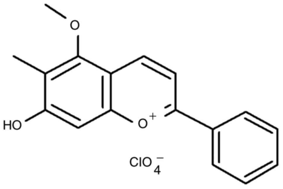
Dracorhodin perchlorate is a synthetic chemical
analog of dracorhodin (Fig. 1)
(10,15-18)
that has been shown to exhibit angiogenic activity on human
umbilical vein endothelial cells (10,19).
Recently, both dracorhodin perchlorate and methanol extracts of
crude dragon blood have demonstrated in vivo pro-angiogenic
activity in zebrafish embryos (10). Additionally, previous studies have
revealed bioactivities of dracorhodin perchlorate in human cancer
cells, where it can induce apoptotic cell death in different types
of cancer cells, including U87MG and T98G glioma cells (20), SK-MES-1 human lung squamous
carcinoma cells (21), MCF-7 human
breast cancer cells (22), SGC-7901
human gastric tumor cells (23),
PC-3 human prostate cancer cells (18), HL-60 leukemia cells (16), A375-S2 human melanoma cells
(24) and HeLa human cervical
cancer cells (15,25). Furthermore, dracorhodin perchlorate
was reported to promote NIH-3T3 fibroblast cell proliferation in
vitro and induce rat wound healing in vivo (26). Dracorhodin perchlorate has also been
shown to accelerate skin wound healing in Wistar rats in
vivo (27). However, the
effects of dracorhodin perchlorate on wound healing in human HaCaT
keratinocytes remain poorly understood. Therefore, in the present
study the underlying mechanism of the wounding healing regulation
and associated signaling pathways mediated by dracorhodin
perchlorate in HaCaT keratinocytes was investigated in
vitro.
Materials and methods
Reagents and chemicals
DMEM, FBS, penicillin/streptomycin and L-glutamine
were purchased from HyClone, Cytiva. All primary antibodies and
anti-mouse/-rabbit immunoglobulin IgG HRP-linked secondary
antibodies were procured from GeneTex International Corporation.
Reagent grade DMSO, thiazolyl blue tetrazolium bromide (MTT), PBS,
propidium iodide (PI), wortmannin (an AKT inhibitor), U0126 (an ERK
inhibitor) and SB203580 (a p38 inhibitor), were obtained from
Sigma-Aldrich, Merck KGaA unless otherwise specified. Dracorhodin
perchlorate (batch no. 110811-201506; purity, 98.6%) was purchased
from the National Institute for the Control of Pharmaceutical and
Biological Products (Beijing, China), which was dissolved in DMSO
and stored at -20˚C freezer before use. The vehicle control was
kept below 0.1% DMSO in culture medium.
Cell culture
Human skin HaCaT keratinocytes were obtained from
CLS Cell Lines Service GmbH and cultured in DMEM supplemented with
10% FBS, 100 U/ml penicillin, 100 µg/ml streptomycin and 2 mM
L-glutamine in a humidified atmosphere at 37˚C in 5%
CO2/95% air as previously described (28,29).
HaCaT cells were cultured until passage 40 (20 weeks) and did not
exceed 40 generations.
Dynamic cell migration assay
HaCaT cells (1x10⁴ cells/well) were
seeded into a 96-well plate overnight before the wound area
(700-800-µm wide) was created using Incucyte 96-Well Woundmaker
Tool (cat. no. 4563; Essen BioScience). The scratched cells were
then exposed to 0, 1 and 2 µg/ml dracorhodin perchlorate in
serum-free DMEM (HyClone; Cytiva). The cell migration experiment
was conducted over 24 h with data collection every 3 h at 37˚C. The
cell images and wound width were monitored using the IncuCyte ZOOM
System instrument by light microscopy (magnification, x100)
[(Incucyte S3 Live-Cell Analysis System and Incucyte Scratch Wound
Analysis Software Module (cat. no. 9600-0012; Essen BioScience)] as
previously described (30,31).
Cell viability assay
HaCaT cells (2.5x10⁵ cells/well) were
plated into 24-well plates and treated with or without 0.5, 1 and 2
µg/ml dracorhodin perchlorate for 24 h at 37˚C. Cell viability was
determined using MTT assay as previously described (32-34).
In brief, MTT solution (0.5 mg/ml) was added to each well for 2 h
at 37˚C before the blue formazan crystals were dissolved in DMSO
(500 µl/well) by constant shaking for 10 min. The absorbance of
each well was measured using an ELISA plate reader at a test
wavelength of 570 nm with a reference wavelength of 620 nm.
For the PI exclusion assay, 2.5x10⁵
cells/well were harvested and plated into 24-well plates and
resuspended in 500 µl of 5 µg/ml PI for 5 min at 4˚C. The numbers
of viable cells and dead cells were determined using a BD
FACSCalibur™ Flow Cytometry System and BD CellQuest Pro Software
version 6.0 (both BD Biosciences), where the PI dye solution was
applied to specifically stain dead cells. Cell viability was
calculated as follows: % Cell viability = (PIem of test)/(PIem of
control) x100%. Where, PIem = gated cells of PI fluorescence
emission (<102 fluorescence).
Western blot analysis
HaCaT cells (5x10⁶ cells per 75T flask)
were incubated with or without 1 and 2 µg/ml dracorhodin
perchlorate for 12 h at 37˚C. Cell samples were lysed in Trident
RIPA Lysis Buffer (GeneTex International Corporation) and collected
as previously described (35,36).
Protein concentration was determined using the Pierce bicinchoninic
acid protein assay kit (Thermo Fisher Scientific, Inc.). Equal
amounts of the protein sample (40 µg) were prepared, and 10-12%
SDS-PAGE was performed. Proteins were transferred onto an
Immobilon-P polyvinylidene difluoride transfer membrane prior to
blocking with Trident Universal Protein Blocking Reagent (GeneTex
International Corporation) for 1 h at room temperature. The
membrane was subsequently incubated with primary antibodies against
β-catenin (cat. no. GTX101435), phosphorylated (p)-AKT (Ser473)
(cat. no. GTX28932), AKT (cat. no. GTX121937), p-ERK (cat. no.
GTX59568), ERK (cat. no. GTX59618), p38 (cat. no. GTX110720), p-p38
(cat. no. GTX48614) and β-actin (cat. no. GTX109639) at a dilution
of 1:1,000 at 4˚C overnight. The membranes were then incubated with
the appropriate anti-mouse (cat. no. GTX213111-01) and anti-rabbit
(cat. no. GTX213110-01) IgG HRP-linked secondary antibodies at a
dilution of 1:10,000 for 1 h at room temperature. Blot
visualization was performed using the Immobilon Western
Chemiluminescent HRP Substrate (Merck KGaA) and all bands of
immunoblots were normalized to the densitometric value of β-actin.
These experiments were conducted in duplicate. The bands were
quantified by densitometry using ImageJ software version 1.41
(National Institutes of Health) as previously described (33,37-39).
Sandwich ELISA assay for p-protein
kinases
HaCaT cells (5x10⁶ cells per T75 flask)
were treated with or without 0.5, 1 and 2 µg/ml dracorhodin
perchlorate for 12 h at 37˚C. The cells were harvested and total
proteins were collected using Trident RIPA Lysis Buffer (GeneTex
International Corporation). The samples were incubated in
microwells coated with the appropriate antibody for 2 h at 37˚C
(for p-ERK) or overnight at 4˚C (for p-Akt) according to the
manufacturer's protocols of the PathScan Sandwich ELISA kits [p-Akt
(Thr308; cat. no. 7252C) and p-ERK (Thr202/Tyr204; cat. no. 7177C);
Cell Signaling Technology, Inc.]. The secondary antibodies and the
reagents used for the chemical reactions were provided in these
kits. Absorbance was measured using an ELISA reader (Anthos 2001;
Anthos Labtec Instruments GmbH) at a wavelength of 450 nm, as
previously described (38-40).
Wound healing assay
HaCaT cells were placed in a six-well tissue culture
plates for 24 h at 37˚C and cultured to 90% confluence. Individual
wells were then scratched with a 200-µl micropipette tip to create
a denuded zone of constant width (1 mm). Cells were then cultured
in serum-free DMEM (HyClone; Cytiva) and incubated with or without
dracorhodin perchlorate (1 µg/ml) or with specific protein kinase
inhibitors [10 µM wortmannin (AKT inhibitor), 10 µM U0126 (ERK
inhibitor) or 10 µM SB203580 (p38 inhibitor)] and 0.1% DMSO as a
control for 24 h at 37˚C. To determine cell migration, cell images
were captured after 24 h treatment under phase-contrast microscopy
(magnification, x100; Leica DMIL microscope; type 090-135.001;
Leica Microsystems GmbH), as previously described (38). Image analysis was performed by using
AxioVision LE64 software (version 4.9.1.0; Carl Zeiss AG). The
ability of cell migration was calculated as follows: Cell migration
(% of control) = [Gw (0 h) of test-Gw (24 h) of test]/[Gw (0 h) of
control-Gw (24 h) of control] x100%. Where, Gw (0 h) = gap width at
0 h, and Gw (24 h) = gap width at 24 h.
Statistical analysis
All results are expressed as the mean ± standard
deviation (three experimental repeats per assay). Differences among
groups were determined by one-way analysis of variance followed by
Tukey's or Dunnett's post hoc test with SPSS software version 16.0
(SPSS, Inc.). P<0.05 or P<0.001 was considered to indicate a
statistically significant difference.
Results
Dracorhodin perchlorate promotes cell
migration in HaCaT keratinocytes
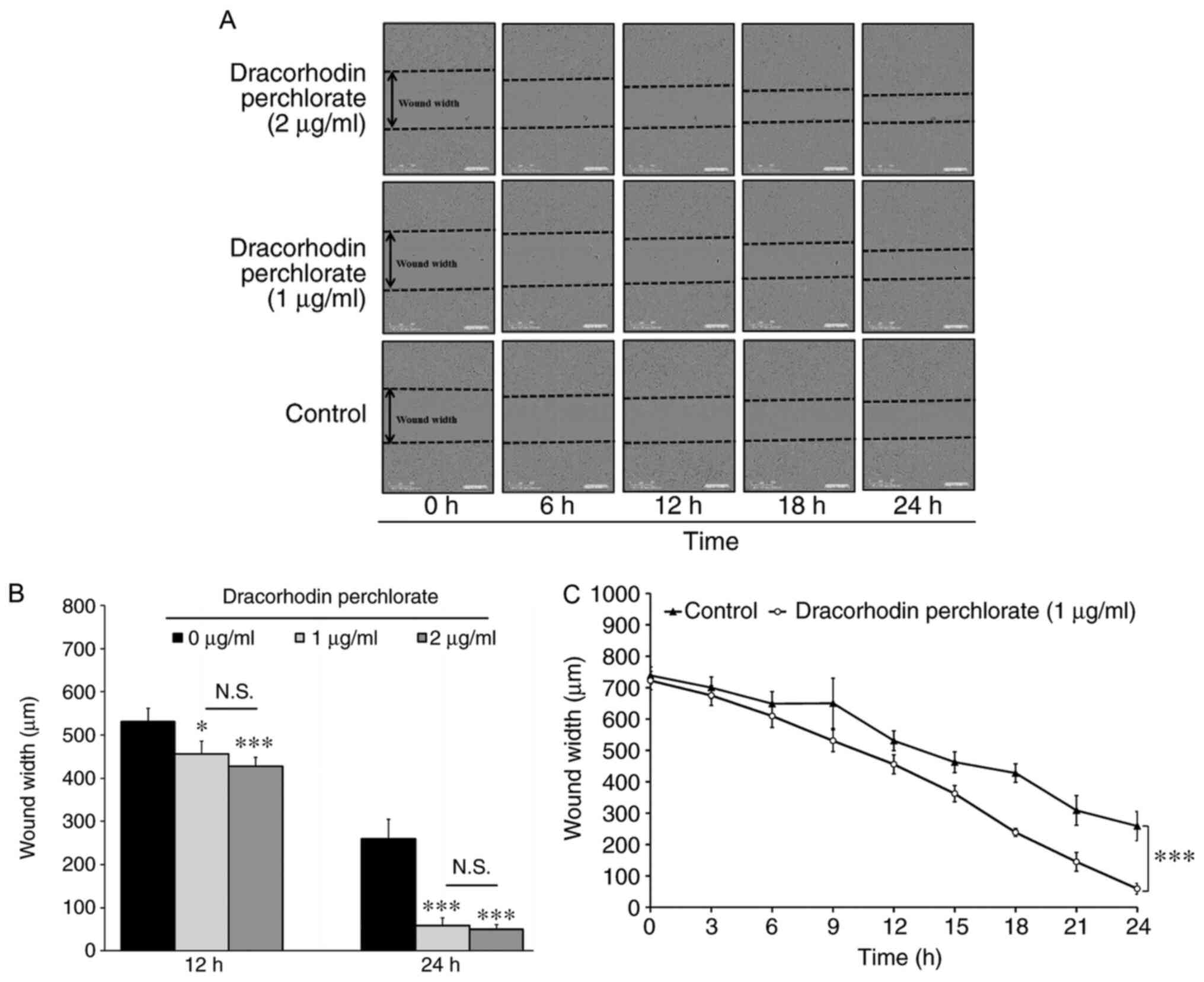
Wound healing activity was first investigated, which
mimics the migration of keratinocytes toward wound margins in
vitro. As shown in Fig. 2A, the
wound was closing progressively following treatment with
dracorhodin perchlorate in HaCaT cells. No significant difference
was found between 1 and 2 µg/ml groups. Therefore, the optimal
dracorhodin perchlorate concentration to monitor wound healing
activity was found to be 1 µg/ml (Fig.
2B). Quantitative data indicated that dracorhodin perchlorate
at 1 µg/ml significantly reduced the wound width at the time point
of 24 h and markedly promoted HaCaT cell migration compared with
those in control cells (Fig. 2C).
Therefore, this suggests that dracorhodin perchlorate enhanced cell
migration in HaCaT keratinocytes.
Dracorhodin perchlorate does not
enhance cell viability in HaCaT keratinocytes
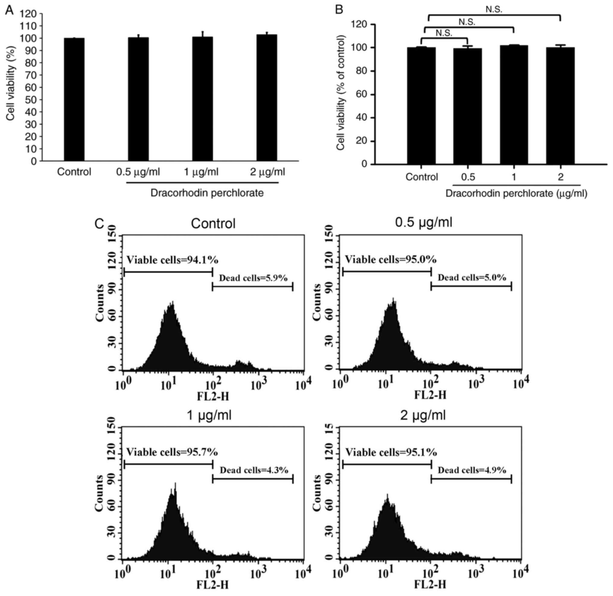
As previously reported, the cell viability of human
primary fibroblast proliferation significantly increased by
dracorhodin perchlorate treatment (41). Therefore, MTT assay and PI exclusion
method were performed to investigate the cell viability and
cytotoxicity of HaCaT keratinocytes after dracorhodin perchlorate
exposure. Dracorhodin perchlorate did not affect the viability of
HaCaT cells at the highest concentration tested in this study,
which was 2 µg/ml (Fig. 3A). As
shown in Fig. 3B and C, viable cells were generally excluded
from PI because of low emission fluorescence. No significant change
was observed in dracorhodin perchlorate-treated HaCaT keratinocytes
(Fig. 3B). Moreover, flow cytometry
results showed that the total counts of dead and viable cells were
<10 and >90%, respectively (Fig.
3C). Thus, the migration ability of HaCaT cells caused by
dracorhodin perchlorate was not due to the cell viability of HaCaT
keratinocytes.
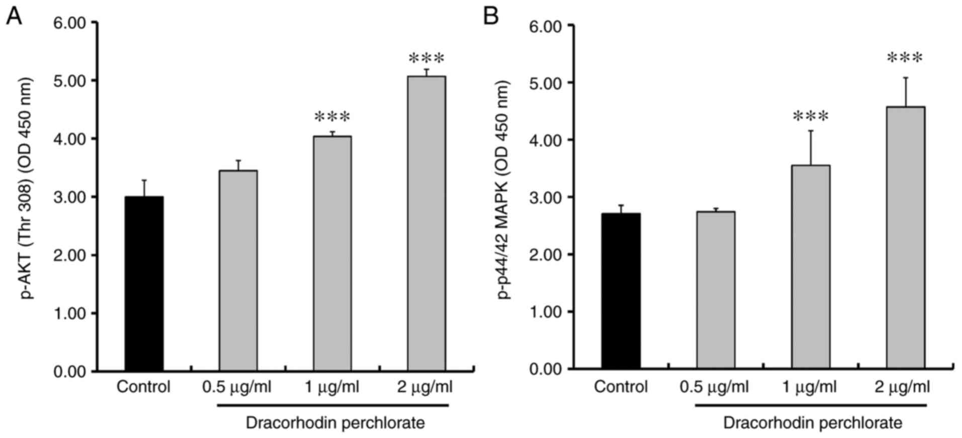
Dracorhodin perchlorate increases the
protein expression of β-catenin and phosphorylation of Akt, p38 and
ERK in HaCaT keratinocytes
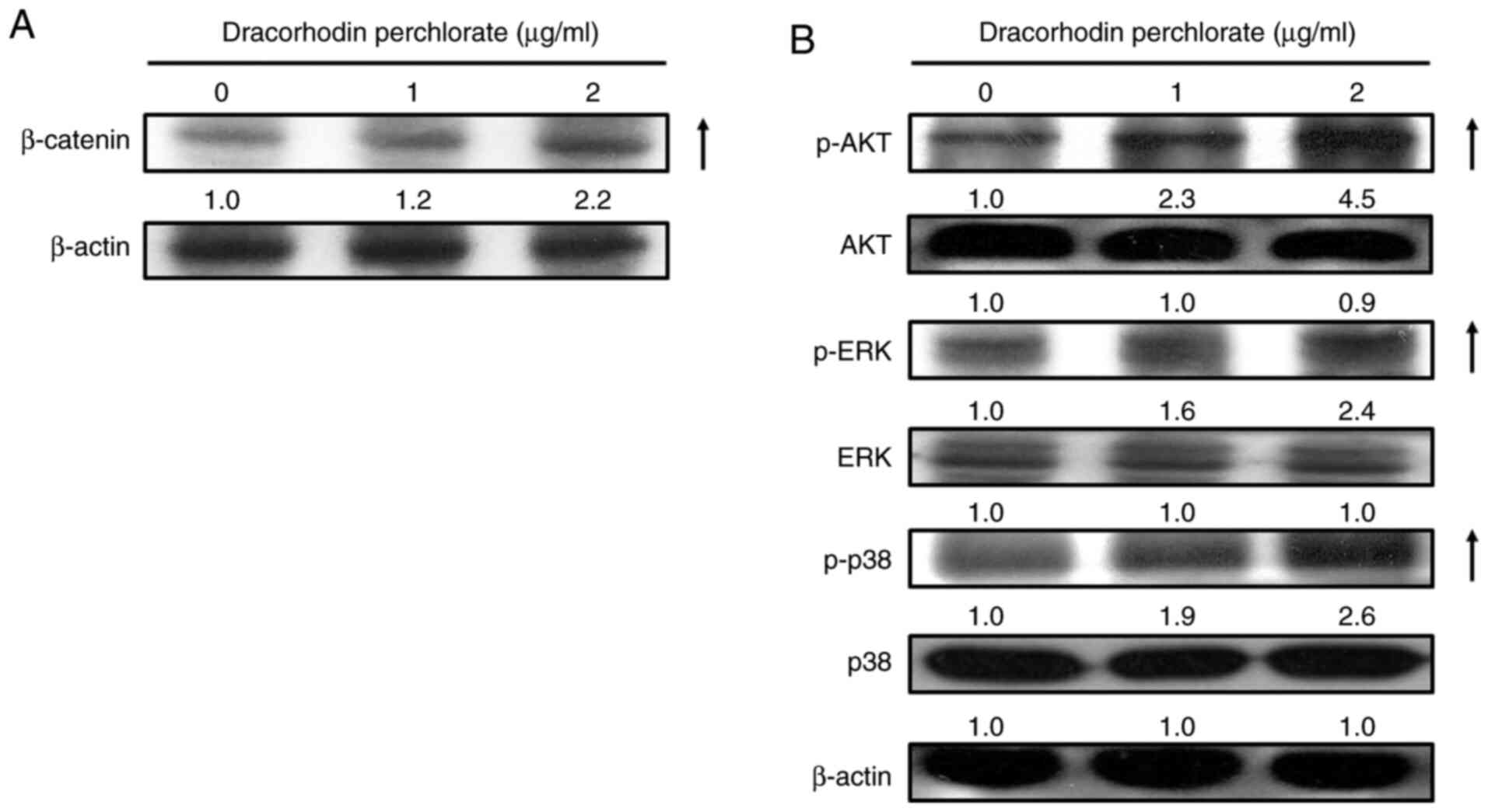
Previous studies have suggested that wound healing
is closely associated with β-catenin signaling (42,43),
where keratinocyte mobility can be stimulated through the MAPK and
AKT signaling pathways (44-46).
Western blotting experiments were therefore performed to
investigate the effects of dracorhodin perchlorate treatment on the
protein expression of β-catenin and both phosphorylation of ERK and
AKT. Dracorhodin perchlorate treatment (2 µg/ml for 12 h) markedly
upregulated the protein levels of β-catenin, phosphorylated ERK,
phosphorylated Akt and phosphorylated p38 in HaCaT keratinocytes
compared with those in control cells (Fig. 4). This was subsequently verified
using sandwich ELISA. The levels of both phosphorylated AKT and ERK
significantly increased after dracorhodin perchlorate treatment in
a dose-dependent manner (Fig.
5).
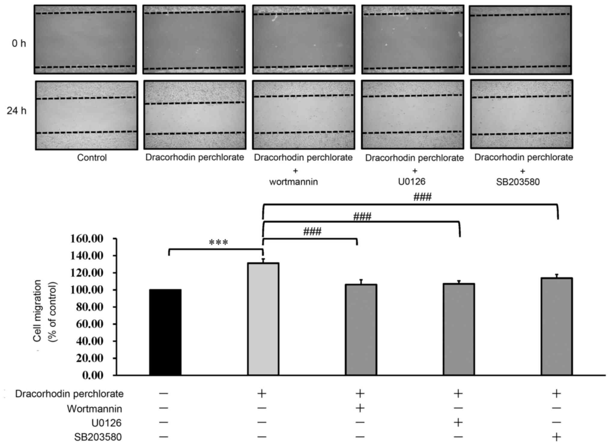
To confirm further that the mechanism of wound
healing underlying the effects of dracorhodin perchlorate,
wortmannin, U0126 and SB203580, inhibitors of AKT, ERK and p38
MAPK, respectively, were used. As shown in Fig. 6, inhibitors of AKT, ERK and p38 MAPK
could all significantly inhibit the migration of dracorhodin
perchlorate-treated keratocytes. Therefore, it could be concluded
that dracorhodin perchlorate activated wound healing by
upregulating the ERK, MAPK and AKT signaling pathways in human
HaCaT keratinocytes in vitro.
Discussion
Dracorhodin perchlorate is a new synthetic chemical
analog of dracorhodin (10,15-18).
Dracorhodin is originally the fruit extract of dragon blood, which
can be used as a TCM (2,10). Dragon blood confers a number of
bioactivities, including antibacterial, anti-diarrheal,
anti-inflammatory, anti-oxidant, antitumor, anti-ulcer, antiviral,
immune-modulatory and wound healing functions (1). Previous studies have demonstrated that
dragon blood can promote the proliferation of fibroblasts, increase
the ratio of cells in the S-phase of the cell cycle and collagen
formation, in addition to enhancing wound healing (1,11,12).
In the present study, dracorhodin perchlorate was found to
significantly promote wound healing in human HaCaT keratinocytes.
However treatment with dracorhodin perchlorate (0.5-2 µg/ml) in
HaCaT keratinocytes exerted no statistically significant effects on
keratinocyte viability. To the best of our knowledge, the present
study is the first time an in vitro scratch assay was
utilized using dynamic analysis facilitated by the IncuCyte Kinetic
Live Cell Imaging System to evaluate the effects on wound closure
in dracorhodin perchlorate-treated HaCaT cells. Furthermore,
visible gaps were observed in the monolayer of cells at the start
of the assay. In accordance with this result, the cell number did
not increase.
Previous studies have shown that activation of the
Wnt/β-catenin signaling pathway serves an important role in the
wound healing process of human keratinocytes (47). During the wound healing of
keratinocytes, cell migration and invasion are regulated by the
activation of the PI3K/AKT, ERK, p38/MAPK and Wnt/β-catenin
signaling cascades (48-51).
Additionally, activation of the ERK and p38/MAPK pathways in cell
migration is crucial for wound healing (52,53).
In accordance with these previous findings aforementioned, results
from the present study showed that dracorhodin perchlorate
treatment markedly increased the phosphorylation of AKT, ERK and
p38 and kinase activities in HaCaT cells. In addition, dracorhodin
perchlorate-treated HaCaT cells were selectively incubated with the
AKT inhibitor wortmannin, the ERK inhibitor U0126 or the p38
inhibitor SB203580 for 24 h. Cell migration was found to be
inhibited by all three inhibitors, suggesting that the ERK, p38
MAPK and AKT signaling pathways were all involved. All these
results indicated that AKT, ERK and p38 MAPK were activated by
dracorhodin perchlorate treatment during the wound healing process
in HaCaT cells in a dose-dependent manner within the concentration
range of 0 and 2 µg/ml. However, the optimal dracorhodin
perchlorate concentration for wound healing activity was found in
the present study to be 1 µg/ml rather than 2 µg/ml. Therefore,
other mechanisms may be involved in this dracorhodin
perchlorate-induced wound healing process in HaCaT cells.
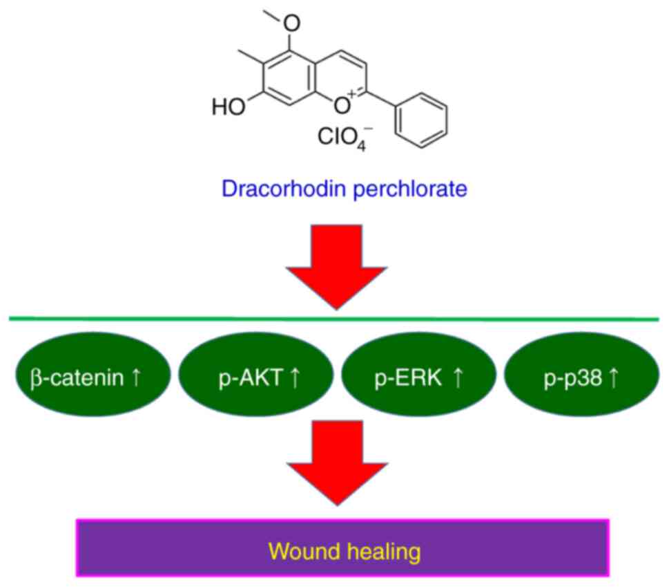
A proposed summary of the wound healing mechanism
underlying the effects mediated by dracorhodin perchlorate in HaCaT
cells is shown in Fig. 7. It was
found that dracorhodin perchlorate promoted the wound healing
activity of HaCaT keratinocytes through the β-catenin, ERK, p38
MAPK and AKT signaling pathways. These findings provide novel
insights into the mechanistic information in the wound healing
activity of dracorhodin perchlorate in human HaCaT
keratinocytes.
Acknowledgements
The authors would like to acknowledge the work of
Mr. Meng-Jou Liao and Mr. Chin-Chen Lin (Tekon Scientific
Corporation, Taipei, Taiwan) for their assistance and equipment
support on this study. The authors would also like to thank Mr.
Chang-Wei Li (AllBio Science, Inc., Taichung, Taiwan) for his
excellent technical support.
Funding
Funding: Financial support was provided by the China Medical
University Hospital (grant no. DMR-106-179) and the China Medical
University Beigang Hospital (grant no. CMUBH R106-005).
Availability of data and materials
The datasets used and/or analyzed during the current
study are available from the corresponding author on reasonable
request.
Authors' contributions
Conceptualization and study design: TJH and HPC.
Cell migration, cell viability, western blotting and protein kinase
activity: CCL, JSY and YJC. Protein kinase inhibition: CCL and YNJ.
Wound healing assay and acquisition of data: YMH, MCY and FJT.
Statistical analysis of all data and interpretation of results:
FJT, TJH and HPC. CCL and HPC confirm the authenticity of all the
raw data. All authors read and approved the final manuscript.
Ethics approval and consent to
participate
Not applicable.
Patient consent for publication
Not applicable.
Competing interests
The authors declare that they have no competing
interests.
References
|
1
|
Pona A, Cline A, Kolli SS, Taylor SL and
Feldman SR: Review of future insights of Dragon's Blood in
dermatology. Dermatol Ther. 32(e12786)2019.PubMed/NCBI View Article : Google Scholar
|
|
2
|
Sousa MM, Melo MJ, Parola AJ, Seixas de
Melo JS, Catarino F, Pina F, Cook FE, Simmonds MS and Lopes JA:
Flavylium chromophores as species markers for dragon's blood resins
from Dracaena and Daemonorops trees. J Chromatogr A.
1209:153–161. 2008.PubMed/NCBI View Article : Google Scholar
|
|
3
|
Edwards HG, de Oliveira LF and Prendergast
HD: Raman spectroscopic analysis of dragon's blood resins-basis for
distinguishing between Dracaena (Convallariaceae),
Daemonorops (Palmae) and Croton (Euphorbiaceae).
Analyst. 129:134–138. 2004.PubMed/NCBI View
Article : Google Scholar
|
|
4
|
Wu C, Cai XQ, Chang Y, Chen CH, Ho TJ, Lai
SC and Chen HP: Rapid identification of dragon blood samples from
Daemonorops draco, Dracaena cinnabari and Dracaena
cochinchinensis by MALDI-TOF mass spectrometry. Phytochem Anal.
30:720–726. 2019.PubMed/NCBI View
Article : Google Scholar
|
|
5
|
Al-Okaishi A: Exploring the historical
distribution of Dracaena cinnabari using ethnobotanical
knowledge on Socotra Island, Yemen. J Ethnobiol Ethnomed.
17(22)2021.PubMed/NCBI View Article : Google Scholar
|
|
6
|
Wu KC: Quality evaluation of herbal
medicine Ercha, Xuejie, Ruxiang and Moyao. Master Thesis, China
Medical University, 2011.
|
|
7
|
Department of Health, Hong Kong Special
Administrative Region: Hong Kong Chinese Materia Medica strandards.
The People's Republic of China: Department of Health, Hong Kong
Special Administrative Region, 362-368, 2005.
|
|
8
|
State Pharmacopoeia Commission of the PRC:
Pharmacopoeia of the People's Republic of China. 1 Beijing,
People's Medical Publishing House, p142, 2015.
|
|
9
|
Taiwan Herbal Pharmacopeia Editorial Panel
Committee. Taiwan Herbal Pharmacopeia, version 2. Taipei: Ministry
of Health and Welfare, Executive Yuan, pp102-103, 2013.
|
|
10
|
Krishnaraj P, Chang Y, Ho TJ, Lu NC, Lin
MD and Chen HP: In vivo pro-angiogenic effects of dracorhodin
perchlorate in zebrafish embryos: A novel bioactivity evaluation
platform for commercial dragon blood samples. J Food Drug Anal.
27:259–265. 2019.PubMed/NCBI View Article : Google Scholar
|
|
11
|
Namjoyan F, Kiashi F, Moosavi ZB, Saffari
F and Makhmalzadeh BS: Efficacy of Dragon's blood cream on wound
healing: A randomized, double-blind, placebo-controlled clinical
trial. J Tradit Complement Med. 6:37–40. 2016.PubMed/NCBI View Article : Google Scholar
|
|
12
|
Li D, Hui R, Hu Y, Han Y and Guo S:
Effects of extracts of Dragon's blood on fibroblast proliferation
and extracellular matrix hyaluronic acid. Zhonghua Zheng Xing Wai
Ke Za Zhi. 31:53–57. 2015.PubMed/NCBI(In Chinese).
|
|
13
|
Xin N, Yang FJ, Li Y, Li YJ, Dai RJ, Meng
WW, Chen Y and Deng YL: Dragon's blood dropping pills have
protective effects on focal cerebral ischemia Rats model.
Phytomedicine. 21:68–74. 2013.PubMed/NCBI View Article : Google Scholar
|
|
14
|
Ho TJ, Jiang SJ, Lin GH, Li TS, Yiin LM,
Yang JS, Hsieh MC, Wu CC, Lin JG and Chen HP: The in vitro and in
vivo Wound Healing Properties of the Chinese Herbal medicine
‘Jinchuang Ointment’. Evid Based Complement Alternat Med.
2016(1654056)2016.PubMed/NCBI View Article : Google Scholar
|
|
15
|
Xia M, Wang D, Wang M, Tashiro S, Onodera
S, Minami M and Ikejima T: Dracorhodin perchlorate induces
apoptosis via activation of caspases and generation of reactive
oxygen species. J Pharmacol Sci. 95:273–283. 2004.PubMed/NCBI View Article : Google Scholar
|
|
16
|
Xia MY, Wang MW, Cui Z, Tashiro SI,
Onodera S, Minami M and Ikejima T: Dracorhodin perchlorate induces
apoptosis in HL-60 cells. J Asian Nat Prod Res. 8:335–343.
2006.PubMed/NCBI View Article : Google Scholar
|
|
17
|
Wang Y, Wang Q, Liu J, Zhang L and Zhang
B: Dracorhodin perchlorate inhibit high glucose-induced connective
tissue growth factor formation in human mesangial cells. Zhongguo
Zhong Yao Za Zhi. 34:896–899. 2009.PubMed/NCBI(In Chinese).
|
|
18
|
He Y, Ju W, Hao H, Liu Q, Lv L and Zeng F:
Dracorhodin perchlorate suppresses proliferation and induces
apoptosis in human prostate cancer cell line PC-3. J Huazhong Univ
Sci Technolog Med Sci. 31(215)2011.PubMed/NCBI View Article : Google Scholar
|
|
19
|
Li F, Jiang T, Liu W, Hu Q and Yin H: The
angiogenic effect of dracorhodin perchlorate on human umbilical
vein endothelial cells and its potential mechanism of action. Mol
Med Rep. 14:1667–1672. 2016.PubMed/NCBI View Article : Google Scholar
|
|
20
|
Chen X, Luo J, Meng L, Pan T, Zhao B, Tang
ZG and Dai Y: Dracorhodin perchlorate induces the apoptosis of
glioma cells. Oncol Rep. 35:2364–2372. 2016.PubMed/NCBI View Article : Google Scholar
|
|
21
|
Zhang G, Sun M, Zhang Y, Hua P, Li X, Cui
R and Zhang X: Dracorhodin perchlorate induces G1/G0 phase arrest
and mitochondria-mediated apoptosis in SK-MES-1 human lung squamous
carcinoma cells. Oncol Lett. 10:240–246. 2015.PubMed/NCBI View Article : Google Scholar
|
|
22
|
Yu JH, Zheng GB, Liu CY, Zhang LY, Gao HM,
Zhang YH, Dai CY, Huang L, Meng XY, Zhang WY and Yu XF: Dracorhodin
perchlorate induced human breast cancer MCF-7 apoptosis through
mitochondrial pathways. Int J Med Sci. 10:1149–1156.
2013.PubMed/NCBI View Article : Google Scholar
|
|
23
|
Rasul A, Ding C, Li X, Khan M, Yi F, Ali M
and Ma T: Dracorhodin perchlorate inhibits PI3K/Akt and NF-κB
activation, up-regulates the expression of p53, and enhances
apoptosis. Apoptosis. 17:1104–1119. 2012.PubMed/NCBI View Article : Google Scholar
|
|
24
|
Xia M, Wang M, Tashiro S, Onodera S,
Minami M and Ikejima T: Dracorhodin perchlorate induces A375-S2
cell apoptosis via accumulation of p53 and activation of caspases.
Biol Pharm Bull. 28:226–232. 2005.PubMed/NCBI View Article : Google Scholar
|
|
25
|
Xia MY, Wang MW, Wang HR, Tashiro S and
Ikejima T: Mechanism of dracorhodin perchlorate-induced Hela cell
apoptosis. Yao Xue Xue Bao. 39:966–970. 2004.PubMed/NCBI(In Chinese).
|
|
26
|
Jiang X, Liu L, Qiao L, Zhang B, Wang X,
Han Y and Yu W: Dracorhodin perchlorate regulates fibroblast
proliferation to promote rat's wound healing. J Pharmacol Sci.
136:66–72. 2018.PubMed/NCBI View Article : Google Scholar
|
|
27
|
Jiang XW, Qiao L, Liu L, Zhang BQ, Wang
XW, Han YW and Yu WH: Dracorhodin Perchlorate Accelerates Cutaneous
Wound Healing in Wistar Rats. Evid Based Complement Alternat Med.
2017(8950516)2017.PubMed/NCBI View Article : Google Scholar
|
|
28
|
Boukamp P, Petrussevska RT, Breitkreutz D,
Hornung J, Markham A and Fusenig NE: Normal keratinization in a
spontaneously immortalized aneuploid human keratinocyte cell line.
J Cell Biol. 106:761–771. 1988.PubMed/NCBI View Article : Google Scholar
|
|
29
|
Xia Q, Chiang HM, Yin JJ, Chen S, Cai L,
Yu H and Fu PP: UVA photoirradiation of benzo[a]pyrene metabolites:
Induction of cytotoxicity, reactive oxygen species, and lipid
peroxidation. Toxicol Ind Health. 31:898–910. 2015.PubMed/NCBI View Article : Google Scholar
|
|
30
|
Gelles JD and Chipuk JE: Robust
high-throughput kinetic analysis of apoptosis with real-time
high-content live-cell imaging. Cell Death Dis.
7(e2493)2016.PubMed/NCBI View Article : Google Scholar
|
|
31
|
Lee MR, Lin C, Lu CC, Kuo SC, Tsao JW,
Juan YN, Chiu HY, Lee FY, Yang JS and Tsai FJ: YC-1 induces G0/G1
phase arrest and mitochondria-dependent apoptosis in
cisplatin-resistant human oral cancer CAR cells. Biomedicine
(Taipei). 7(12)2017.PubMed/NCBI View Article : Google Scholar
|
|
32
|
Yuan CH, Horng CT, Lee CF, Chiang NN, Tsai
FJ, Lu CC, Chiang JH, Hsu YM, Yang JS and Chen FA: Epigallocatechin
gallate sensitizes cisplatin-resistant oral cancer CAR cell
apoptosis and autophagy through stimulating AKT/STAT3 pathway and
suppressing multidrug resistance 1 signaling. Environ Toxicol.
32:845–855. 2017.PubMed/NCBI View Article : Google Scholar
|
|
33
|
Chang HP, Lu CC, Chiang JH, Tsai FJ, Juan
YN, Tsao JW, Chiu HY and Yang JS: Pterostilbene modulates the
suppression of multidrug resistance protein 1 and triggers
autophagic and apoptotic mechanisms in cisplatin-resistant human
oral cancer CAR cells via AKT signaling. Int J Oncol. 52:1504–1514.
2018.PubMed/NCBI View Article : Google Scholar
|
|
34
|
Lee HP, Chen PC, Wang SW, Fong YC, Tsai
CH, Tsai FJ, Chung JG, Huang CY, Yang JS, Hsu YM, et al: Plumbagin
suppresses endothelial progenitor cell-related angiogenesis in
vitro and in vivo. J Funct Foods. 52:537–544. 2019.
|
|
35
|
Chiang JH, Yang JS, Lu CC, Hour MJ, Chang
SJ, Lee TH and Chung JG: Newly synthesized quinazolinone HMJ-38
suppresses angiogenetic responses and triggers human umbilical vein
endothelial cell apoptosis through p53-modulated Fas/death receptor
signaling. Toxicol Appl Pharmacol. 269:150–162. 2013.PubMed/NCBI View Article : Google Scholar
|
|
36
|
Ma YS, Weng SW, Lin MW, Lu CC, Chiang JH,
Yang JS, Lai KC, Lin JP, Tang NY, Lin JG and Chung JG: Antitumor
effects of emodin on LS1034 human colon cancer cells in vitro and
in vivo: Roles of apoptotic cell death and LS1034 tumor xenografts
model. Food Chem Toxicol. 50:1271–1278. 2012.PubMed/NCBI View Article : Google Scholar
|
|
37
|
Tsai YF, Chen YF, Hsiao CY, Huang CW, Lu
CC, Tsai SC and Yang JS: Caspase dependent apoptotic death by
gadolinium chloride (GdCl3) via reactive oxygen species production
and MAPK signaling in rat C6 glioma cells. Oncol Rep. 41:1324–1332.
2019.PubMed/NCBI View Article : Google Scholar
|
|
38
|
Chiu YJ, Hour MJ, Jin YA, Lu CC, Tsai FJ,
Chen TL, Ma H, Juan YN and Yang JS: Disruption of IGF-1R signaling
by a novel quinazoline derivative, HMJ-30, inhibits invasiveness
and reverses epithelial-mesenchymal transition in osteosarcoma U-2
OS cells. Int J Oncol. 52:1465–1478. 2018.PubMed/NCBI View Article : Google Scholar
|
|
39
|
Lu CC, Chiang JH, Tsai FJ, Hsu YM, Juan
YN, Yang JS and Chiu HY: Metformin triggers the intrinsic apoptotic
response in human AGS gastric adenocarcinoma cells by activating
AMPK and suppressing mTOR/AKT signaling. Int J Oncol. 54:1271–1281.
2019.PubMed/NCBI View Article : Google Scholar
|
|
40
|
Han J, Lee JD, Bibbs L and Ulevitch RJ: A
MAP kinase targeted by endotoxin and hyperosmolarity in mammalian
cells. Science. 265:808–811. 1994.PubMed/NCBI View Article : Google Scholar
|
|
41
|
Zhang P, Li J, Tang X, Zhang J, Liang J
and Zeng G: Dracorhodin perchlorate induces apoptosis in primary
fibroblasts from human skin hypertrophic scars via participation of
caspase-3. Eur J Pharmacol. 728:82–92. 2014.PubMed/NCBI View Article : Google Scholar
|
|
42
|
Bowley E, O'Gorman DB and Gan BS:
Beta-catenin signaling in fibroproliferative disease. J Surg Res.
138:141–150. 2007.PubMed/NCBI View Article : Google Scholar
|
|
43
|
Lindley LE, Stojadinovic O, Pastar I and
Tomic-Canic M: Biology and biomarkers for wound healing. Plast
Reconstr Surg. 138 (Suppl 3):S18–S28. 2016.PubMed/NCBI View Article : Google Scholar
|
|
44
|
Pereira Beserra F, Xue M, Maia GLA, Leite
Rozza A, Helena Pellizzon C and Jackson CJ: Lupeol, a Pentacyclic
Triterpene, promotes migration, wound closure, and contractile
effect in vitro: Possible involvement of PI3K/Akt and p38/ERK/MAPK
Pathways. Molecules. 23(2819)2018.PubMed/NCBI View Article : Google Scholar
|
|
45
|
Patruno A, Pesce M, Grilli A, Speranza L,
Franceschelli S, De Lutiis MA, Vianale G, Costantini E, Amerio P,
Muraro R, et al: mTOR activation by PI3K/Akt and ERK signaling in
short ELF-EMF exposed human keratinocytes. PLoS One.
10(e0139644)2015.PubMed/NCBI View Article : Google Scholar
|
|
46
|
Bui NT, Ho MT, Kim YM, Lim Y and Cho M:
Flavonoids promoting HaCaT migration: II. Molecular mechanism of
4',6,7-trimethoxyisoflavone via NOX2 activation. Phytomedicine.
21:570–577. 2014.PubMed/NCBI View Article : Google Scholar
|
|
47
|
Yang HL, Tsai YC, Korivi M, Chang CT and
Hseu YC: Lucidone promotes the Cutaneous Wound Healing process via
activation of the PI3K/AKT, Wnt/β-catenin and NF-κB
signaling pathways. Biochim Biophys Acta Mol Cell Res.
1864:151–168. 2017.PubMed/NCBI View Article : Google Scholar
|
|
48
|
Lee SH, Zahoor M, Hwang JK, Min do S and
Choi KY: Valproic acid induces cutaneous wound healing in vivo and
enhances keratinocyte motility. PLoS One. 7(e48791)2012.PubMed/NCBI View Article : Google Scholar
|
|
49
|
Yeh CJ, Chen CC, Leu YL, Lin MW, Chiu MM
and Wang SH: The effects of artocarpin on wound healing: In vitro
and in vivo studies. Sci Rep. 7(15599)2017.PubMed/NCBI View Article : Google Scholar
|
|
50
|
Chen J, Chen Y, Chen Y, Yang Z, You B,
Ruan YC and Peng Y: Epidermal CFTR Suppresses MAPK/NF-κB to Promote
Cutaneous Wound Healing. Cell Physiol Biochem. 39:2262–2274.
2016.PubMed/NCBI View Article : Google Scholar
|
|
51
|
Gazel A, Nijhawan RI, Walsh R and
Blumenberg M: Transcriptional profiling defines the roles of ERK
and p38 kinases in epidermal keratinocytes. J Cell Physiol.
215:292–308. 2008.PubMed/NCBI View Article : Google Scholar
|
|
52
|
Saika S, Okada Y, Miyamoto T, Yamanaka O,
Ohnishi Y, Ooshima A, Liu CY, Weng D and Kao WW: Role of p38 MAP
kinase in regulation of cell migration and proliferation in healing
corneal epithelium. Invest Ophthalmol Vis Sci. 45:100–109.
2004.PubMed/NCBI View Article : Google Scholar
|
|
53
|
Sharma GD, He J and Bazan HE: p38 and
ERK1/2 coordinate cellular migration and proliferation in
epithelial wound healing: Evidence of cross-talk activation between
MAP kinase cascades. J Biol Chem. 278:21989–21997. 2003.PubMed/NCBI View Article : Google Scholar
|