Introduction
Cholelithiasis is a highly prevalent
gastroenterological disease and one of the leading causes of
hospital admissions worldwide (1).
In China, the incidence of cholelithiasis is 8-10%, and the
incidence rate has been gradually increasing in recent years
(2). It was reported that females
of childbearing age are approximately twice more likely than males
to develop cholelithiasis (3).
Furthermore, among the subtypes of cholelithiasis,
cholecystolithiasis accounts for ~85% of cases (4). Cholelithiasis is composed of >90%
cholesterol, and <10% is composed of black and brown pigments,
which are presented in the gallbladder or infected bile ducts
(5). In recent years, gallbladder
hypomotility was also found to promote cholesterol crystallization
and gallstone formation (6). There
is an urgent need to further clarify the possible mechanisms
underlying gallbladder hypomotility in the process of cholesterol
gallstone formation.
Interstitial cells of Cajal (ICCs) were first
identified in 1889(7) and have been
shown to possess the capacity to generate and propagate electrical
slow waves in the digestive tract (8). To date, ICCs have been detected in
various tissues including the pancreas, heart, urinary tract and
blood vessels (7). ICCs were also
found to be distributed in the gallbladder wall where they are
involved in modulating the excitability and motility of gallbladder
smooth muscle (9). It was reported
that the density and location of ICCs in the gallbladder of
patients with cholelithiasis are notably altered (10,11).
Moreover, a previous study reported that the ultrastructure of
gallbladder ICCs was not significantly altered in mouse models of
cholesterol cholelithiasis (12).
The aim of the present study was to elucidate the changes in the
density and ultrastructure of gallbladder ICCs during the
development of cholesterol cholelithiasis in more detail compared
with the existing literature.
Cholecystokinin (CCK) is a classical type of
intestinal hormone that can trigger the contraction of gallbladder
smooth muscle (13). CCK receptors
belong to the G protein-coupled receptor superfamily and have been
classified as cholecystokinin receptor type A (CCK1R) and
gastrin/cholecystokinin type B receptor (14). It has been shown that CCK1R (also
termed CCK-AR) is expressed in gallbladder ICCs and serves a key
role in mediating CCK-induced evoked gallbladder contraction
(15). However, to the best of our
knowledge, no studies have shown whether CCK1R expression in the
gallbladder, particularly in gallbladder ICCs, is altered during
the development of cholesterol cholelithiasis.
The lithogenic diet (LD), which contains a higher
proportion of cholesterol, is widely used to induce cholesterol
cholelithiasis in animals (16,17).
The aim of the present study was to detect the changes in the
density, ultrastructure and CCK1R expression levels in gallbladder
ICCs in LD-fed guinea pigs.
Materials and methods
Animal models
A total of 40 male guinea pigs (4 weeks old; weight,
250-280 g) were purchased from Charles River Laboratories, Inc. and
randomly assigned to four groups (n=10): Standard diet (SD) for 2
weeks; SD for 8 weeks; LD for 2 weeks and LD for 8 weeks. Guinea
pigs in the LD group were fed with LD (chow diet supplemented with
~1.25% cholesterol, 0.5% bile salt and 15% fat) and animals in the
SD group were fed standard chow. All guinea pigs were maintained in
cages with a 12-h light/dark cycle with free access to food and
water. All animal experiments were performed according to the
National Institutes of Health Guide for the Care and Use of
Laboratory Animals and approved by the Institutional Animal Care
and Use Committee of the First Affiliated Hospital of Nanjing
Medical University (Nanjing, China).
Measurement of total cholesterol
(T-CHOL), lipoproteins and CCK octapeptide (CCK8) levels in the
serum and bile
After feeding for 2 or 8 weeks, guinea pigs in the
SD group (weighing 340-800 g) and the LD group (weighing 420-1200
g) were anesthetized by 1.5-3.0% isoflurane, and blood samples were
obtained from the eyes of guinea pigs (~1 ml per guinea pig) and
collected in tubes containing heparin. Plasma was obtained
following centrifugation at 1,789 x g for 15 min at 4˚C. Moreover,
after resecting the gallbladder, bile was collected and diluted
with saline water (1:10), followed by centrifugation at 2,795 x g
for 10 min at 4˚C to remove the granular components. Following
which, all guinea pigs were euthanized by an overdose of
isoflurane. To degrade the bile pigment, bile was illuminated under
a daylight lamp for 12 h at 4˚C. The serum levels of T-CHOL,
high-density lipoprotein cholesterol (HDL-C) and low-density
lipoprotein cholesterol (LDL-C), as well as the T-CHOL levels in
bile were measured using an automatic biochemical analyser
(Hitachi, Ltd.) with the corresponding reagents (T-CHOL, cat. no.
SNM226; HDL-C, cat. no. SNM224; LDL-C, cat. no. SNM222; all
purchased from Beijing Biolab Science & Technology Co., Ltd.).
The concentration of CCK8 in serum was measured using an ELISA kit
(cat. no. abx150409; Abbexa Ltd.) according to the manufacturer's
protocol.
Immunofluorescence staining
After snap-freezing, the resected gallbladders were
cut into 5-µm sections using a freezing microtome. Subsequently,
all sections were fixed in 4% paraformaldehyde for 20 min at room
temperature and washed three times with PBS. To prevent
non-specific binding, sections were incubated with immunostaining
blocking buffer (Beyotime Institute of Biotechnology) for 1 h at
room temperature. Subsequently, sections were incubated overnight
at 4˚C with the following primary antibodies: Anti-C-kit (1:200;
cat. no. ab25022; Abcam) and anti-CCK1R (5 µg/ml; cat. no. ab77269;
Abcam). Following three washes with PBS, sections were incubated
with rabbit anti-rat IgM/Alexa Fluor 488 antibody (1:500; cat. no.
bs-0346R; BIOSS) and rabbit anti-goat IgG/Alexa Fluor 647 antibody
(1:500; cat. no. bs-0294R; BIOSS) for 2 h at room temperature, and
then incubated with DAPI (Beyotime Institute of Biotechnology) for
10 min at room temperature. Finally, sections were mounted using
antifade mounting medium (Beyotime Institute of Biotechnology). For
the negative control, the primary antibodies were omitted. Images
were captured on a laser scanning confocal microscope
(magnification, x200 or x400). C-kit-positive cells excluding those
with a round shape (mast cells) were considered as gallbladder
ICCs. To quantify the number of gallbladder ICCs, three
gallbladders were obtained from each group, three sections were
prepared from each gallbladder and three random fields of view
(magnification, x200) in each section were selected. The number of
gallbladder ICCs in the lamina propria was counted by two
researchers in a blinded manner.
Western blot analysis
The resected gallbladders were washed with PBS and
lysed in RIPA lysis buffer (Thermo Fisher Scientific, Inc.) to
extract total protein. Protein concentration was measured using a
BCA protein assay kit (Beyotime Institute of Biotechnology)
according to the manufacturer's protocol. Subsequently, 50 µg of
protein was loaded on an 8% SDS gel, resolved using SDS-PAGE and
subsequently transferred to a PVDF membrane (EMD Millipore). PVDF
membranes were blocked in TBS (Boster Biological Technology)
containing 5% skimmed milk powder for 2 h at room temperature.
Subsequently, membranes were washed three times with TBS
supplemented with 0.05% Tween-20 and incubated overnight at 4˚C
with one of the following antibodies: Anti-C-kit (1:1,000; cat. no.
ab256345; Abcam), anti-stem cell factor (SCF; 1:1,000; cat. no.
ab64677; Abcam), anti-Connexin (Cx) 43 (1:2,000; cat. no. ab11370;
Abcam), anti-CCK1R (1:500; cat. no. ab75153; Abcam) or anti-GAPDH
(1:1,000; cat. no. AF0006; Beyotime Institute of Biotechnology),
followed by incubation with horseradish peroxidase- conjugated goat
anti-rabbit IgG (1:5,000; cat. no. bs-0295G; BIOSS) for 2 h at room
temperature. Protein signals were detected using an enhanced
chemiluminescence detection kit (Thermo Fisher Scientific Inc.) on
the ChemiDoc XRS+ chemiluminescence imaging system (Bio-Rad
Laboratories. Inc.). Densitometric analysis was performed using
ImageJ v1.8.0 software (National Institutes of Health), and
expression of each protein was normalized to the respective GAPDH
loading control band.
Transmission electron microscopy
Gallbladder specimens were fixed in 2.5%
glutaraldehyde/0.1 M cacodylate buffer for 2 h at 4˚C and 1%
perosmic acid for 2 h at room temperature. Specimens were then
dehydrated using a series of ethanol solutions of increasing
concentration and embedded in Epon ethoxyline resin at 60˚C.
Ultrathin sections with 70-80-nm thickness were obtained using a
Leica EM UC7 ultramicrotome (Leica Microsystems, Inc.) and
subsequently placed on grids prior to staining with 1% uranyl
acetate for 20 min at room temperature. Sections were examined
under a transmission electron microscope (H-7500; Hitachi, Ltd.) at
a magnification of x10,000 at 80 kV.
Statistical analysis
Data were presented as the mean ± standard
deviation. Significant differences between the SD group and LD
group were analysed using SPSS version 22.0 (IBM Corp.) using an
unpaired two-tailed Student's t-test. P<0.05 was considered to
indicate a statistically significant difference.
Results
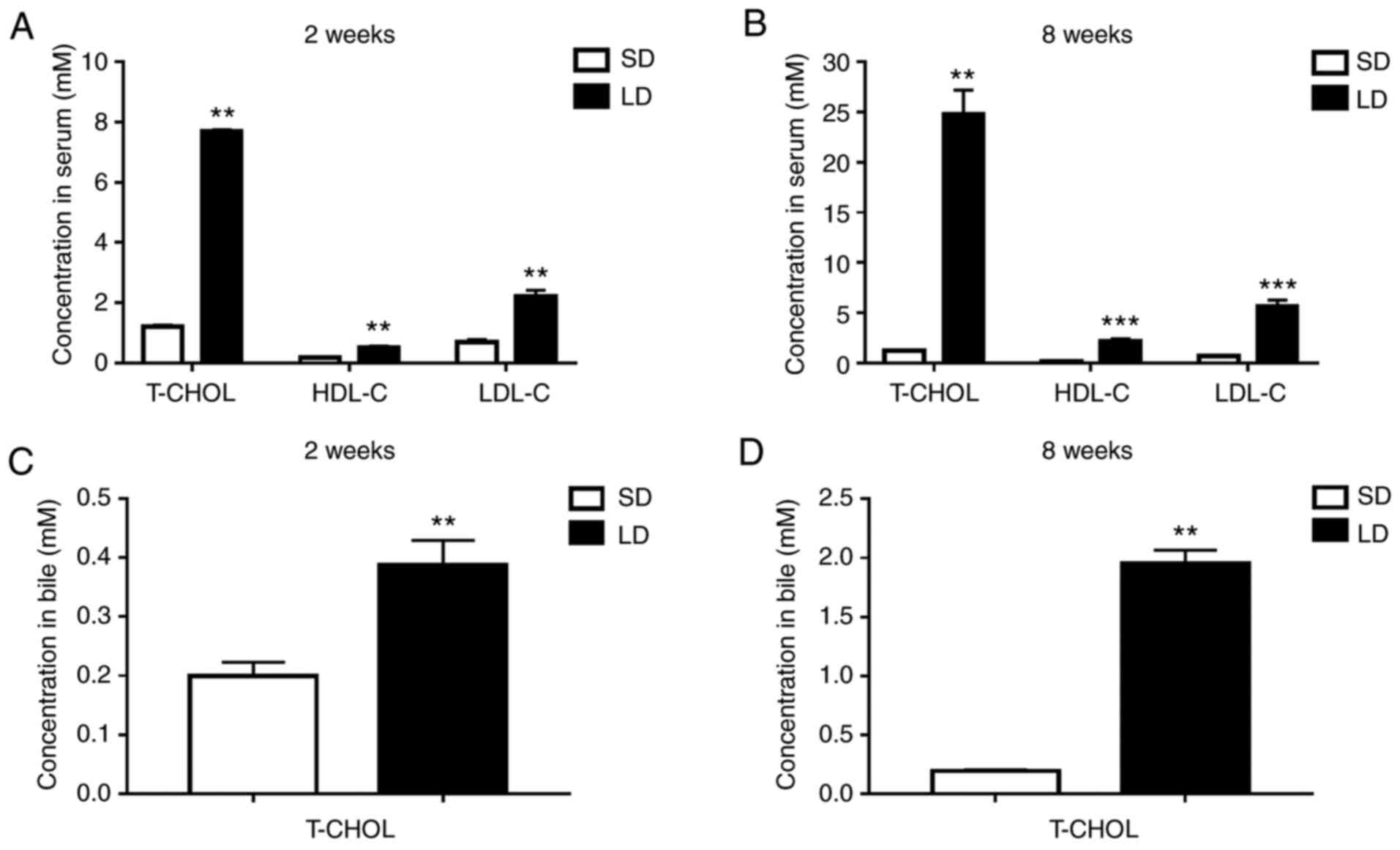
LD significantly increases T-CHOL
levels in the serum and bile and serum levels of HDL-C and
LDL-C
The serum levels of T-CHOL, HDL-C and LDL-C were
significantly increased in guinea pigs fed a LD for 2 weeks
compared with those fed a SD for the same time period (SD/2 weeks
vs. LD/2 weeks: T-CHOL, 1.210±0.051 mM vs. 7.698±0.046 mM; HDL-C,
0.179±0.035 mM vs. 0.513±0.061 mM; LDL-C, 0.693±0.093 mM vs.
2.211±0.210 mM; Fig. 1A). Moreover,
more significant increases in the serum levels of T-CHOL, HDL-C and
LDL-C were observed in guinea pigs fed a LD for 8 weeks (SD/8 weeks
vs. LD/8 weeks: T-CHOL, 1.228±0.065 mM vs. 24.729±2.461 mM; HDL-C,
0.178±0.011 mM vs. 2.175±0.272 mM; LDL-C, 0.699±0.070 mM vs.
5.631±0.651 mM; Fig. 1B).
Furthermore, the bile levels of T-CHOL in guinea pigs fed a LD for
2 weeks was significantly higher compared with those fed a SD for 2
weeks (SD/2 weeks vs. LD/2 weeks: 0.199±0.023 mM vs. 0.388±0.042
mM; Fig. 1C). Similarly, the bile
levels of T-CHOL in guinea pigs fed a LD for 8 weeks were also
significantly increased compared with those fed a SD for 8 weeks
(SD/8 weeks vs. LD/8 weeks: 0.198±0.010 mM vs. 1.955±0.112 mM;
Fig. 1D).
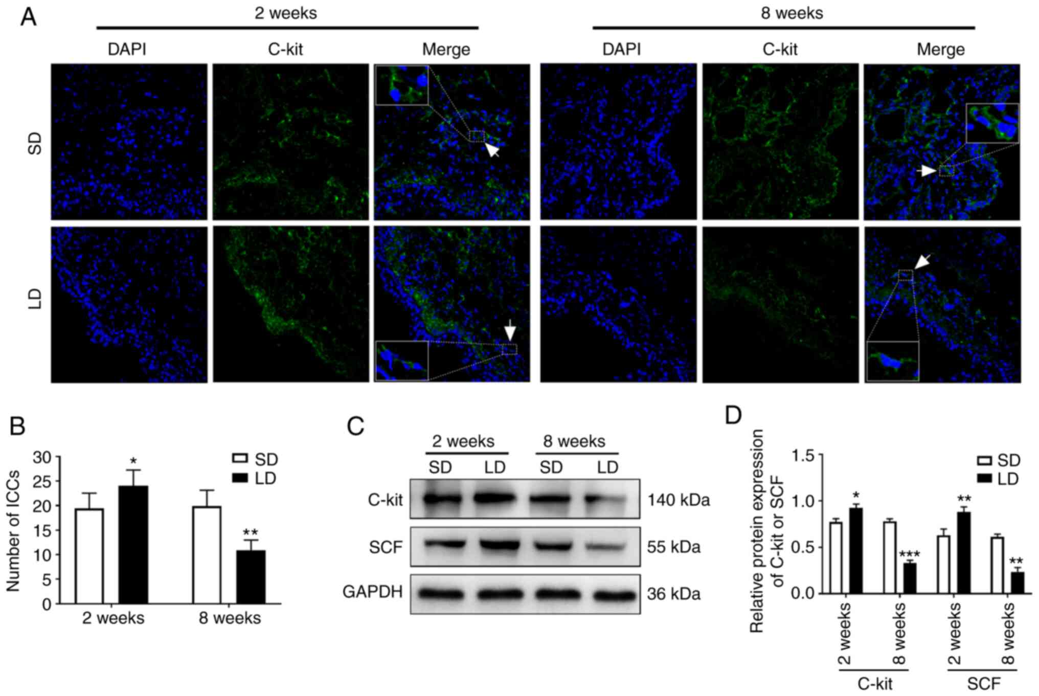
LD significantly alters the number of
ICCs and the protein expression levels of C-kit and SCF in guinea
pig gallbladders
As shown in Fig. 2A,
ICCs in guinea pig gallbladders were labelled using a primary
antibody targeting C-kit (a marker of ICCs, indicated by green
fluorescence in Fig. 2A). The mean
number of ICCs in the lamina propria of guinea pig gallbladders at
x200 magnification was significantly increased in the LD/2 weeks
group compared with the SD/2 weeks group (SD/2 weeks vs. LD/2
weeks: 19.4±3.1 vs. 24.1±3.2), and was significantly decreased in
the LD/8 weeks group compared with the SD/8 weeks group (SD/8 weeks
vs. LD/8 weeks: 19.9±3.2 vs. 10.9±2.1; Fig. 2B). Moreover, western blot analysis
demonstrated that the protein expression levels of C-kit and SCF in
guinea pig gallbladders were significantly increased in the LD/2
weeks group compared with the SD/2 weeks group (C-kit, ~20%; SCF,
~40%), and were significantly reduced in the LD/8 weeks group
compared with the SD/8 weeks group (C-kit, ~58%; SCF, ~62%;
Fig. 2C and D).
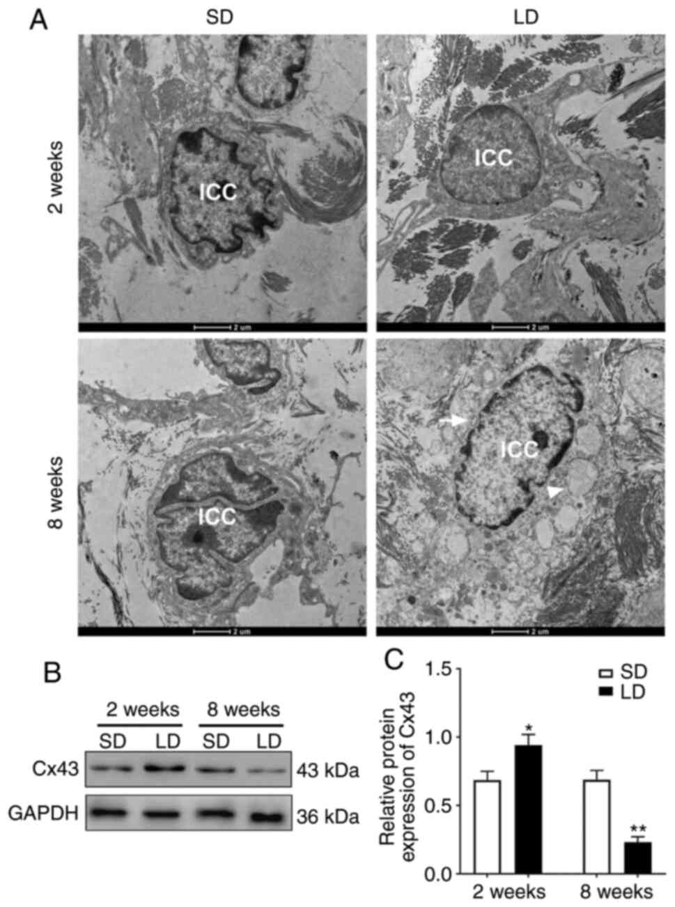
LD significantly alters the
ultrastructure of gallbladder ICCs in guinea pigs fed an LD for 8
weeks and the protein expression levels of Cx43 in guinea pig
gallbladders
Next, the ultrastructure of gallbladder ICCs in
guinea pigs fed a LD was assessed using transmission electron
microscopy. The results showed that gallbladder ICCs in the guinea
pigs fed a SD possessed normal cell nuclei, clear cell contours and
multiple branches. Compared with the SD/2 weeks group, the
ultrastructure of gallbladder ICCs in guinea pigs of the LD/2 weeks
group was not notably altered. However, in guinea pigs of the LD/8
weeks group, gallbladder ICCs exhibited more transparent cell
nuclei, blurred cell outlines, enlarged mitochondria and several
vacuoles in the cytoplasm. Notably, there were no notable branches
observed in gallbladder ICCs in the LD/8 weeks guinea pigs
(Fig. 3A). Furthermore, the protein
expression levels of Cx43 in guinea pig gallbladders were increased
by ~37% in the LD/2 weeks group compared with the SD/2 weeks group,
and was decreased by ~66% in the LD/8 weeks group compared with
that in the SD/8 weeks group (Fig.
3B and C).
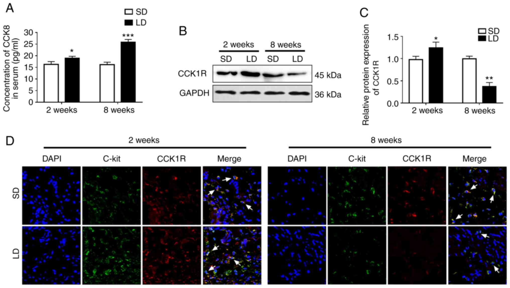
CCK1R expression in gallbladder ICCs
was significantly altered in guinea pigs fed a LD
The effects of LD on CCK1R expression in guinea pig
gallbladders were determined. The serum levels of CCK8 in guinea
pigs of the LD/2 weeks group were significantly increased compared
with the SD/2 weeks group (SD/2 weeks vs. LD/2 weeks: 16.264±1.216
pg/ml vs. 18.934±0.822 pg/ml). Compared with the SD/8 weeks group,
the increase in the serum levels of CCK8 in guinea pigs fed a LD
for 8 weeks were more significant (SD/8 weeks vs. LD/8 weeks:
16.168±1.065 pg/ml vs. 25.812±1.174 pg/ml; Fig. 4A). Moreover, compared with the SD
groups, the total protein expression levels of CCK1R in guinea pig
gallbladders were significantly increased by ~27% in the LD/2 weeks
group, and was significantly decreased by ~63% in the LD/8 weeks
group (Fig. 4B and C). Finally, using double
immunofluorescence staining, CCK1R (red fluorescence) was confirmed
to be expressed in C-kit-positive (green fluorescence) ICCs in
guinea pig gallbladders. Additionally, the red fluorescence
intensity (indicative of CCK1R expression levels) in the
gallbladder ICCs of guinea pigs in the LD/2 weeks group was
moderately higher compared with the SD/2 weeks group; and was
markedly lower in the LD/8 weeks group compared with the SD/8 weeks
group (Fig. 4D).
Discussion
It was previously shown that hypercholesterolemia is
an important risk factor of cholelithiasis (18,19).
Hypercholesterolemia can increase biliary cholesterol
concentrations and consequently lead to cholesterol saturation in
bile, which is also considered a pathogenic factor of
cholelithiasis (20). In the
present study, hypercholesterolemia was successfully induced, and
elevated levels of T-CHOL in the bile from guinea pigs were
observed when fed a LD for 2 or 8 weeks. Several studies have shown
that a LD facilitates the formation of cholesterol gallstones in
guinea pigs (21,22). Hence, in the present study, LD-fed
guinea pigs were used to investigate the possible mechanisms
underlying gallbladder hypomotility in the development of
cholesterol cholelithiasis.
ICCs are pacemaker cells that generate electrical
slow waves in the gastrointestinal tract (23). ICCs have also been shown to serve a
key regulatory role in controlling smooth muscle contractions in
the urinary tract (24). In the
gallbladder, ICCs are involved in the generation and propagation of
spontaneous rhythmicity and the consequent tissue motility
(9). A previous study found that
gallbladder ICCs were redistributed from the muscle layer to the
lamina propria during cholelithiasis (11). According to this result, in the
present study, the changes in density of gallbladder ICCs located
in the lamina propria were investigated. To the best of our
knowledge, the present study is the first to show that a LD for 2
weeks significantly increases the density of gallbladder ICCs in
guinea pigs. In addition, the density of gallbladder ICCs was
significantly decreased in guinea pigs after being fed a LD for 8
weeks. This result was consistent with previous studies where the
density of ICCs was found to be significantly reduced in patients
with cholelithiasis (10) and in
guinea pigs fed a high cholesterol diet for 4 or 8 weeks (12,25).
Based on the aforementioned results, it was hypothesized that
guinea pigs may enhance their gallbladder emptying capacity via a
compensatory increase in the density of ICCs during the early
stages of high cholesterol stimulation, and their gallbladder
motility is considerably impaired at the later stages of
cholesterol cholelithiasis development due to decreases in the
density of ICCs.
Subsequently, the mechanisms underlying the decrease
in the density of gallbladder ICCs were investigated. The SCF/C-kit
pathway is well established as a key regulator of the
differentiation and proliferation of ICCs (26). The present study detected for the
first time that the protein expression levels of SCF and C-kit in
guinea pig gallbladders were significantly increased after feeding
a LD for 2 weeks, indicating increased activation of the SCF/C-kit
pathway. Furthermore, consistent with previous studies in which the
mRNA and protein expression levels of SCF and C-kit were found to
be significantly attenuated in the gallbladders of animal models
fed a high cholesterol diet for 8 weeks (12,27),
the present study also demonstrated that activity of the SCF/C-kit
pathway was significantly suppressed after being fed a LD for 8
weeks. Taken together, it was hypothesized that differential
activity of the SCF/C-kit pathway is observed at various stages of
cholesterol cholelithiasis development, and this may lead to the
corresponding alterations in the density of gallbladder ICCs at
each stage.
A previous study reported that feeding a high
cholesterol diet for 8 weeks did not significantly result in
ultrastructural changes in the gallbladder ICCs in mice (12). However, in the present study,
although the ultrastructure of gallbladder ICCs in guinea pigs was
not notably altered after feeding a LD for 2 weeks, they were
markedly altered after 8 weeks. It was hypothesized that the
differences in outcomes may be attributed to the different
susceptibilities of various species to high cholesterol diets. It
was reported that gallbladder ICCs can form a network with each
other and close contact with smooth muscle cells and nerve endings
through their multiple branches (9). Moreover, it was also shown that ICCs
can propagate their own rhythmic excitation or neural signals to
smooth muscle cells via gap junctions (28). In the present study, an important
change that was observed in the ultrastructure of gallbladder ICCs
at 8 weeks was the loss of their branched morphology. Additionally,
the protein expression levels of the gap junction protein Cx43 in
guinea pig gallbladders was significantly decreased after 8 weeks.
Based on the aforementioned results, it was hypothesized that the
capacity of ICCs for controlling gallbladder contraction via
propagating rhythmic signals to smooth muscle cells may be impaired
at the later stages of cholesterol cholelithiasis progression.
As a classical hormone, CCK can induce gallbladder
contractions via direct action on CCK1R in gallbladder ICCs
(15). In patients with cholesterol
gallstones, induction of gallbladder contraction via exogenous CCK
is impaired, and this phenomenon may be the result of reduced CCK1R
expression and/or CCK binding ability to CCK1R in the gallbladder
(29). Furthermore, a previous
study reported that total CCK1R protein expression in gallbladders
of mice fed with LD for 1 month was significantly reduced compared
with the control mice (30).
Consistently, the results of the present study also showed that
CCK1R protein expression in the gallbladder, particularly in
gallbladder ICCs, was significantly decreased after feeding with LD
for 8 weeks. Although the serum CCK8 levels in guinea pigs were
considerably increased after 8 weeks in the present study, it was
hypothesized that gallbladder contraction was still damaged at this
stage, primarily due to reduced CCK1R expression. In addition, the
results of the present study also demonstrated for the first time
that CCK1R protein expression in the gallbladder, particularly in
the gallbladder ICCs was increased after feeding with LD for 2
weeks. Considering that the serum CCK8 levels in guinea pigs was
also moderately increased after 2 weeks, it was hypothesized that
the synergistic effect of increased serum CCK8 and CCK1R expression
will enhance the emptying capacity of the gallbladder during the
early stages of high cholesterol stimulation.
The present study preliminarily investigated changes
in the density, ultrastructure and CCK1R expression of gallbladder
ICCs during the development of cholesterol cholelithiasis. A
limitation of the present study is that gallbladder contractility
in guinea pigs was not assessed directly using functional
experiments. Additionally, further studies are required to clarify
the possible mechanisms underlying the changes in CCK1R expression,
such as elucidation of the regulation of CCK1R expression by
certain microRNAs, during the development of cholesterol
cholelithiasis.
In conclusion, the present study demonstrated that
the density, ultrastructure and CCK1R expression levels of
gallbladder ICCs were differentially altered at various stages of
cholesterol cholelithiasis progression. These results indicated
that gallbladder ICCs may serve as a potential therapeutic target
for cholesterol cholelithiasis.
Acknowledgements
Not applicable.
Funding
Funding: This study was funded by the Surface Project of Natural
Science Foundation of Jiangsu Province (grant no. BK 20181491).
Availability of data and materials
The datasets used and/or analysed during the current
study are available from the corresponding author on reasonable
request.
Authors' contributions
LH and CD performed the experiments and analysed the
data. LH wrote the manuscript. XS designed the experiments and
revised the manuscript. LH, CD and XS confirm the authenticity of
all the raw data. All authors read and approved the final
manuscript.
Ethics approval and consent to
participate
All animal experiments were performed according to
the National Institutes of Health Guide for the Care and Use of
Laboratory Animals, and approved by the Institutional Animal Care
and Use Committee of the First Affiliated Hospital of Nanjing
Medical University (Nanjing, China).
Patient consent for publication
Not applicable.
Competing interests
The authors declare that they have no competing
interests.
References
|
1
|
Munoz LE, Boeltz S, Bilyy R, Schauer C,
Mahajan A, Widulin N, Gruneboom A, Herrmann I, Boada E, Rauh M, et
al: Neutrophil extracellular traps initiate gallstone formation.
Immunity. 51:443–450.e4. 2019.PubMed/NCBI View Article : Google Scholar
|
|
2
|
Cai JS, Qiang S and Bao-Bing Y: Advances
of recurrent risk factors and management of choledocholithiasis.
Scand J Gastroenterol. 52:34–43. 2017.PubMed/NCBI View Article : Google Scholar
|
|
3
|
Figueiredo JC, Haiman C, Porcel J, Buxbaum
J, Stram D, Tambe N, Cozen W, Wilkens L, Le Marchand L and Setiawan
VW: Sex and ethnic/racial-specific risk factors for gallbladder
disease. BMC Gastroenterol. 17(153)2017.PubMed/NCBI View Article : Google Scholar
|
|
4
|
Chen CH, Lin CL and Kao CH: Association
between Hashimoto's thyroiditis and cholelithiasis: A retrospective
cohort study in Taiwan. BMJ Open. 8(e020798)2018.PubMed/NCBI View Article : Google Scholar
|
|
5
|
Lammert F, Gurusamy K, Ko CW, Miquel JF,
Mendez-Sanchez N, Portincasa P, van Erpecum KJ, van Laarhoven CJ
and Wang DQ: Gallstones. Nat Rev Dis Primers.
2(16024)2016.PubMed/NCBI View Article : Google Scholar
|
|
6
|
Wang HH, Portincasa P, Liu M, Tso P,
Samuelson LC and Wang DQ: Effect of gallbladder hypomotility on
cholesterol crystallization and growth in CCK-deficient mice.
Biochim Biophys Acta. 1801:138–146. 2010.PubMed/NCBI View Article : Google Scholar
|
|
7
|
Pasternak A, Szura M, Gil K and Matyja A:
Interstitial cells of Cajal-systematic review. Folia Morphol.
75:281–286. 2016.PubMed/NCBI View Article : Google Scholar
|
|
8
|
Radenkovic G, Radenkovic D and Velickov A:
Development of interstitial cells of Cajal in the human digestive
tract as the result of reciprocal induction of mesenchymal and
neural crest cells. J Cell Mol Med. 22:778–785. 2018.PubMed/NCBI View Article : Google Scholar
|
|
9
|
Chen L and Yu B: Telocytes and
interstitial cells of Cajal in the biliary system. J Cell Mol Med.
22:3323–3329. 2018.PubMed/NCBI View Article : Google Scholar
|
|
10
|
Pasternak A, Gil K, Matyja A, Gajda M,
Sztefko K, Walocha JA, Kulig J and Thor P: Loss of gallbladder
interstitial Cajal-like cells in patients with cholelithiasis.
Neurogastroenterol Motil. 25:e17–e24. 2013.PubMed/NCBI View Article : Google Scholar
|
|
11
|
Spetana J, Lipinski A and Jelen M:
Comparison of the ICC location in the gallbladder wall in patients
with cholelithiasis and patients with non-calculous changes. Pol J
Pathol. 70:205–209. 2019.PubMed/NCBI View Article : Google Scholar
|
|
12
|
Fan Y, Wu S, Fu B, Weng C and Wang X: The
role of interstitial Cajal-like cells in the formation of
cholesterol stones in guinea pig gallbladder. Hepatol Int.
9:612–620. 2015.PubMed/NCBI View Article : Google Scholar
|
|
13
|
Konno K, Takahashi-Iwanaga H, Uchigashima
M, Miyasaka K, Funakoshi A, Watanabe M and Iwanaga T: Cellular and
subcellular localization of cholecystokinin (CCK)-1 receptors in
the pancreas, gallbladder, and stomach of mice. Histochem Cell
Biol. 143:301–312. 2015.PubMed/NCBI View Article : Google Scholar
|
|
14
|
Rathore RM, Angotzi AR, Jordal AE and
Ronnestad I: Cholecystokinin receptors in Atlantic salmon:
Molecular cloning, gene expression, and structural basis. Physiol
Rep. 1(e00069)2013.PubMed/NCBI View
Article : Google Scholar
|
|
15
|
Xu D, Yu BP, Luo HS and Chen LD: Control
of gallbladder contractions by cholecystokinin through
cholecystokinin-A receptors on gallbladder interstitial cells of
Cajal. World J Gastroenterol. 14:2882–2887. 2008.PubMed/NCBI View Article : Google Scholar
|
|
16
|
Tharp KM, Khalifeh-Soltani A, Park HM,
Yurek DA, Falcon A, Wong L, Feng R, Atabai K and Stahl A:
Prevention of gallbladder hypomotility via FATP2 inhibition
protects from lithogenic diet-induced cholelithiasis. Am J Physiol
Gastrointest Liver Physiol. 310:G855–G864. 2016.PubMed/NCBI View Article : Google Scholar
|
|
17
|
Liu M, Liu C, Chen H, Huang X, Zeng X,
Zhou J and Mi S: Prevention of cholesterol gallstone disease by
schaftoside in lithogenic diet-induced C57BL/6 mouse model. Eur J
Pharmacol. 815:1–9. 2017.PubMed/NCBI View Article : Google Scholar
|
|
18
|
Zheng Y, Xu M, Heianza Y, Ma W, Wang T,
Sun D, Albert CM, Hu FB, Rexrode KM, Manson JE and Qi L: Gallstone
disease and increased risk of mortality: Two large prospective
studies in US men and women. J Gastroenterol Hepatol. 33:1925–1931.
2018.PubMed/NCBI View Article : Google Scholar
|
|
19
|
Lee S, Kweon OK and Kim WH: Associations
between serum leptin levels, hyperlipidemia, and cholelithiasis in
dogs. PLoS One. 12(e0187315)2017.PubMed/NCBI View Article : Google Scholar
|
|
20
|
Di Ciaula A, Garruti G, Fruhbeck G, De
Angelis M, de Bari O, Wang DQ, Lammert F and Portincasa P: The role
of diet in the pathogenesis of cholesterol gallstones. Curr Med
Chem. 26:3620–3638. 2019.PubMed/NCBI View Article : Google Scholar
|
|
21
|
Wu X, Liang X, Du Y, Zhang Y, Yang M, Gong
W, Liu B, Dong J, Zhang N and Zhang H: Prevention of gallstones by
Lidan Granule: Insight into underlying mechanisms using a guinea
pig model. Biomed Rep. 5:50–56. 2016.PubMed/NCBI View Article : Google Scholar
|
|
22
|
Rong ZH, Chen HY, Wang XX, Wang ZY, Xian
GZ, Ma BZ, Qin CK and Zhang ZH: Effects of sphincter of Oddi
motility on the formation of cholesterol gallstones. World J
Gastroenterol. 22:5540–5547. 2016.PubMed/NCBI View Article : Google Scholar
|
|
23
|
Zheng H, Drumm BT, Zhu MH, Xie Y,
O'Driscoll KE, Baker SA, Perrino BA, Koh SD and Sanders KM:
Na+/Ca2+ exchange and pacemaker activity of
interstitial cells of Cajal. Front Physiol. 11(230)2020.PubMed/NCBI View Article : Google Scholar
|
|
24
|
Sanders KM, Ward SM and Koh SD:
Interstitial cells: Regulators of smooth muscle function. Physiol
Rev. 94:859–907. 2014.PubMed/NCBI View Article : Google Scholar
|
|
25
|
Huang ZP, Qiu H and Yu BP: Distribution
changes of interstitial cells of Cajal during cholesterol gallstone
formation in guinea pigs fed a high cholesterol diet. Int J Clin
Exp Pathol. 11:1653–1659. 2018.PubMed/NCBI
|
|
26
|
Chai Y, Huang Y, Tang H, Tu X, He J, Wang
T, Zhang Q, Xiong F, Li D and Qiu Z: Role of stem cell growth
factor/c-Kit in the pathogenesis of irritable bowel syndrome. Exp
Ther Med. 13:1187–1193. 2017.PubMed/NCBI View Article : Google Scholar
|
|
27
|
Fan Y, Wu S, Fu B, Yan X, Wang X and Zhang
W: Decreased expression of stem cell factor mRNA and protein in the
gallbladders of guinea pigs fed on high cholesterol diet. Int J
Clin Exp Med. 8:6379–6383. 2015.PubMed/NCBI
|
|
28
|
Wang L, Liang Y, Chen Q, Ahmed N, Wang F,
Hu B and Yang P: Identification and distribution of the
interstitial cells of Cajal in the abomasum of goats. Cell
Transplant. 27:335–344. 2018.PubMed/NCBI View Article : Google Scholar
|
|
29
|
Wang HH, Portincasa P and Wang DQ: Update
on the molecular mechanisms underlying the effect of
cholecystokinin and cholecystokinin-1 receptor on the formation of
cholesterol gallstones. Curr Med Chem. 26:3407–3423.
2019.PubMed/NCBI View Article : Google Scholar
|
|
30
|
Xu GQ, Xu CF, Chen HT, Liu S, Teng XD, Xu
GY and Yu CH: Association of caveolin-3 and cholecystokinin A
receptor with cholesterol gallstone disease in mice. World J
Gastroenterol. 20:9513–9518. 2014.PubMed/NCBI View Article : Google Scholar
|